. 1997 Nov 25;94(24):12938-43.
doi: 10.1073/pnas.94.24.12938.

Smad8 mediates the signaling of the ALK-2 [corrected] receptor serine kinase

Affiliations

Smad8 mediates the signaling of the ALK-2 [corrected] receptor serine kinase

Y Chen et al. Proc Natl Acad Sci U S A. .

Erratum in

  • Proc Natl Acad Sci U S A 1998 Feb 17;95(4):1968

Abstract

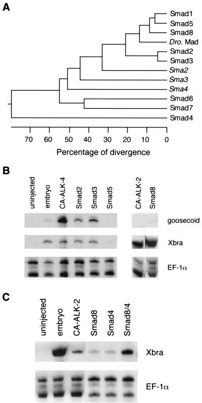
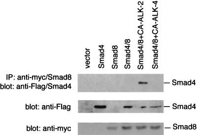
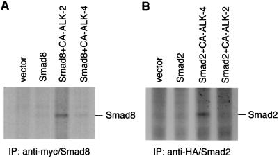
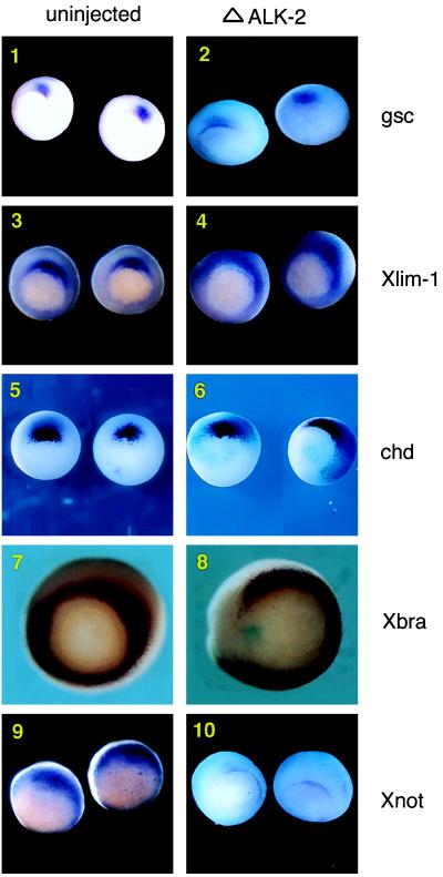
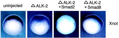
Smad proteins are critical intracellular mediators of signaling by growth and differentiation factors of the transforming growth factor beta superfamily. We have isolated a member of the Smad family, Smad8, from a rat brain cDNA library and biochemically and functionally characterized its ability to transduce signals from serine kinase receptors. In Xenopus embryo, Smad8 is able to transcriptionally activate a subset of mesoderm target genes similar to those induced by the receptor serine kinase, activin receptor-like kinase (ALK)-2. Smad8 can be specifically phosphorylated by a constitutively active ALK-2 but not the related receptor serine kinase, ALK-4. In response to signaling from ALK-2, Smad8 associates with a common regulatory molecule, Smad4, and this association leads to a synergistic effect on gene transcription. Furthermore, Smad8 is able to rescue the expression of mesoderm genes blocked by truncated ALK-2 in the embryo. These results indicate that Smad8 can function as a downstream signaling mediator of ALK-2.


Figures

Figure 1
Figure 1
Smad8 activates Xbra in animal pole explants. (A) Phylogenetic tree of the Smad proteins. The numbers below the tree indicate the percent divergence among the amino acid sequences. The sequences used here include rat Smad1, Smad3, and Smad8, mouse Smad2 and Smad5, human Smad4, Smad6, and Smad7, Drosophila Mad and three Caenorhabditis elegans Mads, sma-2, sma-3 and sma-4. (B) Smad8 induces Xbra in animal pole explants. Xenopus embryos at two-cell stage were injected with 1–2 ng RNA of Smad2, Smad3, Smad5, Smad8 and 10–20 pg of CA ALK-2 or CA ALK-4. The embryos were dissected at stage 9, and the explants were cultured until stage 12. The animal pole explants were analyzed by RNase protection with probes of Xbra, gsc, and EF-1α. The ubiquitously expressed EF-1α was used as a loading control to monitor the total RNA levels among samples. Embryos at stage 12 and the animal pole explants from the uninjected embryos were used as controls. (C) Smad8 synergizes with Smad4 in Xbra activation. Xenopus embryos at two-cell stage were injected with 50 pg of CA ALK2 RNA or 0.2 ng RNA of Smad8 alone, Smad4 alone, or Smad8 together with Smad4. The embryos were dissected at stage 9 and the explants were cultured until stage 12. The animal pole explants were analyzed by RNase protection with probes of Xbra and EF-1α.
Figure 2
Figure 2
CA ALK-2 induces association of Smad8 with Smad4. CHO cells were transiently transfected with control vector (pcDNA3), a Flag-tagged Smad4 (Flag/Smad4) alone, a myc-tagged Smad8 (myc/Smad8) alone, myc/Smad8 and Flag/Smad4, or myc/Smad8 and Flag/Smad4 with CA ALK-2 or CA ALK-4. The transfected cells were lysed, and myc/Smad8 was purified by immunoprecipitation with an anti-myc antibody. The immunoprecipitate was separated on SDS/PAGE, and Flag/Smad4 that complexed with Smad8 was detected by an anti-Flag antibody. The crude cell lysate (1/30th equivalent of the amount used in immunoprecipitation) from above transfection also were separated on SDS/PAGE and detected with anti-Flag or anti-myc antibody to determine the protein expression level of Flag/Smad4 and myc/Smad8.
Figure 3
Figure 3
CA ALK-2 specifically phosphorylates Smad8. (A) CA ALK-2 but not CA ALK-4 phosphorylates Smad8. CHO cells were transiently transfected with control vector (pcDNA3), myc/Smad8, or myc/Smad8 with either CA ALK-2 or CA ALK-4. The transfected cells were labeled with [32P]phosphorus for 3 h before harvesting. The cell lysate was immunoprecipitated with an anti-myc antibody, separated on SDS/PAGE, and detected by autoradiography. The migration of myc/Smad8 on the gel was determined by anti-myc Western blot analysis with the lysate from 32P-unlabeled cells that expressed myc/Smad8 (data not shown). (B) CA ALK-4 but not CA ALK-2 phosphorylates Smad2. 293 cells were transiently transfected with control vector (pcDNA3), a HA-tagged Smad2 (HA/Smad2), and HA/Smad2 with either CA ALK-4 or CA ALK-2. The transfected cells were labeled with [32P]phosphorus, and the cell lysate was immunoprecipitated with an anti-HA antibody followed by separation on SDS/PAGE. The phosphorylation products were detected by autoradiography.
Figure 4
Figure 4
Truncated ALK-2 blocks a subset of mesoderm genes in Xenopus embryo. Albino embryos were injected with ΔALK-2 RNA and a lacZ transcript in the marginal zone at the four-cell stage without dorsal/ventral bias. The lacZ transcript was used in all injections to indicate the distribution of the injected truncated receptor. Embryos were cultured until the 10.5 stage and used in whole-mount in situ hybridization with probes of gsc, Xlim-1, chd, Xbra, and Xnot. Representative embryos are shown here (vegetal view with dorsal side up). (1) gsc expression in uninjected embryos. (2) gsc expression in ΔALK-2 injected embryos. (3) Xlim-1 expression in uninjected embryos. (4) Xlim-1 expression in ΔALK-2 injected embryos. (5) chd expression in uninjected embryos. (6) chd expression in ΔALK-2 injected embryos. (7) Xbra expression in uninjected embryos. (8) Xbra expression in ΔALK-2 injected embryos. (9) Xnot expression in uninjected embryos. (10) Xnot expression in ΔALK-2 injected embryos.
Figure 5
Figure 5
Smad8 rescues the block of Xnot by truncated ALK-2. Albino embryos at the four-cell stage were injected in the marginal zone with in vitro transcribed RNA of ΔALK-2 alone or ΔALK-2 with either Smad2 or Smad8. The embryos were cultured until stage 10.5 and used in whole-mount in situ hybridization with a Xnot probe. The lacZ transcripts was used to indicate the distribution of the injected RNA. Representative embryos are shown (vegetal view with dorsal side up).

References

    1. Mathews L S, Vale W W. Cell. 1991;65:973–982. - PubMed
    1. Lin H Y, Wang X F, Ng-Eaton E, Weinberg R A, Lodish H F. Cell. 1992;68:775–785. - PubMed
    1. Attisano L, Wrana J L, Lopez C F, Massague J. Biochim Biophys Acta. 1994;1222:71–80. - PubMed
    1. Wrana J L, Attisano L, Carcamo J, Zentella A, Doody J, Laiho M, Wang X F, Massague J. Genes Dev. 1996;10:1880–1889. - PubMed
    1. Wieser R, Wrana J L, Massague J. EMBO J. 1995;4:2199–2208. - PMC - PubMed

Publication types

Associated data