. 1997 Nov 25;94(24):13087-92.
doi: 10.1073/pnas.94.24.13087.

Base excision repair deficient mice lacking the Aag alkyladenine DNA glycosylase

Affiliations

Base excision repair deficient mice lacking the Aag alkyladenine DNA glycosylase

B P Engelward et al. Proc Natl Acad Sci U S A. .

Abstract

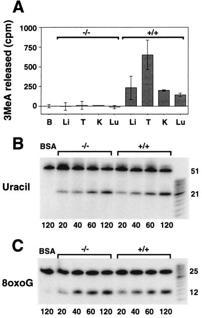
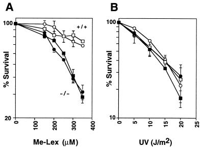
3-methyladenine (3MeA) DNA glycosylases remove 3MeAs from alkylated DNA to initiate the base excision repair pathway. Here we report the generation of mice deficient in the 3MeA DNA glycosylase encoded by the Aag (Mpg) gene. Alkyladenine DNA glycosylase turns out to be the major DNA glycosylase not only for the cytotoxic 3MeA DNA lesion, but also for the mutagenic 1,N6-ethenoadenine (epsilonA) and hypoxanthine lesions. Aag appears to be the only 3MeA and hypoxanthine DNA glycosylase in liver, testes, kidney, and lung, and the only epsilonA DNA glycosylase in liver, testes, and kidney; another epsilonA DNA glycosylase may be expressed in lung. Although alkyladenine DNA glycosylase has the capacity to remove 8-oxoguanine DNA lesions, it does not appear to be the major glycosylase for 8-oxoguanine repair. Fibroblasts derived from Aag -/- mice are alkylation sensitive, indicating that Aag -/- mice may be similarly sensitive.


Figures

Figure 1
Figure 1
Genotyping Aag +/+, +/−, and −/− mice. (A) Schematic representation of a portion of the wild-type and targeted Aag alleles indicating the location of three primers and HindIII (H) sites. Note that Aag exon II is disrupted by insertion of a neo expression cassette to create an Aag null allele. Primers PI and PIII yield a 164-bp fragment in the presence of the wild-type allele. In the presence of the targeted allele, primers PII and PIII yield a 523-bp fragment. HindIII sites are loosely indicated to show that disruption of Aag by neo insertion increases the size of the HindIII fragment (details have been presented elsewhere) (12). (B) (Upper) PCR genotyping of MEFs (lanes 1–5). PCR genotyping of DNA from spleens of mice used in subsequent biochemical analysis (lanes 6–7). (Lower) Southern blot analysis of the DNA used for PCR analysis above. HindIII-digested DNA was probed with a fragment of the Aag gene that lies outside of the targeting construct (12). The Aag null allele (8.4 kb) and the wild-type Aag allele (6.3 kb) are indicated.
Figure 2
Figure 2
3MeA, uracil, and 8-oxoG DNA glycosylase activity in mouse tissue extracts. (A) 1,000 μg (BSA, B; liver, Li; and kidney, K), 500 μg (lung, Lu) or 250 μg (testes, T) of protein extracts from Aag +/+ or Aag −/− tissues were incubated in triplicate reactions for 5 hr at 37°C with [3H]methyl-N-nitrosourea-treated calf thymus DNA. 3MeA was separated from other bases by descending paper chromatography. The cpm associated with 3MeA per mg protein extracts are indicated for the average of two or three independent mice. Error bars represent standard deviations. (B) Time-dependent release of site-specific uracil from 5′ 32P-labeled double-stranded oligonucleotides by Aag −/− and Aag +/+ liver extracts. BSA (100 μg) was incubated with the oligo substrate (first lane). Remaining lanes each contain 100 μg of either Aag −/− or Aag +/+ liver extract. After incubation at 37°C for the times indicated in min beneath each lane, oligonucleotides were chemically cleaved at abasic sites and analyzed by denaturing PAGE. DNA glycosylase activity is indicated by the appearance of a 21-mer. (C) Time-dependent release of site-specific 8oxoG from 5′ 32P-labeled double-stranded oligonucleotides by Aag −/− and Aag +/+ testes extracts. BSA (75 μg) were incubated with the 8oxoG containing oligonucleotides (first lane). Remaining lanes each contain 75 μg of either Aag −/− or Aag +/+ testes extracts. Samples were analyzed as described in B. DNA glycosylase activity is indicated by the appearance of a 12-mer.
Figure 3
Figure 3
Toxicity assay of Aag +/+ or Aag −/− MEFs treated with increasing doses of Me-Lex (A) or UV (B). Percent metabolically active treated cells is expressed relative to untreated control cells. Cells from two Aag −/− embryos (closed symbols) and two Aag +/+ embryos (open symbols) were analyzed.
Figure 4
Figure 4
Aag is the major Hx and ɛA DNA glycosylase in mouse liver, testes, kidney, and lung. (A) Release of ɛA and Hx by partially purified poly-histidine-Aag fusion protein (see Materials and Methods). 5′ 32P-labeled double-stranded oligonucleotides containing site-specific ɛA or Hx were incubated with either polyhistidine-tagged Aag (lanes 2 and 4) or with negative control extracts from E. coli not expressing the Aag cDNA protein (see Materials and Methods) (lanes 1 and 3). After 1 hr incubation at 37°C to allow release of aberrant bases by Aag glycosylase, oligonucleotides were chemically cleaved at the abasic sites. Fragments were separated by 20% denaturing PAGE. Release of Hx or ɛA is indicated by the appearance of either a 14-mer or a 12-mer, respectively. (B) Liver: time-dependent release of Hx (Upper) and ɛA (Lower) for liver extract proteins from Aag +/+ mice (136 μg) and Aag −/− mice (142 μg). The BSA control or extract proteins were incubated at 37°C for the indicated times, chemically cleaved, and analyzed as described above. (C) Testes: 100 μg of extract proteins, prepared from the testes of two Aag −/− and two Aag +/+ mice were incubated for 5 hr with Hx and ɛA oligonucleotide substrates and analyzed as described above. (D) Kidney: 131 μg of extract proteins, prepared from the kidneys of two Aag −/− and two Aag +/+ mice, were incubated for 6 hr with Hx and ɛA containing oligonucleotide substrates and analyzed as described above. (E) Lung: 95 μg of extract proteins, prepared from the lungs of two Aag −/− and Aag +/+ mice, were incubated for 6 hr with Hx and ɛA oligonucleotide substrates and analyzed as described above.

Comment in

  • Life without DNA repair.
    Wilson DM 3rd, Thompson LH. Wilson DM 3rd, et al. Proc Natl Acad Sci U S A. 1997 Nov 25;94(24):12754-7. doi: 10.1073/pnas.94.24.12754. Proc Natl Acad Sci U S A. 1997. PMID: 9398071 Free PMC article. No abstract available.

References

    1. Friedberg E C, Walker G C, Siede W. DNA Repair and Mutagenesis. Washington, DC: Am. Soc. Microbiol.; 1995.
    1. Lindahl T. Nature (London) 1976;259:64–66. - PubMed
    1. Chakravarti D, Ibeanu G C, Tano K, Mitra S. J Biol Chem. 1991;266:15710–15715. - PubMed
    1. O’Connor T R, Laval F. EMBO J. 1990;9:3337–3342. - PMC - PubMed
    1. O’Connor T R, Laval J. Biochem Biophys Res Commun. 1991;176:1170–1177. - PubMed

Publication types

LinkOut - more resources