. 1997 Nov 25;94(24):13152-7.
doi: 10.1073/pnas.94.24.13152.

Btk dosage determines sensitivity to B cell antigen receptor cross-linking

Affiliations

Btk dosage determines sensitivity to B cell antigen receptor cross-linking

A B Satterthwaite et al. Proc Natl Acad Sci U S A. .

Abstract

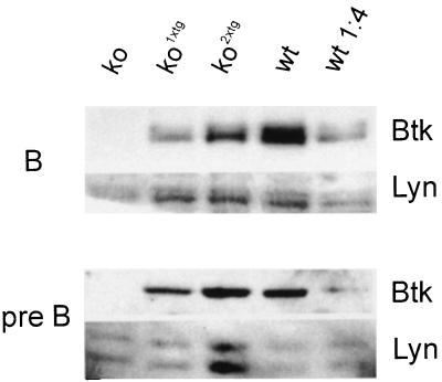
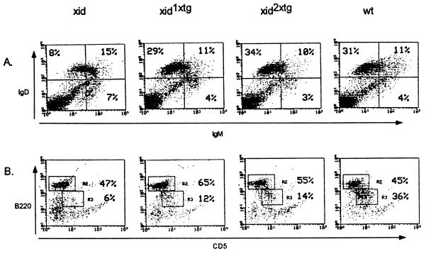
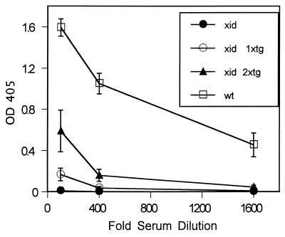
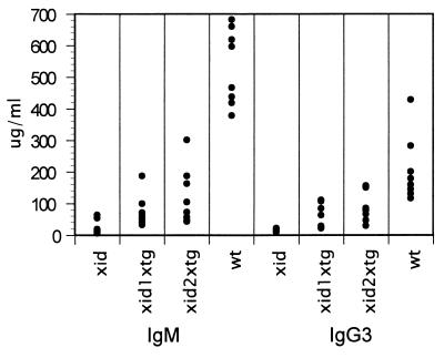
Mutations in Btk result in the B cell immunodeficiencies X-linked agammaglobulinemia (XLA) in humans and X-linked immunodeficiency (xid) in mice. Btk is a critical component of signaling pathways regulating B cell development and function. We used a genetic approach to determine whether Btk is also limiting for these processes. One allele of a murine Btk transgene expressed a dosage of Btk (25% of endogenous levels in splenic B cells) sufficient to restore normal numbers of phenotypically mature conventional B cells in xid mice. 2,4,6-trinitrophenyl-Ficoll response, anti-IgM-induced proliferation, B1 cell development, and serum IgM and IgG3 levels remained significantly impaired in these animals. B cells from Btk -/- transgenic mice also responded poorly to anti-IgM, indicating that the xid mutation does not create a dominant negative form of Btk. Response to 2,4,6-trinitrophenyl-Ficoll and B cell receptor cross-linking were increased 3- to 4-fold in xid mice homozygous for the transgene. These results demonstrate that Btk is a limiting component of B cell antigen receptor signaling pathways and suggest that B cell development and response to antigen may require different levels of Btk activity.


Figures

Figure 1
Figure 1
Transgene expression in kotg mice. Total cell lysates from B220+ spleen cells and v-abl-transformed pre-B cells were subjected to Western blot analysis and probed with an anti-Btk polyclonal antibody. Transgene-derived Btk protein was quantified by comparison with a 1:4 dilution of wild-type cell lysates into Btk-deficient cell lysates. Equal loading of samples was confirmed by Coomassie blue staining of the bottom portion of the gel and reprobing the blots with an anti-lyn polyclonal antibody.
Figure 2
Figure 2
Rescue of B cell development in xidtg mice. (A) Spleen cells from 8- to 12-week-old mice were depleted of red blood cells and stained with anti-IgM FITC (x axis) and anti-IgD PE (y axis). Live cells were gated based on forward and side scatter. The percentage of cells in each quadrant is indicated. (B) Peritoneal cells were depleted of red blood cells and stained with anti-CD5 FITC (x axis) and anti-B220 PE (y axis). Lymphocytes gated based on forward and side scatter are shown. The percentage of B220+CD5 and B220+CD5+ cells in this gate is indicated.
Figure 3
Figure 3
TNP–Ficoll response depends on Btk dosage. Eight- to 12-week-old xid, xid1xtg, xid2xtg, and wild-type (wt) mice were immunized with 10 μg TNP–Ficoll and bled 6 days later. Serum was diluted 1:100, 1:400, and 1:1,600. Anti-TNP IgM was measured by ELISA. OD405 readings are presented as mean ± SD. The number of individual mice tested in each group is as follows: xid, 8; xid1xtg, 8; xid2xtg, 8; wt, 5. All results differed significantly from each other (P < 0.001).
Figure 4
Figure 4
IgM and IgG3 levels are partially restored in xidtg mice. Serum IgM and IgG3 levels from 8- to 12-week-old xid, xid1xtg, xid2xtg, and wild-type (wt) mice were measured by ELISA. Each symbol represents an individual animal (n = 8). xid1xtg and xid2xtg are significantly different from both xid (P < 0.05) and wt (P < 0.001) but not each other.
Figure 5
Figure 5
Anti-IgM response depends on Btk dosage. (A) [3H]thymidine incorporation. Purified B220+ cells from spleen were incubated in triplicate for 48 hr with medium alone, 2 μg/ml anti-IgM, 20 μg/ml anti-IgM, or 10 ng/ml PMA and 1 μM ionomycin, then labeled overnight with [3H]thymidine. Counts per minute (cpm) incorporated in untreated or anti-IgM treated cells were normalized to the PMA and ionomycin response, which was similar for all cell types. The number of mice tested per group is: medium alone, 9; 2 μg/ml anti-IgM, 3; 20 μg/ml anti-IgM, 9; PMA and ionomycin, 9. Data are plotted as mean ± SEM. xid1xtg is significantly different from both wild type (wt) and xid2xtg (P < 0.005). (B) BrdUrd incorporation. Total splenocytes were incubated for 48 hr with medium alone, 2 μg/ml or 20 μg/ml anti-IgM, and labeled with BrdUrd for the final 24 hr. Samples were stained with anti-B220 PE and anti-BrdUrd FITC. The percentage of B220+ cells that incorporated BrdUrd is presented as mean ± SEM. The number of mice tested per group is: xid, 7; xid1xtg, 7; xid2xtg, 7; wt, 10; wt1xtg, 5; wt2xtg, 5. xid1xtg is significantly different from both wt (P < 0.005) and xid2xtg (P < 0.05). For wt vs. wt2xtg, 0.05 < P < 0.1.

References

    1. Goodnow C C. Proc Natl Acad Sci USA. 1996;93:2264–2271. - PMC - PubMed
    1. Carter R H, Fearon D T. Science. 1992;256:105–107. - PubMed
    1. Engel P, Zhou L-J, Ord D C, Sato S, Koller B, Tedder T F. Immunity. 1995;3:39–50. - PubMed
    1. Lund F E, Yu N, Kim K-M, Reth M, Howard M C. J Immunol. 1996;157:1455–1467. - PubMed
    1. Pezzutto A, Rabinovitch P S, Dorken B, Moldenhauer G, Clark E A. J Immunol. 1988;140:1791–1795. - PubMed

Publication types

LinkOut - more resources