. 1997 Nov 25;94(24):13287-92.
doi: 10.1073/pnas.94.24.13287.

Two amyloid precursor protein transgenic mouse models with Alzheimer disease-like pathology

Affiliations

Two amyloid precursor protein transgenic mouse models with Alzheimer disease-like pathology

C Sturchler-Pierrat et al. Proc Natl Acad Sci U S A. .

Abstract

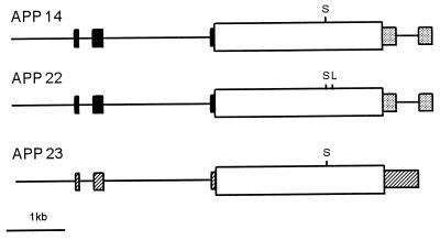
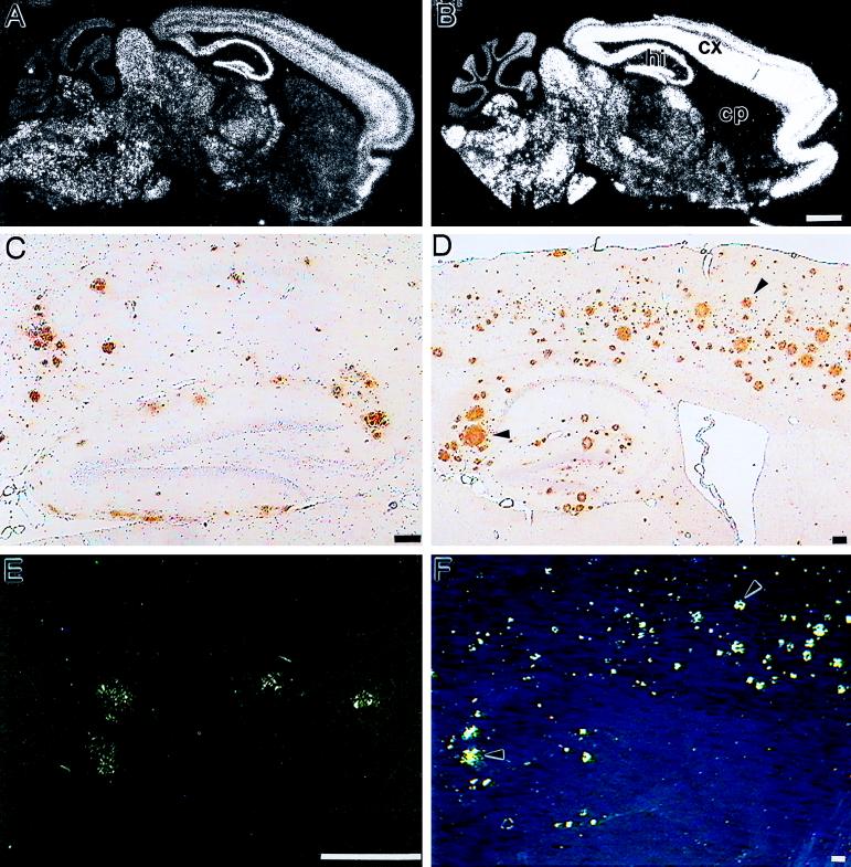
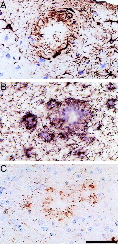
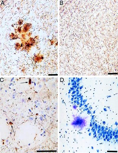
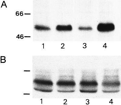
Mutations in the amyloid precursor protein (APP) gene cause early-onset familial Alzheimer disease (AD) by affecting the formation of the amyloid beta (A beta) peptide, the major constituent of AD plaques. We expressed human APP751 containing these mutations in the brains of transgenic mice. Two transgenic mouse lines develop pathological features reminiscent of AD. The degree of pathology depends on expression levels and specific mutations. A 2-fold overexpression of human APP with the Swedish double mutation at positions 670/671 combined with the V717I mutation causes A beta deposition in neocortex and hippocampus of 18-month-old transgenic mice. The deposits are mostly of the diffuse type; however, some congophilic plaques can be detected. In mice with 7-fold overexpression of human APP harboring the Swedish mutation alone, typical plaques appear at 6 months, which increase with age and are Congo Red-positive at first detection. These congophilic plaques are accompanied by neuritic changes and dystrophic cholinergic fibers. Furthermore, inflammatory processes indicated by a massive glial reaction are apparent. Most notably, plaques are immunoreactive for hyperphosphorylated tau, reminiscent of early tau pathology. The immunoreactivity is exclusively found in congophilic senile plaques of both lines. In the higher expressing line, elevated tau phosphorylation can be demonstrated biochemically in 6-month-old animals and increases with age. These mice resemble major features of AD pathology and suggest a central role of A beta in the pathogenesis of the disease.


Figures

Figure 1
Figure 1
Transcription units for APP transgenic mice. The diagrams are linear representations of the injected expression constructs and are labeled accordingly. Exons of the human Thy-1 gene are illustrated as solid boxes, and stippled boxes indicate sequences of simian virus 40 tumor antigen at the 3′ end of APP 14 and APP 22 constructs. Hatched boxes represent the murine Thy-1 exons used in the APP 23 construct. APP751 cDNAs are shown as open boxes with vertical lines marking the positions of the respective Swedish (S) and London (L) FAD mutations.
Figure 2
Figure 2
Expression of APP mRNA and deposition of Aβ in brains of transgenic mice. (A, C, and E) Sections taken from 18-month-old APP 22 mice. (B, D, and F) Sections from 24-month-old APP 23 mice. In situ hybridization on sagittal sections of brains from mouse line APP 22 (A) and APP 23 (B) was performed with an oligonucleotide probe specific for human APP. cp, Caudate putamen; cx, cortex; hi, hippocampus. (Bars = 1 mm.) Immunohistochemistry (C and D) and Congo Red staining (E and F) of sagittal sections from brains of transgenic mice. Immunostaining was performed with an antibody raised against Aβ8–17 (Dako). (Bars = 100 μm.) Arrowheads point to identical deposits in D and F.
Figure 3
Figure 3
Characterization of inflammatory processes in 12-month-old APP 23 transgenic mice. Immunostaining with a GFAP-specific antibody (A) indicates hypertrophic astrocytes. Activated microglia are visualized by immunostaining with a MAC-1 antibody (B) and a phosphotyrosine antibody (C). Both glia cell types are intimately associated with Aβ deposits. (Bar = 50 μm.)
Figure 4
Figure 4
Degenerative processes in APP 23 mice. (A and B) Staining for acetylcholinesterase in 12-month-old transgenic and control mice, respectively. A local distortion of cholinergic fibers in the plaque vicinity (A) compared with normal animals (B) can be noted. Neuritic spheroids (arrows) are stained with the neurofilament antibody NF200 (C). The loss of pyramidal neurons in the vicinity of Aβ deposits in area CA3 is shown in D by toluidine blue staining. (Bars = 50 μm.)
Figure 5
Figure 5
Phosphorylation of tau in the brains of 12-month-old APP 23 transgenic mice. Immunocytochemistry with antibody AT8 was performed on free floating sections (40 μm) (A and B). Immunostaining reveals fiber-like structures resembling distorted neurites. Qualitatively similar patterns are obtained with the Alz50 antibody (C). [Bars = 100 μm (A) and 10 μm (B and C).]
Figure 6
Figure 6
Biochemical detection of hyperphosphorylated tau protein. Western blots of brain extracts from APP 23 mice, 6 months (lane 2) and 15 months (lane 4) of age and littermate controls (lanes 1 and 3) are shown in A and B. Blots were stained with antibodies AT8 (A) and tau7 (B). Numbers indicate molecular weights of marker proteins in kDa.

References

    1. Probst A, Langui D, Ulrich J. Brain Pathol. 1991;1:229–239. - PubMed
    1. Selkoe D J. Annu Rev Cell Biol. 1994;10:373–403. - PubMed
    1. Yankner B A. Neuron. 1996;16:921–932. - PubMed
    1. Goedert M. Trends Neurosci. 1993;16:460–465. - PubMed
    1. Trojanowski J Q, Schmidt M L, Shin R-W, Bramblett G T, Rao D, Lee V M-Y. Clin Neurosci. 1993;1:184–191.

Publication types