. 1997 Nov 25;94(24):13305-10.
doi: 10.1073/pnas.94.24.13305.

A homeodomain gene Ptx3 has highly restricted brain expression in mesencephalic dopaminergic neurons

Affiliations

A homeodomain gene Ptx3 has highly restricted brain expression in mesencephalic dopaminergic neurons

M P Smidt et al. Proc Natl Acad Sci U S A. .

Abstract

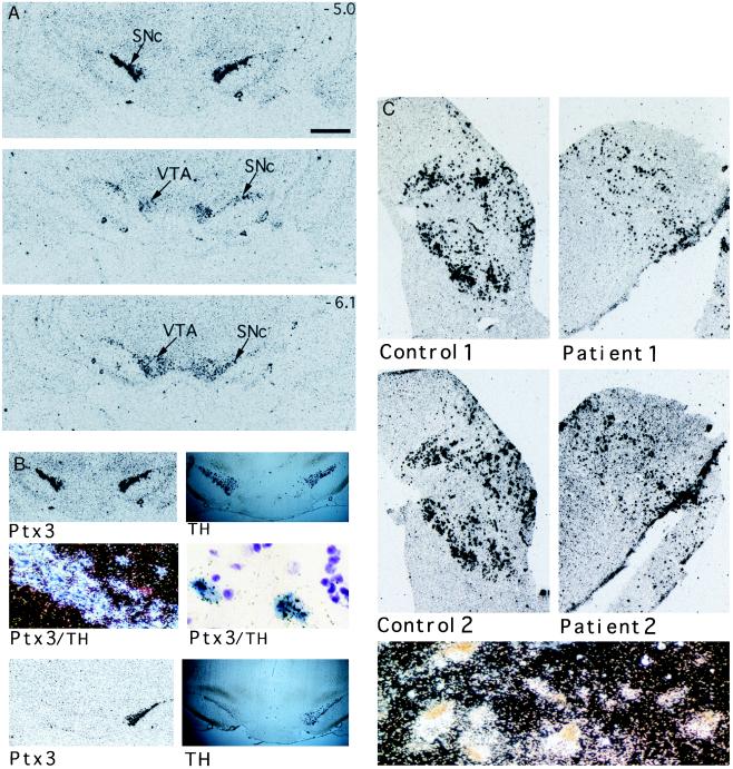
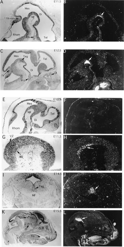
The mesencephalic dopaminergic (mesDA) system regulates behavior and movement control and has been implicated in psychiatric and affective disorders. We have identified a bicoid-related homeobox gene, Ptx3, a member of the Ptx-subfamily, that is uniquely expressed in these neurons. Its expression starting at E11.5 in the developing mouse midbrain correlates with the appearance of mesDA neurons. The number of Ptx3-expressing neurons is reduced in Parkinson patients, and these neurons are absent from 6-hydroxydopamine-lesioned rats, an animal model for this disease. Thus, Ptx3 is a unique transcription factor marking the mesDA neurons at the exclusion of other dopaminergic neurons, and it may be involved in developmental determination of this neuronal lineage.


Figures

Figure 1
Figure 1
Structure and properties of PTX3. (A) Primary structure of Ptx3 deduced from the ORF of the Ptx3 cDNA and alignment to Ptx1 (–13) and Ptx2 (RGS) (14). The homeodomain is underlined, with the bicoid-type-specific lysine at position 9 of the third helix double-underlined (23). A consensus nuclear localization signal is shown in boldface, italic type (24). The conserved C- terminal domain (14) is in boldface type. (B) Binding of Ptx3 to the CE3 element of the rat POMC promoter in gel shift analysis. Ptx3 binds to the CE3 element of the rat POMC promoter as was found for the related factors Ptx1, Otx1, and Otx2. Competition experiments using mutant recognition sites (11) within (M1) and outside (M3) the bicoid core of the CE3 element (200-fold molar excess) show that the ineffective mutant M3 and the wild type compete for Ptx3 binding, because the effective mutant M1 cannot. (C) Transactivational properties of ptx3 in transient transfection assays. The activity of Ptx3 on an artificial promoter construct based on three copies of the POMC-CE3 element is comparable to that of Ptx1 (11), whereas Otx1 and Otx2 are clearly less active. When using the M1 mutated binding site, the activity is lost.
Figure 2
Figure 2
Northern blot analysis of Ptx3 expression in the adult rat brain and peripheral organs. Blots containing 20 μg of total RNA of dissected rat brain regions and selected peripheral organs were hybridized with a full-length Ptx3 cDNA probe. GapDh was used as a control. The positions of 18S and 28S ribosomal RNAs are indicated by arrows.
Figure 3
Figure 3
Expression of Ptx3 in the mesencephalic dopaminergic system of rat and human. (A) The expression pattern of Ptx3 in rat brain starting anterior at the substantia nigra compacta (SNc) to posterior at the ventral tegmental area (VTA). The size bar (Top) represents a distance of 1 mm. The rostral–caudal positions of the Top and Bottom are indicated as millimeters relative to bregma in the upper right corner. (B) Double in situ hybridizations using a DIG-labeled tyrosine hydroxylase (TH) cRNA probe and a 35S-labeled Ptx3 cRNA probe show a similar hybridization pattern for both probes in the tegmentum of the rat brain (Top Left and Top Right). Microscopic examination (Middle) shows TH-positive cells in blue and Ptx3 labeled by silver grains (Middle Left, dark-field image; Middle Right, higher magnification, bright-field image). A complete overlap is found. In situ hybridization for TH and Ptx3 expression of animals with a unilateral lesion of dopaminergic neurons within the mesDA system. (Lower) The animals displayed increased turning behavior in the direction of the lesioned side. Both TH and Ptx3 expression was lost at the lesioned side. (C) Ptx3 expression in the human substantia nigra of two healthy controls (Control 1 and 2) and two Parkinson patients (Patient 1 and 2). The sections of the patients show a lower density of Ptx3-expressing cells, which correlates with the loss of dopaminergic neurons within this region. (Lower) A dark-field image showing Ptx3 expression (silver grains) colocalizing with typical brown-pigmented dopaminergic neurons of the human substantia nigra. Material was obtained from the Netherlands Brain Bank. Patient material was diagnosed clinically and pathologically.
Figure 4
Figure 4
In situ hybridization of Ptx3 in embryonic mouse brain sections between E10.5 and E15.5. Micrographs of bright-field and dark-field images of the same sections are shown. (A and B) At E11.5, expression is seen in a layer of postmitotic cells (arrow) in the mesencephalon (Mes) lining the mesencephalic flexure (MF). (C and D) At E12.5 the layer of expressing cells has thickened as part of the developing tegmentum (Teg). Extraneural expression is seen in the tongue (To) but not in the developing pituitary (Pit). (E and F) Expression of Ptx3 was not detected in the brain at E10.5. Lack of expression in Rathke’s pouch (RP), a site of Ptx1 and Ptx2 expression (–14), confirms the specificity of the hybridization reaction. (G and H) Higher magnification of the tegmental region expressing Ptx3 at E11.5. Expression is detected ventrally in the marginal zone (MZ) of the neuroepithelium. (I and J) A coronal section of an E14.5 mouse brain shows the lateral extent of Ptx3 expression in the developing tegmentum (Teg). (K and L) A sagittal section of an E15.5 mouse head shows strong expression of Ptx3 in the ventral tegmentum (vTeg). In contrast to other hybridizations that were performed with 5′ or 3′ Ptx3 probes, the probe used for this experiment contained the homeodomain, and there is weak cross-reactivity with other members of the Ptx family outside the brain, e.g., the pituitary (Pit) (–14). Mes, mesencephalon; Di, diencephalon; Tel, Telencephalon; Rhom, rhombencephalon; MF, mesencephalic flexure; RP, Rathke’s pouch; Is, isthmus; Pit, pituitary; To, tongue; Ol, olfactory epithelium; MZ, marginal zone; VZ, ventricular zone; Str, striatum; vTeg, ventral tegmentum; ct, cartilage of the throat; uLi, upper lip, lLi, lower lip.

References

    1. Lumsden A, Krumlauf R. Science. 1996;274:1109–1115. - PubMed
    1. Tanabe Y, Jessell T M. Science. 1996;274:1115–1123. - PubMed
    1. Specht L A, Pickel V M, Joh T H, Reis D. J Comp Neurol. 1981;199:233–253. - PubMed
    1. Voorn P, Kalsbeek A, Jorritsma-Byham J, Groenewegen H J. Neuroscience. 1988;25:857–887. - PubMed
    1. Solberg Y, Silverman W, Pollack Y. Dev Brain Res. 1993;73:91–97. - PubMed

Publication types

Associated data