. 1997 Oct;110(4):379-89.
doi: 10.1085/jgp.110.4.379.

Structural regions of the cardiac Ca channel alpha subunit involved in Ca-dependent inactivation

Affiliations

Structural regions of the cardiac Ca channel alpha subunit involved in Ca-dependent inactivation

B Adams et al. J Gen Physiol. 1997 Oct.

Abstract

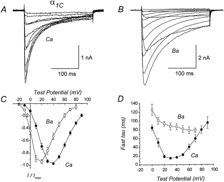
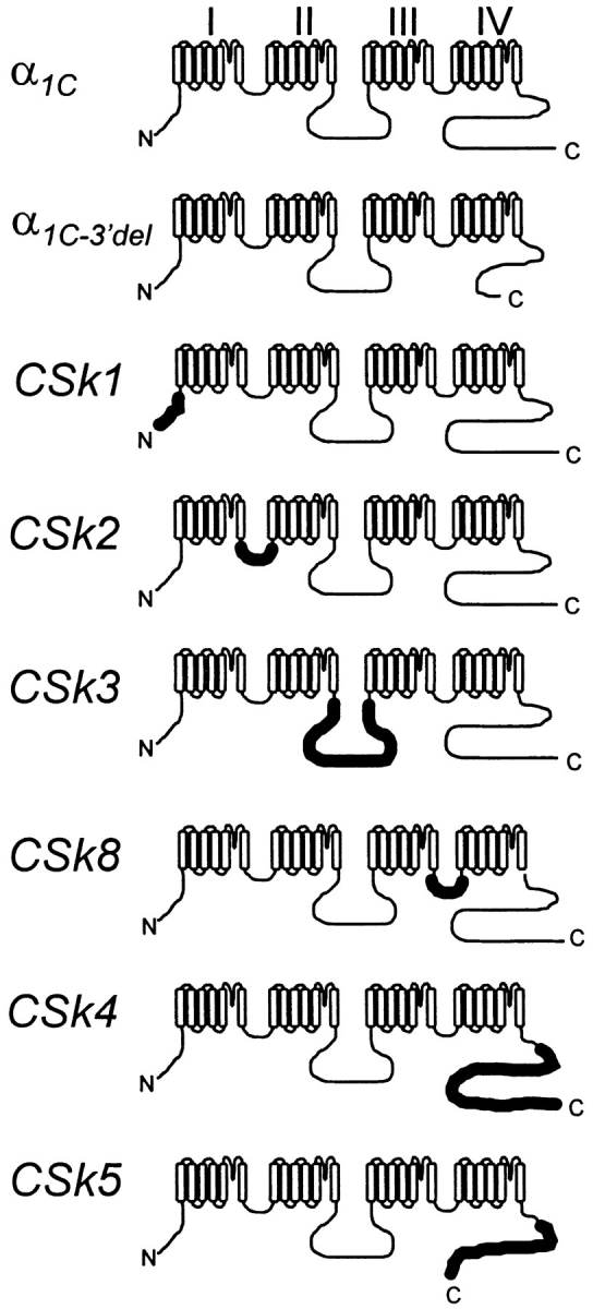
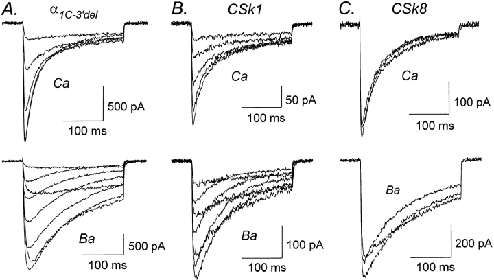
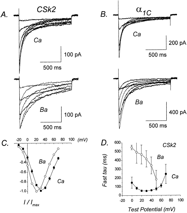
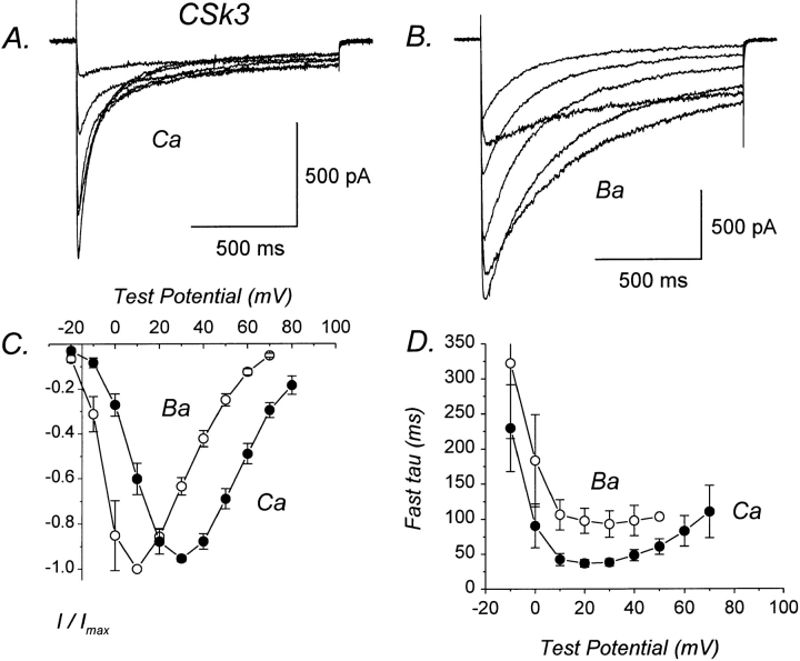
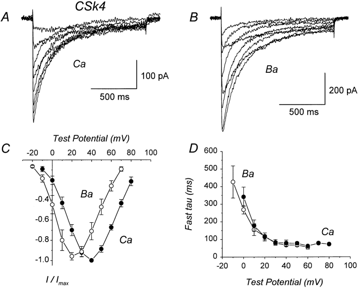
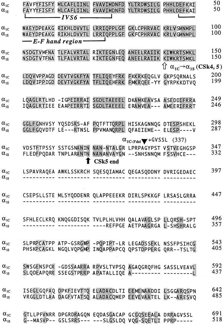
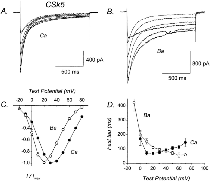
We investigated the molecular basis for Ca-dependent inactivation of the cardiac L-type Ca channel. Transfection of HEK293 cells with the wild-type alpha or its 3' deletion mutant (alpha) produced channels that exhibited prominent Ca-dependent inactivation. To identify structural regions of alpha involved in this process, we analyzed chimeric alpha subunits in which one of the major intracellular domains of alpha was replaced by the corresponding region from the skeletal muscle alpha subunit (which lacks Ca-dependent inactivation). Replacing the NH terminus or the III-IV loop of alpha with its counterpart from alpha had no appreciable effect on Ca channel inactivation. In contrast, replacing the I-II loop of alpha with the corresponding region from alpha dramatically slowed the inactivation of Ba currents while preserving Ca-dependent inactivation. A similar but less pronounced result was obtained with a II-III loop chimera. These results suggest that the I-II and II-III loops of alpha may participate in the mechanism of Ca-dependent inactivation. Replacing the final 80% of the COOH terminus of alpha with the corresponding region from alpha completely eliminated Ca-dependent inactivation without affecting inactivation of Ba currents. Significantly, Ca-dependent inactivation was restored to this chimera by deleting a nonconserved, 211-amino acid segment from the end of the COOH terminus. These results suggest that the distal COOH terminus of alpha can block Ca-dependent inactivation, possibly by interacting with other proteins or other regions of the Ca channel. Our findings suggest that structural determinants of Ca-dependent inactivation are distributed among several major cytoplasmic domains of alpha.


Figures

Figure 1
Figure 1
Ca-dependent inactivation of wild-type α1C Ca channels coexpressed in HEK293 cells with skeletal muscle α2δa and β1a subunits. (A) Representative whole-cell Ca currents mediated by α1C. The illustrated currents were evoked by depolarizations from −10 to +90 mV. The compensated series resistance (RS) was 4.2 MΩ. Linear cell capacitance (C) = 41 pF. File 96506016. (B) Representative Ba currents mediated by α1C. Test pulses −20 to +90 mV. Same cell as in A. RS = 2.1 MΩ. File 96506018. (C) Average I–V relations for Ca and Ba currents mediated by α1C. Each plotted point represents the mean (±SEM) of 13 (Ca) and 6 (Ba) different cells. (D) Time constants for inactivation of Ca or Ba currents mediated by α1C. Currents were evoked by 250-ms test pulses and were fit with either a single or double exponential function. For currents requiring two exponentials, only the fastest component was included in the analysis. Each plotted point represents the mean (±SEM) of 2–14 (Ca) and 3–7 (Ba) different cells. Experiments summarized in this figure were done using the TEACl-based external solution.
Figure 2
Figure 2
Schematic representations of the expressed α1 subunits. Thinner lines denote regions having amino acid sequence corresponding to the rabbit cardiac α1C subunit (Mikami et al., 1989); thicker lines denote regions corresponding to the rabbit skeletal muscle α1S subunit (Tanabe et al., 1987). The amino acid composition of α1C−3′del and construction of its cDNA has been previously described; the final five amino acid residues from the COOH terminus of α1C (GVSSL) are retained in α1C−3′del (Zong et al., 1994). The amino acid composition and cDNA construction of CSk1, CSk2, CSk3, and CSk4 are described in Tanabe et al. (1990b). The compositions of CSk5 and CSk8 and the constructions of their cDNAs are described in materials and methods.
Figure 3
Figure 3
Ca-dependent inactivation is normal in α1C−3′del, CSk1, and CSk8. (A) Representative Ca (top) and Ba (bottom) currents mediated by α1C−3′del, a deletion mutant of α1C lacking amino acid residues 1813–2166 from the COOH terminus. Test pulses from 0 to +40 mV (Ca) and −10 to +60 mV (Ba). Files 95929002, 95929006. C = 42 pF. RS = 3.8 MΩ. (B) Representative Ca and Ba currents mediated by CSk1, a chimera in which the NH2 terminus of α1C was replaced by the corresponding region from α1S. Test pulses from 0 to +40 mV (Ca) and 0 to +60 mV (Ba). Files 95D01016, 95D01018. C = 21 pF. RS = 6.3 MΩ. (C) Representative Ca and Ba currents mediated by chimera CSk8, in which the III–IV loop of α1C was replaced by the corresponding region from α1S. Test pulses from +30 to +50 mV (Ca) and +20 to +40 mV (Ba). Files 96118045, 96118053. C = 15 pF. RS = 4.9 MΩ. Experiments summarized in this figure were done using the TEACl-based external solution.
Figure 4
Figure 4
CSk2 retains Ca-dependent inactivation, but Ba current inactivation is dramatically slowed. (A) Representative Ca and Ba currents mediated by CSk2. Test pulses from −10 to +60 mV (Ca) and −10 to +50 mV (Ba). Files 97428010, 97428014. C = 17 pF. RS = 2.2 MΩ. (B) Representative Ca and Ba currents mediated by α1C. Test pulses from −10 to +40 mV (Ca) and −10 to +50 mV (Ba). Files 97502005, 97502012. C = 31 pF. RS = 2.3 MΩ. (C) I–V relations for CSk2 currents. Plotted points represent the mean (±SEM) of 4–10 different cells. (D) Voltage dependence of fast time constants for inactivation of CSk2. Plotted points represent mean (±SEM) of four to five different cells. Experiments summarized in this figure were done using the NaCl-based external solution.
Figure 5
Figure 5
CSk3 exhibits Ca-dependent inactivation. (A) Representative Ca currents mediated by CSk3. Test pulses from −10 to +40 mV. File 97512062. C = 41 pF. RS = 4.0 MΩ. (B) Representative Ba currents mediated by CSk3. Test pulses from −10 to +40 mV. File 97512067. Same cell as in A. (C) I–V relations for CSk3 currents. Plotted points represent the mean (±SEM) of three to eight different cells. (D) Voltage dependence of fast time constants for inactivation of CSk3. Plotted points represent mean (±SEM) of three to six different cells. Experiments summarized in this figure were done using the NaCl-based external solution.
Figure 6
Figure 6
CSk4 lacks Ca-dependent inactivation. (A) Representative Ca currents mediated by CSk4, in which the distal 80% of the COOH terminus of α1C has been replaced by the corresponding region from α1S. File 97501003. C = 26 pF. RS = 3.6 MΩ. (B) Representative Ba currents mediated by CSk4. File 97501008. Same cell as in A. For the illustrated currents, inactivation appears to be slightly slower for Ba than for Ca currents, but this is only apparent: the Ba currents are larger and therefore less contaminated by outward currents. (C) I–V relations for CSk4 currents. Each point represents the mean (±SEM) of four different cells. (D) Voltage dependence of fast time constants for inactivation of CSk4 currents. Each point represents the mean (±SEM) of four different cells. Experiments summarized in this figure were done using the NaCl-based external solution.
Figure 7
Figure 7
Sequence alignment of the COOH-terminal regions of the rabbit cardiac α1C (Mikami et al., 1989) and rabbit skeletal muscle α1S subunits (Tanabe et al., 1987). The sequences begin just before the last predicted transmembrane segment (S6) of domain IV (first bracket). The putative EF hand motif is indicated (second bracket), as is the junctional site within CSk4 and CSk5 where the sequence changes from α1C to α1S (white arrow). The termination of mutant α1C−3′del is indicated by a filled triangle; this construct retains the final five residues (GVSSL) of the wild-type COOH terminus. The termination point of chimera CSk5 is indicated (black arrow). Shaded amino acids are identical between α1C and α1S; dashes indicate gaps in the alignment.
Figure 8
Figure 8
Ca-dependent inactivation is restored in CSk5. (A) Representative Ca currents mediated by CSk5, which is identical to CSk4 except that the nonconserved region (211 amino acids) has been deleted from the end of the COOH terminus. File 97522014. C = 43 pF. RS = 3.0 MΩ. (B) Representative Ba currents mediated by CSk5. File 97522021. Same cell as in A. (C) I–V relations for CSk5 currents. Each plotted point represents the mean (±SEM) of eight different cells. (D) Voltage dependence of fast time constants for inactivation of CSk5 currents. Each plotted point represents the mean (±SEM) of five to six different cells. Experiments summarized in this figure were done using the NaCl-based external solution.

References

    1. Adams B, Tanabe T. Calcium-dependent inactivation of heterologously expressed cardiac L-type calcium channel. Biophys J. 1996;70:238a. . (Abstr.) - PubMed
    1. Beam KG, Knudson CM. Calcium currents in embryonic and neonatal mammalian skeletal muscle. J Gen Physiol. 1988;91:781–798. - PMC - PubMed
    1. Beam KG, Adams BA, Niidome T, Numa S, Tanabe T. Function of a truncated dihydropyridine receptor as both voltage sensor and calcium channel. Nature (Lond) 1992;360:169–171. - PubMed
    1. Boyett MR, Harrison SM, Janvier NC, McMorn SO, Owen JM, Shui Z. A list of vertebrate cardiac ionic currents: nomenclature, properties, function and cloned equivalents. Cardiovasc Res. 1996;32:455–481. - PubMed
    1. de Leon M, Wang Y, Jones L, Perez-Reyes E, Wei XY, Soong TW, Snutch TP, Yue DT. Essential Ca2+-binding motif for Ca2+-sensitive inactivation of L-type Ca2+channels. Science (Wash DC) 1995;270:1502–1506. - PubMed

Publication types