. 1997 Sep 30;94(20):10547-53.
doi: 10.1073/pnas.94.20.10547.

Structure at 2.7 A resolution of the Paracoccus denitrificans two-subunit cytochrome c oxidase complexed with an antibody FV fragment

Affiliations

Structure at 2.7 A resolution of the Paracoccus denitrificans two-subunit cytochrome c oxidase complexed with an antibody FV fragment

C Ostermeier et al. Proc Natl Acad Sci U S A. .

Abstract

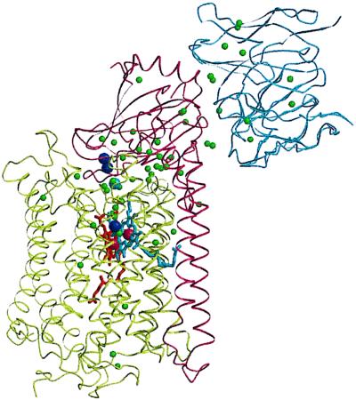
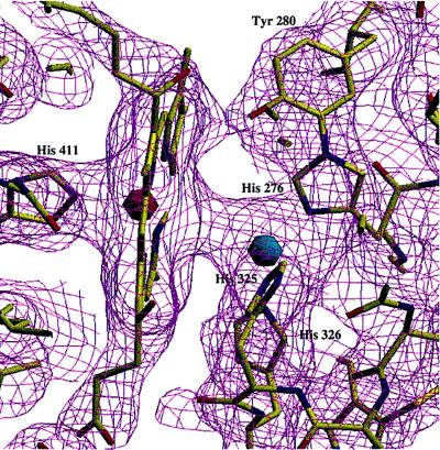
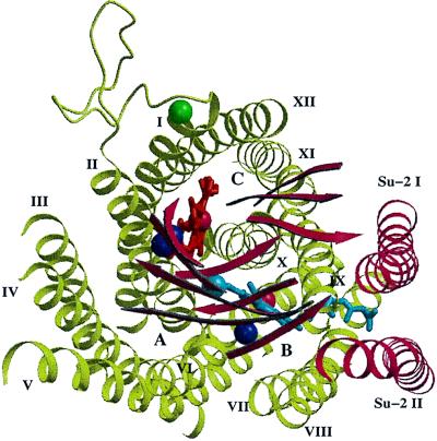
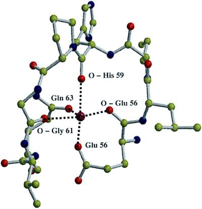
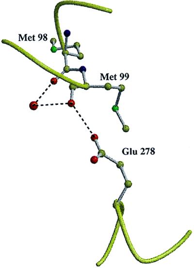
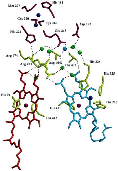
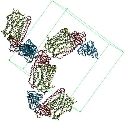
The aa3 type cytochrome c oxidase consisting of the core subunits I and II only was isolated from the soil bacterium Paracoccus denitrificans and crystallized as complex with a monoclonal antibody Fv fragment. Crystals could be grown in the presence of a number of different nonionic detergents. However, only undecyl-beta-D-maltoside and cyclohexyl-hexyl-beta-D-maltoside yielded well-ordered crystals suitable for high resolution x-ray crystallographic studies. The crystals belong to space group P212121 and diffract x-rays to at least 2.5 A (1 A = 0.1 nm) resolution using synchrotron radiation. The structure was determined to a resolution of 2.7 A using molecular replacement and refined to a crystallographic R-factor of 20.5% (Rfree = 25.9%). The refined model includes subunits I and II and the 2 chains of the Fv fragment, 2 heme A molecules, 3 copper atoms, and 1 Mg/Mn atom, a new metal (Ca) binding site, 52 tentatively identified water molecules, and 9 detergent molecules. Only four of the water molecules are located in the cytoplasmic half of cytochrome c oxidase. Most of them are near the interface of subunits I and II. Several waters form a hydrogen-bonded cluster, including the heme propionates and the Mg/Mn binding site. The Fv fragment binds to the periplasmic polar domain of subunit II and is critically involved in the formation of the crystal lattice. The crystallization procedure is well reproducible and will allow for the analysis of the structures of mechanistically interesting mutant cytochrome c oxidases.


Figures

Figure 1
Figure 1
Ribbon representation of the structure of the two-subunit cytochrome c oxidase from P. denitrificans complexed with the antibody Fv fragment 7E2. Subunit I, olive green; subunit II, dark red; Fv fragment, blue; heme a, red; heme a3, blue; copper atoms, dark blue spheres; water molecules, green spheres. The programs molscript (29) and raster 3d (30) were used to prepare the figure.
Figure 2
Figure 2
“Unbiased” electron density map at the 1σ level of the binuclear heme a3-CuB site and the atomic model. A simulated annealing omit map (31), omitting all residues (including heme a3) with an atom closer than 4.5 Å to CuB, was calculated. The heme a3 iron atom is shown in red, CuB in blue, nitrogen atoms in blue, oxygen atoms in red, carbon atoms in yellow, and the electron density in purple. The figure was prepared using the program setor (32).
Figure 3
Figure 3
View down onto the two-subunit cytochrome c oxidase from the periplasmic side showing the localization of the new metal binding site. Only the transmembrane helices, the loop connecting transmembrane helices I and II of subunit I, the β-strands of subunit II, the prosthetic group, and the metals are shown. The metal of the new metal binding site is shown in green. Subunit I, olive green; subunit II, red; heme a, red; heme a3, blue; copper atoms, dark blue spheres; iron atoms, red spheres. The transmembrane helices are indicated by roman numerals, the positions of the three “pores” are indicated by A, B, and C. The figure was created using the programs molscript (29) and raster 3d (30).
Figure 4
Figure 4
Ball and stick model of the new metal binding site. Oxygen atoms are shown in red, nitrogen atoms in blue, carbon atoms in yellow, and the new metal in purple. The programs molscript (29) and raster 3d (30) were used to prepare the figure.
Figure 5
Figure 5
Model of Glu-278 (subunit I) and its hydrogen bond to Met-99. A water molecule (larger red sphere) connects the backbone oxygen atoms of Met-98 and Met-99. The figure was prepared using the programs molscript (29) and raster 3d (30).
Figure 6
Figure 6
The hydrogen bonded network between the heme groups and CuA. Residues of subunit I are shown in olive green, those of subunit II in dark red, copper atoms are dark blue spheres, iron atoms dark red spheres, the Mg/Mn atom is a blue sphere, water molecules are green spheres, heme a is red, and heme a3 is blue. The figure was produced using the programs molscript (29) and raster 3d (30).
Figure 7
Figure 7
The crystal packing of the two-subunit cytochrome c oxidase complexed with the antibody Fv fragment (backbone wire models). Subunit I is shown in olive green, subunit II in red, and the Fv fragment in dark blue. The figure was prepared with the program setor (32).

References

    1. Saraste M. Q Rev Biophys. 1990;23:331–366. - PubMed
    1. Babcock G T, Wikström M. Nature (London) 1992;356:301–309. - PubMed
    1. Calhoun M W, Thomas J W, Gennis R B. Trends Biochem Sci. 1994;19:325–330. - PubMed
    1. Malatesta F, Antonini G, Sarti P, Brunori M. Biophys Chem. 1995;54:1–33. - PubMed
    1. Ferguson-Miller S, Babcock G T. Chem Rev. 1996;96:2889–2907. - PubMed

Publication types

Associated data

LinkOut - more resources