. 1997 Sep 30;94(20):10594-9.
doi: 10.1073/pnas.94.20.10594.

Evidence for two nonidentical drug-interaction sites in the human P-glycoprotein

Affiliations

Evidence for two nonidentical drug-interaction sites in the human P-glycoprotein

S Dey et al. Proc Natl Acad Sci U S A. .

Abstract

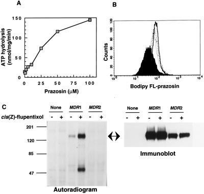
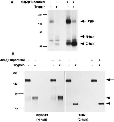
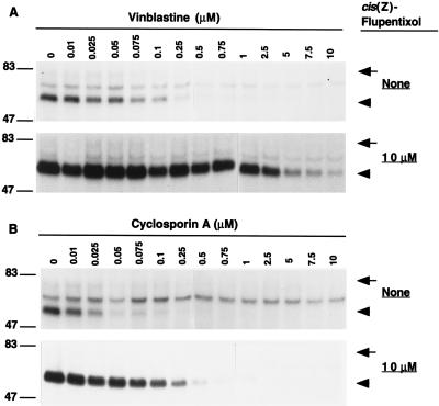
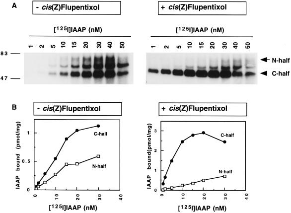
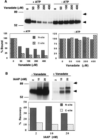
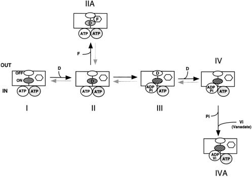
Human P-glycoprotein (Pgp) confers multidrug resistance to cancer cells by ATP-dependent extrusion of a great many structurally dissimilar hydrophobic compounds. The manner in which Pgp recognizes these different substrates is unknown. The protein shows internal homology between its N- and C-terminal halves, each comprised of six putative transmembrane helices and a consensus ATP binding/utilization site. Photoactive derivatives of certain Pgp substrates specifically label two regions, one on each half of the protein. In this study, using [125I]iodoarylazidoprazosin ([125I]IAAP), a photoactive analog of prazosin, we have demonstrated the presence of two nonidentical drug-interaction sites within Pgp. Taking advantage of a highly susceptible trypsin cleavage site in the linker region of Pgp, we characterized the [125I]IAAP binding to the N- and C-terminal halves. cis(Z)-Flupentixol, a modulator of Pgp function, preferentially increased the affinity of [125I]IAAP for the C-terminal half of the protein (C-site) by reducing the Kd from 20 to 6 nM without changing the labeling or affinity (Kd = 42-46 nM) of the N-terminal half (N-site). Also, the concentration of vinblastine (Pgp substrate) and cyclosporin A (Pgp modulator) required for 50% inhibition of [125I]IAAP binding to the C-site was increased 5- to 6-fold by cis(Z)-flupentixol without any effect on the N-site. In addition, [125I]IAAP binding to the N-site was less susceptible than to C-site to inhibition by vanadate which blocks ATP hydrolysis and drug transport. These data demonstrate the presence of at least two nonidentical substrate interaction sites in Pgp.


Figures

Figure 1
Figure 1
Interaction of prazosin and its analogs with Pgp. (A) Stimulation of Pgp-mediated ATP hydrolysis by prazosin. The vanadate-sensitive Pgp-ATPase activity in the presence of indicated concentration of prazosin in High Five cell membranes was measured as described. (B) Pgp-mediated extrusion of Bodipy FL-prazosin from NIH-MDR1 cells. Accumulation of Bodipy FL-prazosin was measured in drug-sensitive NIH 3T3 and drug-resistant NIH-MDR1 cells. After incubating in glucose containing DMEM with 0.5 μM of Bodipy FL-prazosin in the presence or absence of cyclosporin A, cells were pelleted and analyzed for intracellular accumulation (fluorescence intensity) of the prazosin derivative by fluorescence-activated cell sorter. NIH 3T3 and NIHMDR1 cells in the absence (… … . , filled area) and presence (---, ——) of cyclosporin A. (C) Photoaffinity labeling of Pgp in insect cell membranes by [125I]IAAP. Membranes isolated from High Five insect cells, infected with recombinant virus BV-MDR1(H6) or BV-MDR2 were photoaffinity labeled with 2 nM of [125I]IAAP in presence and absence of 25 μM cis(Z)-flupentixol as described. (Left) Autoradiogram of 3 μg of membrane protein/lane separated in an 8% gel by SDS/PAGE. (Right) Immunoblot analysis of the same samples using Pgp-specific mAb C219.
Figure 2
Figure 2
Distribution of [125I]IAAP label between the N- and C-terminal halves of Pgp. (A) Insect cell membranes (10 μg) containing recombinant Pgp were photoaffinity labeled with 5 nM [125I]IAAP in the presence and absence of 50 μM cis(Z)-flupentixol. Photoaffinity-labeled membranes were incubated with 7.5 μg of trypsin as described. A total of 2 μg of each sample were run on a 8% SDS/PAGE, dried, and exposed to x-ray film. (B) The identity of the two halves of the Pgp molecule was determined by immunoblot analysis using polyclonal antibodies PEPG 13 and 4007, specific for the N- and C-terminal halves of Pgp, respectively. The arrow with long tail, the arrowhead with short tail, and the arrowhead show the position of Pgp, N-half, and C-half, respectively.
Figure 4
Figure 4
Effect of vinblastine and cyclosporin A on [125I]IAAP labeling of the N- and C-sites of Pgp in the absence and presence of cis(Z)-flupentixol. (A) Photoaffinity labeling of Pgp containing crude insect cell membranes were carried out with 5 nM of [125I]IAAP and with varying concentrations (0–10 μM) of vinblastine in the presence (Lower) and absence (Upper) of 10 μM cis(Z)-flupentixol. N- and C-terminal halves of Pgp were separated in an 8% gel by SDS/PAGE, and [125I]IAAP labeling was quantified by measuring radioactivity associated with each fragment, as described. (B) Membranes were photoaffinity labeled as described above except with varying concentrations (0–10 μM) of cyclosporin A in the presence (Lower) and absence (Upper) of cis(Z)-flupentixol. Concentration of vinblastine and cyclosporin A required for half maximal inhibition (IC50) was calculated from their respective inhibition curves.
Figure 3
Figure 3
Concentration-dependent binding of [125I]IAAP to the N- and the C-terminal halves of Pgp in the presence and in absence of cis(Z)-flupentixol. (A) Pgp-containing insect cell membranes (10 μg of membrane protein) were photoaffinity labeled with concentrations of [125I]IAAP ranging from 1 to 50 nM with (Right) or without (Left) pre-incubation with 50 μM of cis(Z)-flupentixol. Photoaffinity labeled membranes were then treated with trypsin as described in the legend to Fig. 2, and samples were analyzed in an 8% gel by SDS/PAGE. (B) Radioactive bands corresponding to the N- (70 kDa) and C-terminal (60 kDa) halves were cut out, and bound [125I]IAAP (pmol/mg of membrane protein) was determined as described. Nonlinear regression analysis were done using graphics program graph pad.
Figure 5
Figure 5
Effect of vanadate trapping on [125I]IAAP photoaffinity labeling of the N- and C-terminal halves of Pgp. (A) Pgp-containing insect cell membranes were incubated at 37°C for 10 min with 4 nM [125I]IAAP, 10 μM cis(Z)-flupentixol, 5 mM MgCl2, and varying concentrations of vanadate in presence and absence of 2.5 mM ATP prior to UV exposure for 10 min. Photoaffinity labeled membranes were trypsinized to generate the N- and C-terminal fragments and analyzed by SDS/PAGE. (Lower) Radioactivity associated with the N- and C-halves in the presence and absence of ATP is shown. (B) Insect cell membranes were incubated at 37°C for 10 min with 10 μM cis(Z)-flupentixol, 2.5 mM ATP, and 5 mM MgCl2 in presence and absence of 400 μM of vanadate. After incubation, [125I]IAAP was added to final concentrations of 2, 10, and 20 nM, and Pgp was photoaffinity labeled the same way as above. (Lower) Recovery of radioactivity in both halves is shown. For other details, see legend to Fig. 2.
Figure 6
Figure 6
Schematic representation of the Pgp catalytic cycle and the possible functional role of the N- and the C-sites in drug translocation. In this model, the two TM domains and the ATP binding sites are represented by squares and circles, respectively. The model shows one cycle of ATP hydrolysis where the shaded circle represents the noncatalytic state. The two drug-interacting sites are along the drug-translocating pathway and are designated by two ellipses (shaded for ON-site and open for OFF-site). The cis(Z)-flupentixol (modulator) interaction site is depicted by the hexagon. The hatched ellipse indicates a conformational change in the drug-interaction site closer to the cytosolic phase of the lipid bilayer. The substrate molecule, cis(Z)-flupentixol and vanadate are shown as “D,” “F,” and “Vi,” respectively. The dark arrows represent favored reaction. Various states of Pgp during the catalytic/drug-translocation cycle are as follows: I, Pgp·MgATP; II, PgpMgATP·DON; IIA, PgpMgATP·DON·F; III, PgpMgADP·Pi·DOFF; IV, PgpMgADP·Pi; IVA, PgpMgADP·Vi.

References

    1. Gottesman M M, Pastan I. Annu Rev Biochem. 1993;62:385–427. - PubMed
    1. Gottesman M M, Pastan I, Ambudkar S V. Curr Opin Genet Dev. 1996;6:610–617. - PubMed
    1. Horio M, Gottesman M M, Pastan I. Proc Natl Acad Sci USA. 1988;85:3580–3584. - PMC - PubMed
    1. Gottesman M M, Hrycyna C A, Schoenlein P V, Germann U A, Pastan I. Annu Rev Genet. 1995;29:607–649. - PubMed
    1. Chen C-J, Chin J E, Ueda K, Clark D P, Pastan I, Gottesman M M, Roninson I B. Cell. 1986;47:381–389. - PubMed

MeSH terms

LinkOut - more resources