. 1997 Sep 30;94(20):10624-9.
doi: 10.1073/pnas.94.20.10624.

B cell receptor-associated protein alpha4 displays rapamycin-sensitive binding directly to the catalytic subunit of protein phosphatase 2A

Affiliations

B cell receptor-associated protein alpha4 displays rapamycin-sensitive binding directly to the catalytic subunit of protein phosphatase 2A

K Murata et al. Proc Natl Acad Sci U S A. .

Abstract

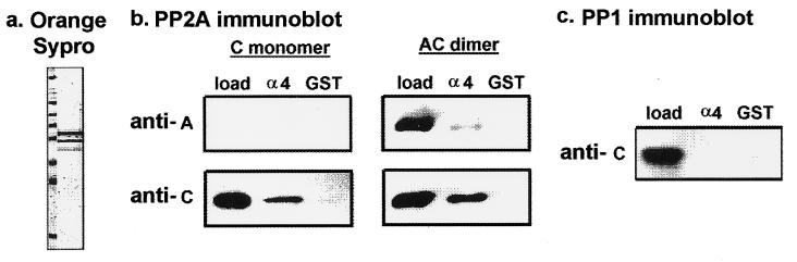
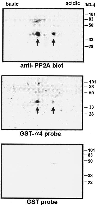
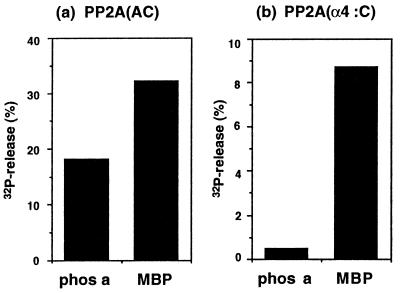
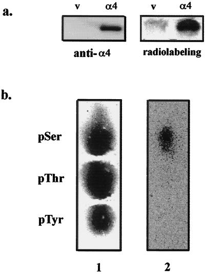
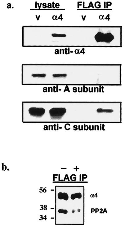
Recently, TAP42 was isolated as a high copy suppressor of sit4-, a yeast phosphatase related to protein phosphatase 2A (PP2A). TAP42 is related to the murine alpha4 protein, which was discovered independently by its association with Ig-alpha in the B cell receptor complex. Herein we show that a glutathione S-transferase (GST)-alpha4 fusion protein bound the catalytic subunit (C) of human PP2A from monomeric or multimeric preparations of PP2A in a "pull-down" assay. In an overlay assay, the GST-alpha4 protein bound to the phosphorylated and unphosphorylated forms of C that were separated in two-dimensional gels and immobilized on filters. The results show direct and exclusive binding of alpha4 to C. This is unusual because all known regulatory B subunits, or tumor virus antigens, bind stably only to the AC dimer of PP2A. The alpha4-C form of PP2A had an increased activity ratio compared with the AC form of PP2A when myelin basic protein phosphorylated by mitogen-activated protein kinase and phosphorylase a were used as substrates. Recombinant alpha4 cleaved from GST was phosphorylated by p56(lck) tyrosine kinase and protein kinase C. A FLAG-tagged alpha4 expressed in COS7 cells was recovered as a protein containing phosphoserine and coimmunoprecipitated with the C but not the A subunit of PP2A. Treatment of cells with rapamycin prevented the association of PP2A with FLAG-alpha4. The results reveal a novel heterodimer alpha4-C form of PP2A that may be involved in rapamycin-sensitive signaling pathways in mammalian cells.


Figures

Figure 1
Figure 1
Binding of GST–α4 with purified PP2A. (a) Bacterial GST–α4 fusion protein was purified, analyzed by SDS/PAGE, and visualized by fluorescent protein staining with Orange Sypro (Molecular Probes). Molecular mass markers shown in the left lane are 205, 116, 97, 84, 66, 55, 45, and 30 kDa. (b) Immunoblotting with anti-PP2A subunit antibodies was used to assay binding of PP2A to GST–α4. Human PP2A (C monomer or AC dimer) was incubated with equimolar amounts of either GST–α4 protein (α4) bound to glutathione-agarose or GST alone (GST) bound to glutathione-agarose. After washing and elution, 50% of the eluted proteins were analyzed by immunoblotting with antibodies against A subunit (anti-A) or C subunit (anti-C) of PP2A. For visual comparison, 5% of the PP2A added to the assay (load) was stained. (c) The C subunit of PP1 was incubated with GST–α4 (α4) or GST alone (GST), as described above. Immunoblotting used anti-peptide antibody against the PP1 C subunit (anti-C).
Figure 2
Figure 2
Binding of 32P-labeled GST–α4 probe to PP2A from fibroblasts. Cell lysates of mouse 10T1/2 fibroblasts were subjected to two-dimensional gel electrophoresis and transferred onto nitrocellulose filters. The filters were immunoblotted with antibody against C subunit of PP2A (Top). Unphosphorylated and phosphorylated PP2A was detected as two forms (arrows) at 36 kDa, separated by isoelectric focusing. By using duplicate filters, one was incubated for 30 min with 32P-labeled GST–α4 probe (Middle) and the other was incubated with 32P-labeled GST probe (Bottom). Molecular mass standards are shown on the right.
Figure 3
Figure 3
Effects of α4 binding on PP2A phosphatase activity. Phosphatase activity was measured by using 32P-labeled phos a and 32P-labeled MBP as substrates. (a) The AC dimer of PP2A was incubated with 15 μM 32P-labeled phos a or 1 μM 32P-labeled MBP for 1 h at 30°C. (b) The AC dimer of PP2A was bound to GST–α4 on agarose beads or beads with GST alone, as described in text. The PP2A bound to GST–α4 was assayed as the difference in activity between the GST–α4 and GST alone. Results shown are the average of two experiments.
Figure 4
Figure 4
FLAG-α4 in COS7 cells is phosphorylated exclusively on serine. (a) Cells transfected with empty vector (lanes v) or FLAG-tagged α4 (lanes α4) were metabolically labeled with 32P. After immunoprecipitation with anti-FLAG M2 affinity gel, proteins were analyzed by immunoblotting using anti-α4 antiserum (Left) and for 32P by PhosphorImaging (Right). FLAG-α4 migrated as a protein of 47 kDa. Results were replicated in three independent experiments. (b) The FLAG-α4 protein was excised from the gel and digested with trypsin, and the tryptic peptides were acid-hydrolyzed to yield phosphoamino acids that were analyzed with internal standards by high-voltage electrophoresis (24, 25). The standards, phosphoserine (pSer), phosphothreonine (pThr), and phosphotyrosine (pTyr) were stained with ninhydrin (lane 1) and radioactivity in the same lane was detected by phosphorimaging (lane 2), showing that the FLAG-α4 only contained radiolabeled phosphoserine.
Figure 5
Figure 5
Binding of FLAG-α4 to PP2A in COS7 cells is inhibited by rapamycin. (a) Cells were transfected with empty vector (lanes v) or FLAG-tagged α4 (lanes α4). Lysates (5% of total, left lanes) and immunoprecipitates with anti-FLAG M2 affinity gel (50% of total, right lanes) were analyzed by immunoblotting using anti-α4 antiserum (Top), anti-A subunit of PP2A antibody (Middle), and anti-C subunit of PP2A antibody (Bottom). Expression of FLAG-α4 in these cells did not alter the level of endogenous A (60 kDa) and C (36 kDa) subunits of PP2A. Results were replicated in several experiments. (b) Cells were transfected and processed as in above, except 100 nM rapamycin (lane +) or vehicle alone (lane −) was added in serum-free medium for 30 min before harvesting. The anti-FLAG precipitates were resolved by SDS/PAGE and immunoblotted with a mixture of anti-α4 and anti-C subunit antibodies to detect both proteins in a single step. Immunoblotting with each antibody separately confirmed the specificity of staining. Cells transfected with empty vector gave no signals as shown above. The results were replicated in independent experiments.

References

    1. Mumby M C, Walter G. Physiol Rev. 1993;73:673–699. - PubMed
    1. Wera S, Hemmings B A. Biochem J. 1995;311:17–29. - PMC - PubMed
    1. Kamibayashi C, Estes R, Slaughter C, Mumby M C. J Biol Chem. 1991;266:13251–13260. - PubMed
    1. Kamibayashi C, Lickteig R L, Estes R, Walter G, Mumby M C. J Biol Chem. 1992;267:21864–21872. - PubMed
    1. Kamibayashi C, Estes R, Lickteig R L, Yang S I, Craft C, Mumby M C. J Biol Chem. 1994;269:20139–20148. - PubMed

Publication types