. 1997 Sep 30;94(20):10663-8.
doi: 10.1073/pnas.94.20.10663.

Strain-specific differences in mouse hepatic wound healing are mediated by divergent T helper cytokine responses

Affiliations

Strain-specific differences in mouse hepatic wound healing are mediated by divergent T helper cytokine responses

Z Shi et al. Proc Natl Acad Sci U S A. .

Abstract

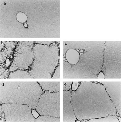
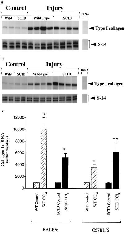
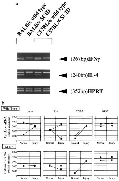
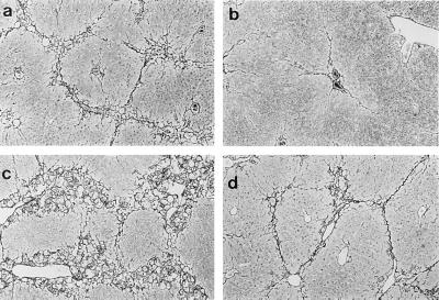
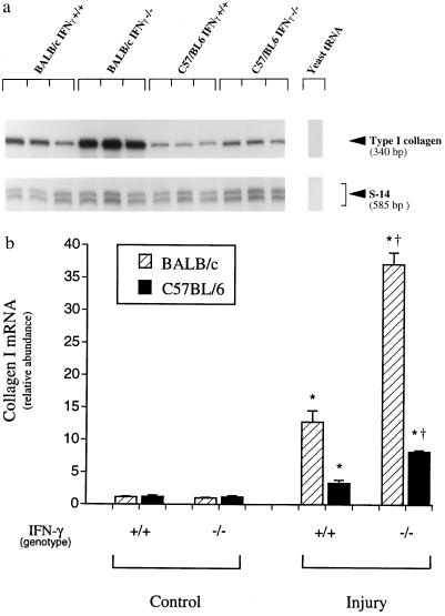
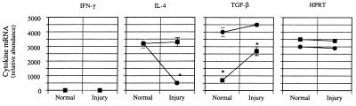
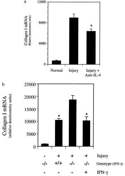
Hepatic fibrosis represents the generalized response of the liver to injury and is characterized by excessive deposition of extracellular matrix. The cellular basis of this process is complex and involves interplay of many factors, of which cytokines are prominent. We have identified divergent fibrosing responses to injury among mouse strains and taken advantage of these differences to examine and contrast T helper (Th)-derived cytokines during fibrogenesis. Liver injury was induced with carbon tetrachloride, fibrosis was quantitated, and Th1/Th2 cytokine mRNAs measured. Liver injury in BALB/c mice resulted in severe fibrosis, whereas C57BL/6 mice developed comparatively minimal fibrosis. Fibrogenesis was significantly modified in T and B cell-deficient BALB/c and C57BL/6 severe combined immunodeficient (SCID) mice compared with wild-type counterparts, suggesting a role of Th subsets. Fibrogenic BALB/c mice exhibited a Th2 response during the wounding response, whereas C57BL/6 mice displayed a Th1 response, suggesting that hepatic fibrosis is influenced by different T helper subsets. Moreover, mice lacking interferon gamma, which default to the Th2 cytokine pathway, exhibited more pronounced fibrotic lesions than did wild-type animals. Finally, shifting of the Th2 response toward a Th1 response by treatment with neutralizing anti-interleukin 4 or with interferon gamma itself ameliorated fibrosis in BALB/c mice. These data support a role for immune modulation of hepatic fibrosis and suggest that Th cytokine subsets can modulate the fibrotic response to injury.


Figures

Figure 1
Figure 1
Hepatic extracellular matrix expression is differentially regulated in wild-type and SCID mice BALB/c and C57BL/6 mice. Liver fibrosis was induced by gavage with CCl4 (3.5 ml/kg, mixed 1:1 with corn oil administered at 7 day intervals). Seven days after the 4th dose of CCl4, animals were killed, and livers were fixed in 10% formalin and stained with Sirius Red as described. Collagen is stained by Sirius Red and appears black. (a) Normal control BALB/c receiving corn oil vehicle alone (identical to C57BL/6; data not shown). (b) Wild-type BALB/c. (c) Wild-type C57BL/6. (d) SCID BALB/c. (e) SCID C57BL/6. Representative photomicrographs are shown. (×90.)
Figure 2
Figure 2
Type I collagen mRNA expression in wild-type and SCID mice BALB/c and C57BL/6 mice. Liver fibrosis was induced by gavage with CCl4 (3.5 ml/kg, mixed 1:1 with corn oil, was administered at 7 day intervals by gavage). Seven days after the 4th dose of CCl4, animals were killed and total cellular RNA extracted as described. RNase protection assay with probes complimentary to type I collagen and S-14 (an internal standard mRNA) using 10 μg of RNA was performed. Representative RNase protection assays are shown in a and b [BALB/c and C57BL/6 mice, respectively]. Specific bands were quantitated and normalized to the signal for S-14 in c. (n = 4, ∗, P < 0.05 for CCl4 compared with control for each individual strain, †, P < 0.05 for SCID compared with wild type.
Figure 3
Figure 3
Hepatic cytokine mRNA expression in fibrotic liver injury in SCID mice. Liver fibrosis was induced as in Fig. 1. Seven days after the 4th dose of CCl4, animals were killed, total cellular RNA extracted, and competitive PCR using primers for IFN-γ, IL-4, TGF-β, and HPRT performed as described. A representative gel is shown in a. Scanned, quantitated, and normalized data are presented graphically in b. Circles represent C57BL/6 mice, and squares represent BALB/c animals. ∗, P < 0.05 for C57BL/6 compared with BALB/c mice (n = 4 for group).
Figure 4
Figure 4
Hepatic extracellular matrix production in IFN-γ-deficient mice after liver injury. Liver fibrosis was induced by gavage with CCl4 (3.5 ml/kg, mixed 1:1 with corn oil, was administered at 7 day intervals by gavage). Seven days after the 4th dose of CCl4, animals were killed, and livers were fixed in 10% formalin and Sirius Red as described. Collagen is stained with Sirius Red and appears black. (a) Wild-type BALB/c. (b) Wild-type C57BL/6. (c) IFN-γ−/− BALB/c. (d) IFN-γ−/− C57BL/6. Representative photomicrographs are shown. (×60.)
Figure 5
Figure 5
Type I collagen mRNA expression after toxin-induced fibrosis in IFN-γ-deficient BALB/c and C57BL/6 mice. Liver fibrosis was induced by gavage with CCl4 (3.5 ml/kg, mixed 1:1 with corn oil, was administered at 7 day intervals by gavage). Seven days after the 4th dose of CCl4, animals were killed and total cellular RNA extracted as described. RNase protection assay with probes complimentary to type I collagen, and S-14 (an internal standard mRNA) using 10 μg of RNA was performed. A representative RNase protection assay is shown in a. Specific bands were quantitated, normalized to the signal for S-14, and displayed graphically in b. The value for type I collagen mRNA in normal BALB/c mice was arbitrarily set to 1. ∗, P < 0.05 compared with control (either IFN-γ+/+ or IFN-γ−/−). †, P < 0.05 for IFN-γ−/− compared with IFN-γ+/+ mice receiving CCl4.
Figure 6
Figure 6
Hepatic cytokine mRNA expression in fibrotic liver injury in IFN-γ-deficient mice. Liver fibrosis was induced as in Fig. 1. Seven days after the 4th dose of CCl4, animals were killed, total cellular RNA extracted, and competitive PCR using primers for IFN-γ, IL-4, TGF-β, and HPRT performed as described. Scanned, quantitated, and normalized data are presented graphically. Circles represent C57BL/6 mice and squares BALB/c animals. ∗, P < 0.05 for C57BL/6 compared with BALB/c mice (n = 3 for each group).
Figure 7
Figure 7
Effect of anti-IL-4 or IFN-γ on hepatic fibrogenesis. Liver injury and fibrosis were induced with CCl4 as in Fig. 1. (a) Anti-IL-4 (5 mg) [hybridoma cells producing neutralizing anti-IL-4 (clone 11B11) were a kind gift of B. Paul (National Institutes of Health)] was administered intraperitoneally 48 h after each dose of CCl4 to wild-type BALB/c mice. Livers were harvested and RNase protection assay for type I collagen and S-14 mRNAs was performed as described. Data presented represent those after quantitation of specific bands and normalization as in Fig. 2. ∗, P < 0.05 compared with CCl4 alone (n = 4). (b) Recombinant mouse IFN-γ (25,000 units; Genentech) was begun 48 h after the initial dose of CCl4 and continued daily in IFN-γ−/− BALB/c mice. After four doses of CCl4, type I collagen and S-14 mRNAs were detected and quantitated, and type I collagen mRNA abundance expressed graphically. ∗, P < 0.05 compared with IFN-γ−/− animals not receiving IFN-γ (n = 3).

References

    1. Friedman S L. N Engl J Med. 1993;328:1828–1835. - PubMed
    1. Matsuoka M, Pham N T, Tsukamoto H. Liver. 1989;9:71–78. - PubMed
    1. Nakatsukasa H, Nagy P, Evarts R P, Hsia C C, Marsden E, Thorgeirsson S S. J Clin Invest. 1990;85:1833–1843. - PMC - PubMed
    1. Czaja M J, Weiner F R, Flanders K C, Giambrone M A, Wind R, Biempica L, Zern M A. J Cell Biol. 1989;108:2477–2482. - PMC - PubMed
    1. Czaja M J, Weiner F R, Takahashi S, Giambrone M A, van der Meide P H, Schellekens H, Biempica L, Zern M A. Hepatology. 1989;10:795–800. - PubMed

Publication types

LinkOut - more resources