. 1997 Sep 30;94(20):10821-6.
doi: 10.1073/pnas.94.20.10821.

Copolymer 1 induces T cells of the T helper type 2 that crossreact with myelin basic protein and suppress experimental autoimmune encephalomyelitis

Affiliations

Copolymer 1 induces T cells of the T helper type 2 that crossreact with myelin basic protein and suppress experimental autoimmune encephalomyelitis

R Aharoni et al. Proc Natl Acad Sci U S A. .

Abstract

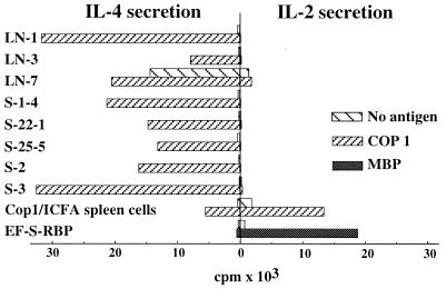
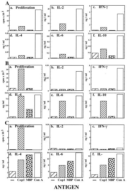
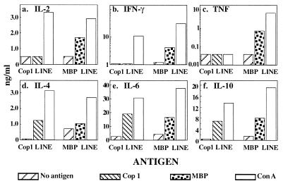
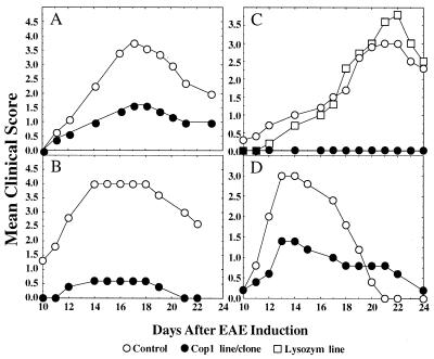
The synthetic amino acid copolymer copolymer 1 (Cop 1) suppresses experimental autoimmune encephalomyelitis (EAE) and is beneficial in multiple sclerosis. To further understand Cop 1 suppressive activity, we studied the cytokine secretion profile of various Cop 1-induced T cell lines and clones. Unlike T cell lines induced by myelin basic protein (MBP), which secreted either T cell helper type 1 (Th1) or both Th1 and Th2 cytokines, the T cell lines/clones induced by Cop 1 showed a progressively polarized development toward the Th2 pathway, until they completely lost the ability to secrete Th1 cytokines. Our findings indicate that the polarization of the Cop 1-induced lines did not result from the immunization vehicle or the in vitro growing conditions, but rather from the tendency of Cop 1 to preferentially induce a Th2 response. The response of all of the Cop 1 specific lines/clones, which were originated in the (SJL/JxBALB/c)F1 hybrids, was restricted to the BALB/c parental haplotype. Even though the Cop 1-induced T cells had not been exposed to the autoantigen MBP, they crossreacted with MBP by secretion of interleukin (IL)-4, IL-6, and IL-10. Administration of these T cells in vivo resulted in suppression of EAE induced by whole mouse spinal cord homogenate, in which several autoantigens may be involved. Secretion of anti-inflammatory cytokines by Cop 1-induced suppressor cells, in response to either Cop 1 or MBP, may explain the therapeutic effect of Cop 1 in EAE and in multiple sclerosis.


Figures

Figure 1
Figure 1
IL-2 and IL-4 secretion by T cell lines/clones. Eight different Cop 1-specific T cell lines and clones, spleen cells of mice injected with Cop 1 in ICFA, and a rat MBP-specific line (EF-S-RBP) were cultured with Cop 1 (10 μg/culture) or rat MBP (20 μg/culture). The presence of IL-2 and IL-4 in the supernatants was determined by their ability to support IL-2-dependent CTLD and IL-4-dependent CT4S cell lines. Results are expressed as mean cpm of thymidine incorporation for triplicate cultures. SDs were under 20% of the mean cpm. Results are from one representative experiment of more than 10 performed.
Figure 3
Figure 3
Proliferation and cytokine secretion profile of S-2 line during its development. (A) Initial response of spleen cells. (B) After 6 weeks in culture. (C) After 6 months in culture. Cells were cultured with no antigen, Cop 1 (50 μg/ml), MBP (100 μg/ml), and ConA (5 μg/ml). Proliferation was measured by thymidine incorporation for triplicate cultures. Cytokine concentration was measured by quantitative ELISA in duplicate wells for each one of duplicate culture supernatants. SDs were under 20% of the mean. Results represent one of three independent experiments.
Figure 2
Figure 2
Comparison between cytokine secretion by Cop 1- and MBP-induced Ts lines. Quantative ELISA of supernatants of two T cell lines established from spleens of mice, which had been rendered unresponsive to EAE by injection of Cop 1 or MBP in ICFA, stimulated by no antigen, Cop 1 (50 μg/ml), MBP (100 μg/ml), and ConA (5 μg/ml). Results are expressed as mean cytokine concentration of duplicate wells. SDs for both assays were under 20% of the mean.
Figure 4
Figure 4
Inhibition of MSCH-induced EAE by T cell lines and clones. (A) Cop 1-specific line S-2 after 2 months in culture (20 × 106/mouse). (B) S-2 line after 6 months in culture (10 × 106/mouse). (C) Cop 1-specific clone S-22–1 and lysozyme-specific line Lys-1 after 6 months in culture (15 × 106/mouse). (D) Cop 1-specific clone LN-3 after 6 months in culture (20 × 106/mouse). Cells were injected intravenously 3 days after stimulation with Cop 1 to (SJL/J×BALB/c)F1 mice 5–10 in a group, followed by EAE induction by MSCH. Control mice were induced with EAE alone. Results are expressed as mean daily clinical score of 5–10 mice in a group. Statistical analysis by Student’s t test: (A) S-2 after 2 months in culture, P < 0.04. (B) S-2 after 6 months in culture, P < 0.01. (C) l-22–1, P < 0.002, Lys-1, P < 0.7. (D) LN-3 clone, P < 0.09.

References

    1. Mosmann T R, Cherwinski H, Bond M W, Gieldin M A, Coffman R L. J Immunol. 1986;136:2348–2357. - PubMed
    1. Mosmann T R, Coffman R L. Annu Rev Immunol. 1989;7:145–173. - PubMed
    1. Abbas A K, Murphy K M, Sher A. Nature (London) 1996;383:787–793. - PubMed
    1. Liblau R S, Singer S M, McDevitt H O. Immunol Today. 1995;16:34–38. - PubMed
    1. Nicholson L B, Kuchroo V K. Curr Opin Immunol. 1996;8:837–842. - PubMed

Publication types

MeSH terms

LinkOut - more resources