. 1997 Sep 30;94(20):10868-72.
doi: 10.1073/pnas.94.20.10868.

The mechanism of cancer-mediated conversion of plasminogen to the angiogenesis inhibitor angiostatin

Affiliations

The mechanism of cancer-mediated conversion of plasminogen to the angiogenesis inhibitor angiostatin

S Gately et al. Proc Natl Acad Sci U S A. .

Abstract

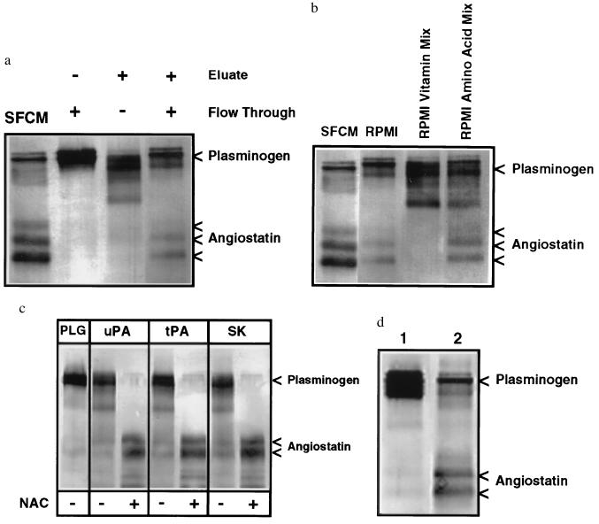
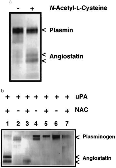
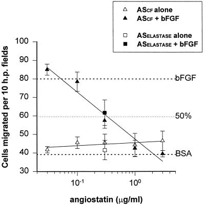
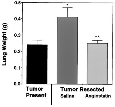
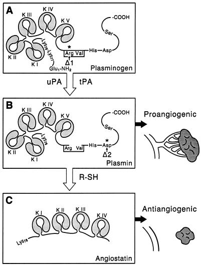
Angiostatin, a potent naturally occurring inhibitor of angiogenesis and growth of tumor metastases, is generated by cancer-mediated proteolysis of plasminogen. Human prostate carcinoma cells (PC-3) release enzymatic activity that converts plasminogen to angiostatin. We have now identified two components released by PC-3 cells, urokinase (uPA) and free sulfhydryl donors (FSDs), that are sufficient for angiostatin generation. Furthermore, in a defined cell-free system, plasminogen activators [uPA, tissue-type plasminogen activator (tPA), or streptokinase], in combination with one of a series of FSDs (N-acetyl-L-cysteine, D-penicillamine, captopril, L-cysteine, or reduced glutathione] generate angiostatin from plasminogen. An essential role of plasmin catalytic activity for angiostatin generation was identified by using recombinant mutant plasminogens as substrates. The wild-type recombinant plasminogen was converted to angiostatin in the setting of uPA/FSD; however, a plasminogen activation site mutant and a catalytically inactive mutant failed to generate angiostatin. Cell-free derived angiostatin inhibited angiogenesis in vitro and in vivo and suppressed the growth of Lewis lung carcinoma metastases. These findings define a direct mechanism for cancer-cell-mediated angiostatin generation and permit large-scale production of bioactive angiostatin for investigation and potential therapeutic application.


Figures

Figure 1
Figure 1
Western blot analysis of angiostatin generation. (a) The angiostatin-generating activity of PC-3 SFCM required two distinct fractions from Reactive Red 120-agarose chromatography. Angiostatin was generated when plasminogen was incubated with SFCM. No angiostatin generation was detected by incubation of plasminogen with the Reactive Red 120-agarose flow-through or 1.0 M KCl eluate dialyzed against TBS. When the flow-through and dialyzed eluate were combined, however, angiostatin generation was restored. (b) A cofactor for angiostatin generation was present in unconditioned RPMI 1640 medium and its amino acid mixture. Plasminogen incubated with PC-3 SFCM generates angiostatin. Plasminogen incubated with the dialyzed Reactive Red 120-agarose eluate supplemented with unconditioned RPMI 1640 medium also generates angiostatin. The cofactor activity of RPMI 1640 medium, necessary for angiostatin generation, was not present in the RPMI 1640 vitamin mixture but was present in the RPMI 1640 amino acid mixture. (c) Angiostatin was generated in a cell-free system consisting of human 0.2 μM plasminogen (PLG), uPA, tPA, or streptokinase and the sulfhydryl donor N-acetyl-l-cysteine (NAC). Human plasminogen incubated with uPA (0.2 nM), tPA (1.0 nM), or streptokinase (8.0 nM) generated angiostatin only in the presence of NAC (100 μM). (d) Human PC-3 prostate carcinoma cells, cultured for 24 h in the sulfhydryl-depleted RPMI 1640 medium, secreted sufficient FSDs and uPA to generate angiostatin from plasminogen. Human plasminogen was incubated with sulfhydryl-depleted RPMI 1640 medium and uPA (lane 1) or with identical sulfhydryl-depleted RPMI 1640 medium conditioned by human PC-3 prostate carcinoma cells (lane 2).
Figure 2
Figure 2
(a) Plasmin is converted to angiostatin in the presence of a FSD. Human plasminogen was converted to plasmin by incubation with uPA-Sepharose. Plasmin was only converted to angiostatin in the presence of 100 μM NAC. (b) Plasmin generation and catalytic activity is essential for angiostatin generation. Plasma-derived human plasminogen (0.2 μM), incubated with uPA (0.2 nM) and NAC (NAC) generates angiostatin (lane 1). The recombinant wild-type plasminogen (lanes 2 and 3) is also converted to angiostatin by the addition of uPA and NAC. The R561A activation site mutant (lanes 4 and 5), not susceptible to activation by plasminogen activators, failed to generate angiostatin when incubated with uPA and NAC. The D646E catalytically inactive mutant (lanes 6 and 7) also failed to generate angiostatin, demonstrating the requirement for plasmin catalytic activity.
Figure 3
Figure 3
Inhibition of bovine capillary endothelial cell migration by angiostatin produced in a cell-free system (ASCF). Endothelial cell migration in a Boyden chamber toward a range of concentrations of angiostatin was measured in the presence (solid symbol) or absence (open symbol) of stimulatory bFGF. As a control, single points using elastase-generated angiostatin (ASElastase) are shown (squares). These data demonstrate that ASCF inhibits bFGF-induced endothelial cell migration in a dose-dependent manner, with an ED50 of 0.33 μg/ml.
Figure 4
Figure 4
Angiostatin produced in a cell-free system suppresses the growth Lewis lung carcinoma lung metastases after resection of the primary tumor. The presence of the primary subcutaneous Lewis lung tumor suppressed the expansion of lung metastases (tumor present control). By contrast resection of the Lewis lung tumor and administration of saline resulted in a significant increase in the mean lung mass compared with the tumor present control, confirming primary tumor-mediated suppression of metastatic tumor growth (∗, P < 0.01). Subcutaneous administration of angiostatin after removal of the primary tumor, significantly suppressed the expansion of lung metastases to levels comparable to the tumor control group (angiostatin compared with saline; ∗∗, P < 0.01).
Figure 5
Figure 5
Conversion of the proangiogenic proteinase plasmin to the angiogenesis inhibitor angiostatin. (A) The zymogen plasminogen is converted to the active proteinase plasmin by cleavage of the Arg560-Val561 peptide bond by plasminogen activators such as uPA and tPA. (B) Plasmin is a proangiogenic proteinase capable of degrading a variety of extracellular matrix proteins, facilitating endothelial cell migration and angiogenesis. (C) Plasmin in the presence of a FSD is converted to the angiogenesis inhibitor angiostatin. The plasminogen activation site mutant R561A, indicated by Δ1, is not cleaved by plasminogen activators, preventing conversion of plasminogen to the plasmin intermediate required for angiostatin generation. The plasminogen mutant D646E, indicated by Δ2, is cleaved by plasminogen activators, but the resulting two-chain plasmin is inactive due to the substitution of a catalytically essential aspartic acid residue in the serine proteinase catalytic triad. In the presence of a FSD, the inactive D646E mutant plasmin is not converted to angiostatin, demonstrating the requirement for plasmin catalytic activity.

References

    1. Folkman J. J Natl Cancer Inst. 1990;82:4–6. - PubMed
    1. Folkman J, Watson K, Ingber D, Hanahan D. Nature (London) 1989;339:58–61. - PubMed
    1. Weidner N, Semple J P, Welch W R, Folkman J. N Engl J Med. 1991;324:1–8. - PubMed
    1. Fotsis T, Zhang Y, Pepper M S, Adlercreutz H, Montesano R, Nawroth P P, Schweigerer L. Nature (London) 1994;368:237–239. - PubMed
    1. Kim K J, Li B, Winer J, Armanini M, Gillett N, Phillips H S, Ferrara N. Nature (London) 1993;362:841–844. - PubMed

Publication types

MeSH terms