. 1997 Dec 1;139(5):1183-95.
doi: 10.1083/jcb.139.5.1183.

Functional dissection of COP-I subunits in the biogenesis of multivesicular endosomes

Affiliations

Functional dissection of COP-I subunits in the biogenesis of multivesicular endosomes

F Gu et al. J Cell Biol. .

Abstract

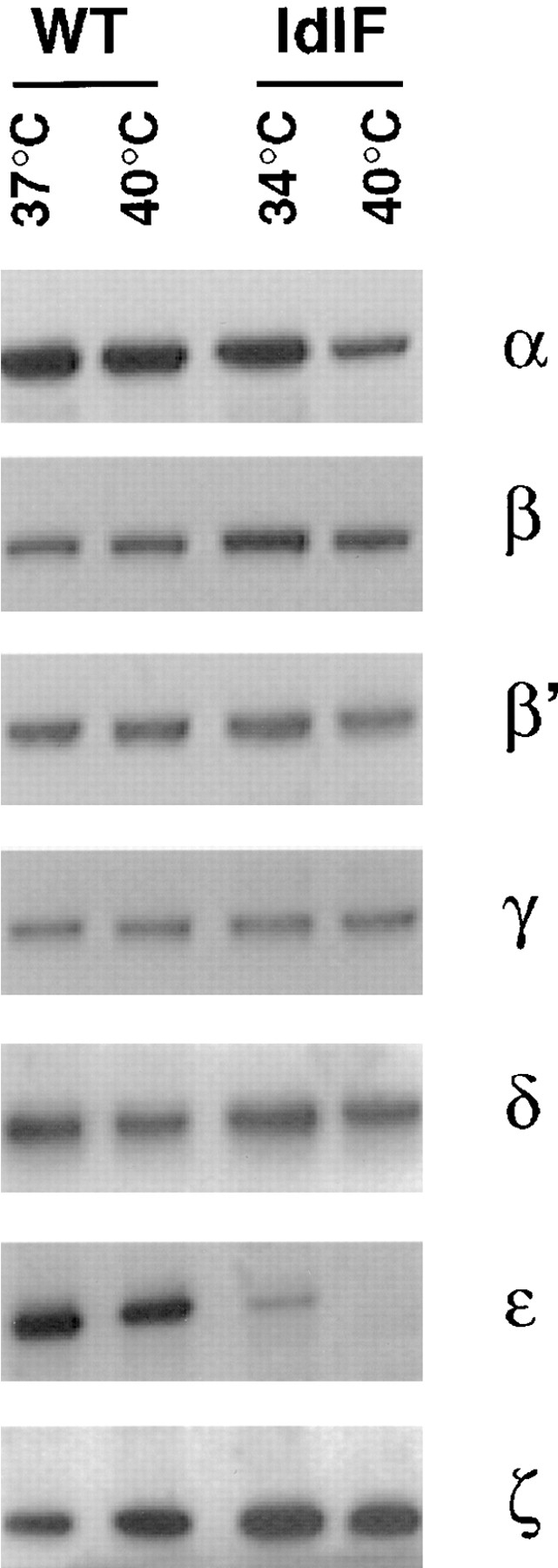
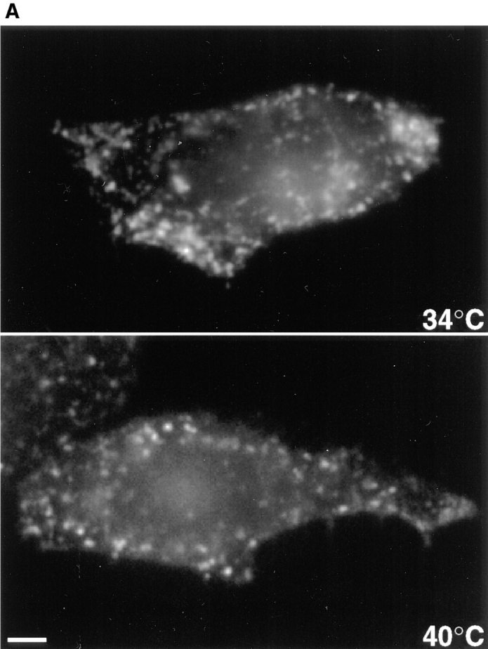
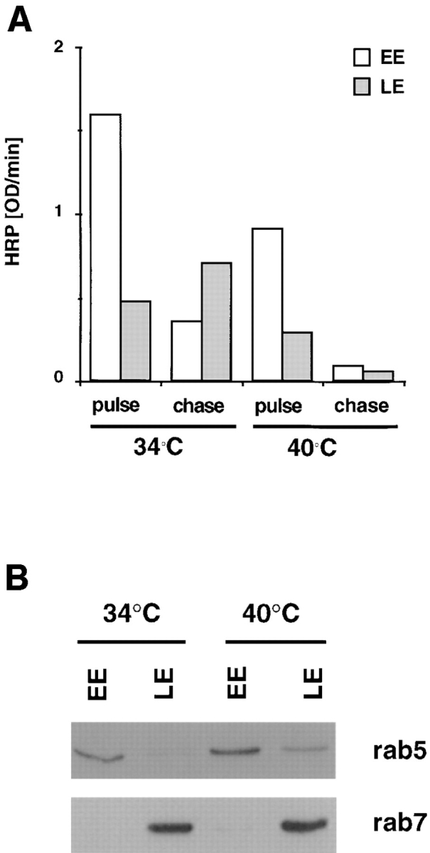
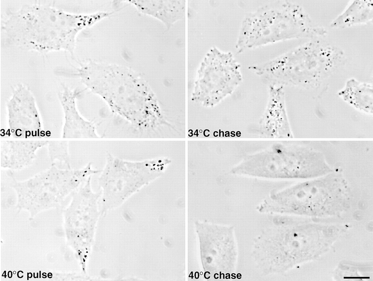
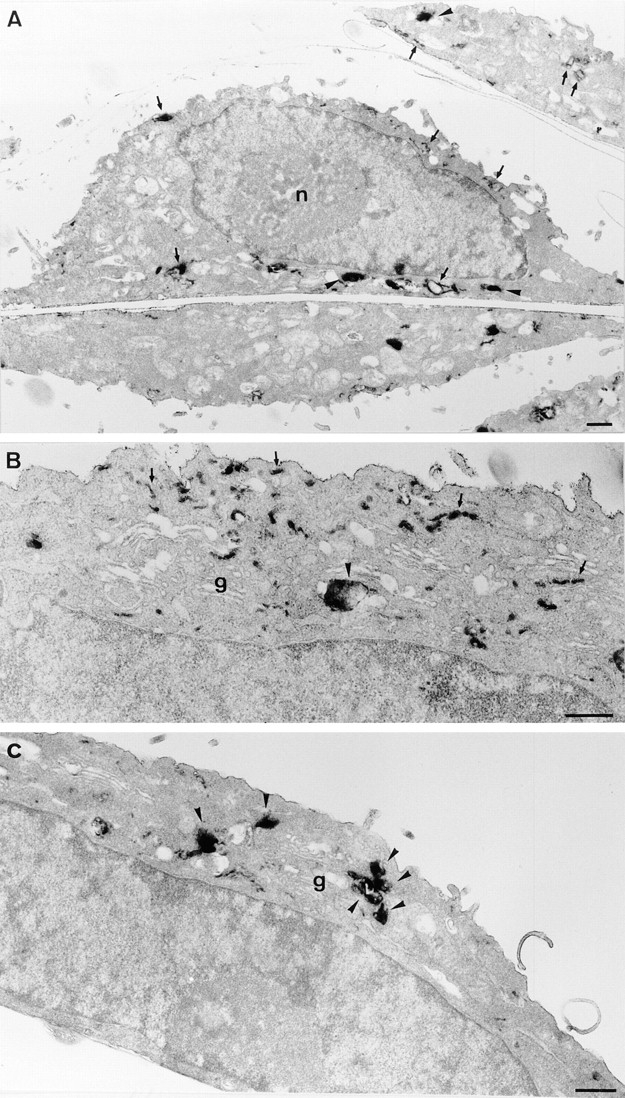
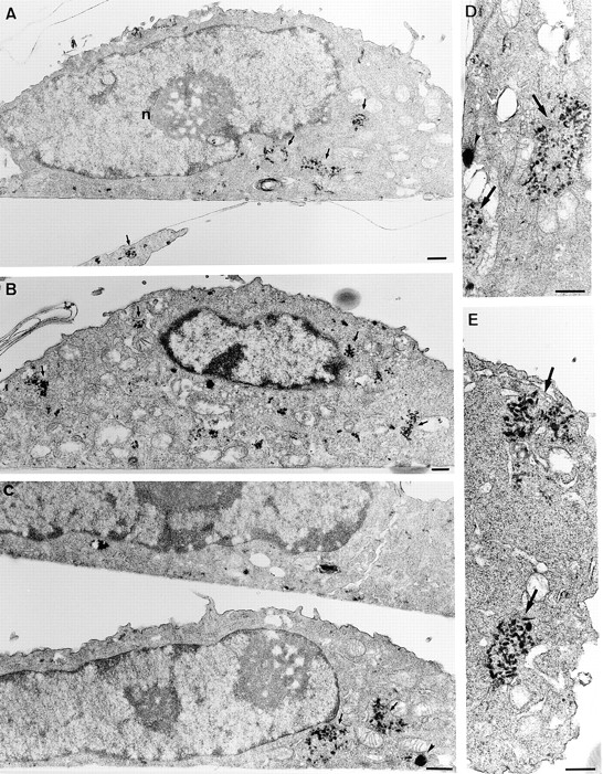
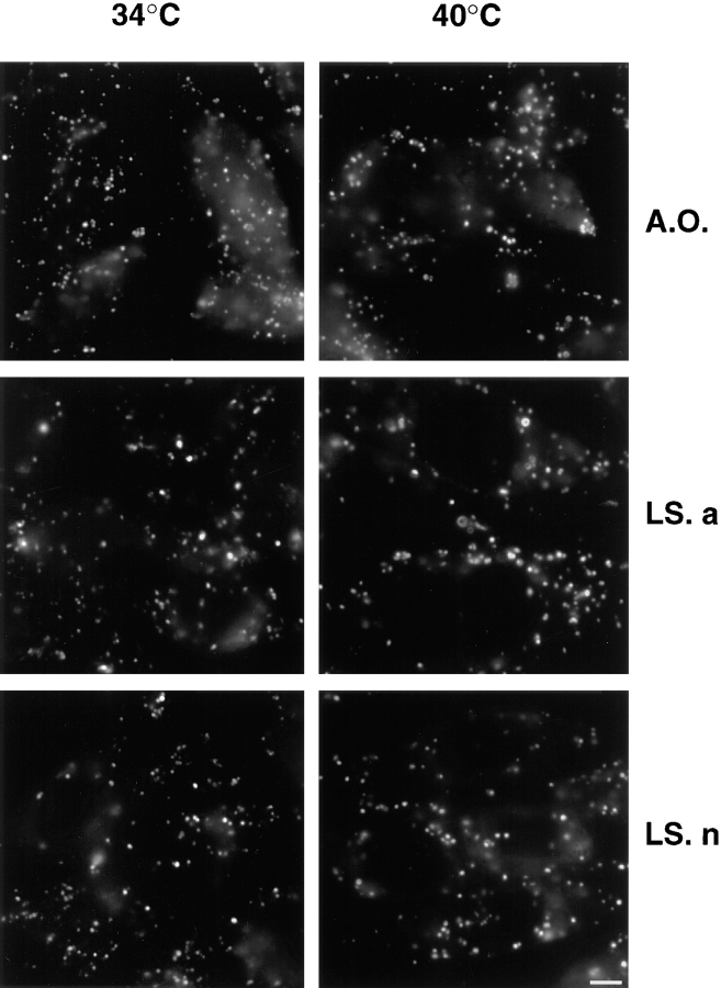
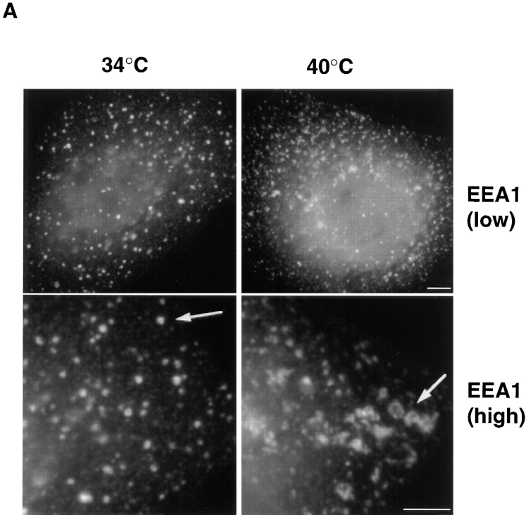
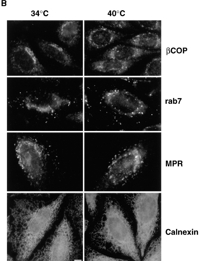
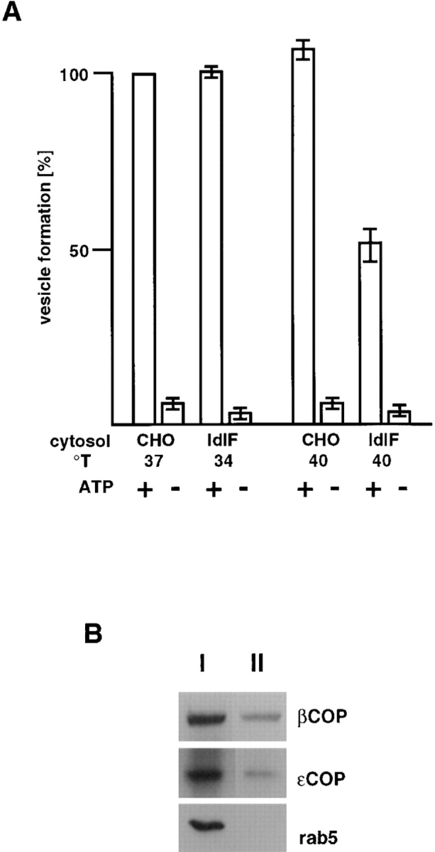
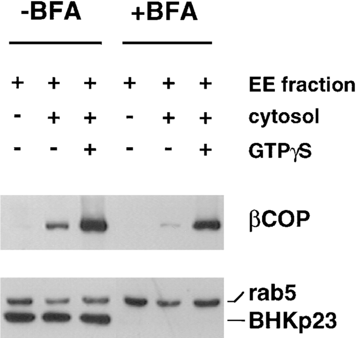
In the present paper, we show that transport from early to late endosomes is inhibited at the restrictive temperature in a mutant CHO cell line (ldlF) with a ts-defect in epsilon coatomer protein (epsilonCOP), although internalization and recycling continue. Early endosomes then appear like clusters of thin tubules devoid of the typical multivesicular regions, which are normally destined to become vesicular intermediates during transport to late endosomes. We also find that the in vitro formation of these vesicles from BHK donor endosomes is inhibited in cytosol prepared from ldlF cells incubated at the restrictive temperature. Although epsilonCOP is rapidly degraded in ldlF cells at the restrictive temperature, cellular amounts of the other COP-I subunits are not affected. Despite the absence of epsilonCOP, we find that a subcomplex of beta, beta', and zetaCOP is still recruited onto BHK endosomes in vitro, and this binding exhibits the characteristic properties of endosomal COPs with respect to stimulation by GTPgammaS and sensitivity to the endosomal pH. Previous studies showed that gamma and deltaCOP are not found on endosomes. However, alphaCOP, which is normally present on endosomes, is no longer recruited when epsilonCOP is missing. In contrast, all COP subunits, except obviously epsilonCOP itself, still bind BHK biosynthetic membranes in a pH-independent manner in vitro. Our observations thus indicate that the biogenesis of multivesicular endosomes is coupled to early endosome organization and depends on COP-I proteins. Our data also show that membrane association and function of endosomal COPs can be dissected: whereas beta, beta', and zetaCOP retain the capacity to bind endosomal membranes, COP function in transport appears to depend on the presence of alpha and/or epsilonCOP.


Figures

Figure 1
Figure 1
Distribution of COP-I subunits in CHO and ldlF cells. Cytosols were prepared from ldlF cells incubated at the permissive (34°C) or restrictive (40°C) temperature. For comparison, cytosols were also prepared from WT CHO cells incubated at 37 or 40°C. The COP-I composition of each cytosol was analyzed by SDS-PAGE followed by Western blotting using antibodies against each of the COP-I components. 20 μg protein was loaded per lane.
Figure 2
Figure 2
Endocytosis in ldlF cells. Cells were incubated at the permissive (34°C) or restrictive (40°C) temperature for 6 h. (A) Transferrin internalization. Cells had been transiently transfected with the cDNA encoding for the human transferrin receptor before incubation at 34 or 40°C. Transferrin internalization was visualized by fluorescence microscopy after 5 min incubation with 50 μg/ml rhodamine-transferrin at the corresponding temperature. (B) Continuous internalization of HRP. Cells were incubated with 3 mg/ml HRP at the corresponding temperature for 5, 15, 30, 60, or 120 min. The amounts of endocytosed HRP were quantified and expressed as OD U/min/mg cellular protein. (C) Recycling of internalized HRP. Cells were incubated with 0.5 mg/ ml HRP for 5 min at the corresponding temperature, washed, and then reincubated for 10, 20, 30, or 40 min. At each time point, cells and the media were collected. At each time point, HRP remaining associated to the monolayer is expressed as a percentage of the total (cell-associated and regurgitated). In B and C, each panel shows the mean of two representative series of experiments. Bar, 5 μm.
Figure 2
Figure 2
Endocytosis in ldlF cells. Cells were incubated at the permissive (34°C) or restrictive (40°C) temperature for 6 h. (A) Transferrin internalization. Cells had been transiently transfected with the cDNA encoding for the human transferrin receptor before incubation at 34 or 40°C. Transferrin internalization was visualized by fluorescence microscopy after 5 min incubation with 50 μg/ml rhodamine-transferrin at the corresponding temperature. (B) Continuous internalization of HRP. Cells were incubated with 3 mg/ml HRP at the corresponding temperature for 5, 15, 30, 60, or 120 min. The amounts of endocytosed HRP were quantified and expressed as OD U/min/mg cellular protein. (C) Recycling of internalized HRP. Cells were incubated with 0.5 mg/ ml HRP for 5 min at the corresponding temperature, washed, and then reincubated for 10, 20, 30, or 40 min. At each time point, cells and the media were collected. At each time point, HRP remaining associated to the monolayer is expressed as a percentage of the total (cell-associated and regurgitated). In B and C, each panel shows the mean of two representative series of experiments. Bar, 5 μm.
Figure 4
Figure 4
Subcellular fractionation of ldlF endosomes. (A) Cells prepared as in Fig. 3 were incubated at the permissive (34°C) or restrictive (40°C) temperature with medium containing 4 mg/ml HRP for 5 min (pulse) or subsequently reincubated for 40 min in the absence of marker (chase). Early (EE) and late (LE) endosomes were then separated by subcellular fractionation. The total HRP content of the fractions was quantified using a colorimetric reaction and is expressed as OD U/min. HRP recovery in the fractions corresponded to ∼40% of the total, latent cell-associated activity (∼50% lost to the nuclear pellet after gentle homogenization) and HRP latency after homogenization was always >70%. (B) The recovery of rab5 and rab7 in fractions (EE or LE at each temperature) prepared as in A was analyzed by SDS gel electrophoresis, followed by Western blotting with indicated antibodies. Each lane contained 20% of the total protein in the corresponding fraction.
Figure 3
Figure 3
Distribution of endocytosed HRP in ldlF cells. Cells were maintained at the permissive (34°C) or restrictive (40°C) temperature for 6 h. Then, they were incubated at the corresponding temperature in the presence of 10 mg/ml high activity HRP for 5 min to label early endosomes (pulse). Late endosomes were labeled after reincubation of the cells for 30 min in the absence of HRP, at the corresponding temperature (chase). After cell fixation, intracellular HRP distribution was revealed using a cytochemical reaction and analyzed by phase contrast light microscopy. Bar, 10 μm.
Figure 7
Figure 7
Ultrastructure of ldlF endosomes at the permissive temperature. Cells cultured at 34°C were incubated with HRP for 5 min to label early endosomes and then either fixed immediately (A and B) or further incubated at 34°C for 30 min to label late endosomes (C). Cells were then processed for plastic sections, and semithick (∼150 nm; A and C) or ultrathin sections (50 nm; B) were prepared. Early endosomal compartments (A and B) are comprised of tubular and cisternal regions (arrows) and vesicular domains (arrowheads), as in other cells. B shows a higher magnification view of the Golgi (g) area. After further incubation for 30 min (C), HRP was rarely observed within tubular domains. As expected, it was distributed within larger multivesicular elements concentrated in the Golgi area (arrowheads), which presumably correspond to late endosomes. Bars, 0.5 μm.
Figure 8
Figure 8
Ultrastructure of ldlF endosomes at the restrictive temperature. Cells cultured at 40°C for 6 h were incubated with HRP for 5 min and then either fixed immediately (A and B) or further incubated at 40°C for 30 min (C–E). Semithick (A and C) or ultrathin (B, D, and E) sections of the cell pellet were prepared as in Fig. 8. Early endosomal compartments (A and B) were composed of small tubular and vesicular elements that were predominantly in discrete clusters (arrows). Few large vesicular profiles were evident (compare with Fig. 7, A and B). After further incubation for 30 min, little HRP remained in the cells (see Figs. 2–4). However, when detected (C–E), the bulk of HRP was still observed within clusters of tubular and vesicular elements (arrows), which appeared identical to those labeled after the 10 min pulse. Few vesicular elements were labeled (arrowhead). Note the clear difference when compared with cells incubated for the same time at the permissive temperature (Fig. 7 C). As shown at higher magnification (D and E), labeled elements comprise vesicles and short tubules, which appear discontinuous from the analysis of both semithick (C) and thin (D and E) sections. Bars, 0.5 μm.
Figure 5
Figure 5
Acidification in ldlF cells. Cells prepared as in Fig. 3 at the permissive or restrictive temperature were treated with acridine orange (A.O.), LysoSensor acidic (detection range pH 4.5–6; LS. a) and LysoSensor neutral (detection range pH 6.5–8; LS. n) for 10 min to reveal acidic compartments. The intrinsic fluorescence of each dye after accumulation within acidic endosomes and lysosomes was observed by fluorescence microscopy. Bar, 5 μm.
Figure 6
Figure 6
Distribution of different markers in ldlF cells. (A) Cells prepared as in Fig. 3 were processed for immunofluorescence microscopy using human antiserum against EEA1. Arrows point at changes in the appearance of early endosomal elements at 40°C, when compared with 34°C. low, low magnification; high, high magnification. (B) Cells were processed for immunofluorescence microscopy as in A using the maD antibody against βCOP or a rabbit antiserum against either the Man6P-R, rab7, or calnexin. The maD antibody does not label endosomal βCOP to any significant extent under these conditions. Bars, 5 μm.
Figure 6
Figure 6
Distribution of different markers in ldlF cells. (A) Cells prepared as in Fig. 3 were processed for immunofluorescence microscopy using human antiserum against EEA1. Arrows point at changes in the appearance of early endosomal elements at 40°C, when compared with 34°C. low, low magnification; high, high magnification. (B) Cells were processed for immunofluorescence microscopy as in A using the maD antibody against βCOP or a rabbit antiserum against either the Man6P-R, rab7, or calnexin. The maD antibody does not label endosomal βCOP to any significant extent under these conditions. Bars, 5 μm.
Figure 9
Figure 9
In vitro formation of ECV/MVBs. (A) BHK early endosomes containing HRP internalized for 5 min at 37°C were prepared by flotation on a sucrose gradient and used as donor membranes in the assay (Aniento et al., 1996). Donor endosomes were incubated with (+) or without (−) an ATP-regenerating system and in the presence of cytosol. Cytosols were prepared, as indicated, from ldlF cells incubated at the permissive (34°C) or restrictive (40°C) temperature, or from WT CHO cells incubated at 37 or 40°C. Vesicles formed in vitro were then separated from donor membranes by flotation in a gradient, and the HRP content of both fractions quantified. In the assay, ∼10% of HRP originally internalized into early endosomes was entrapped within vesicles formed in vitro, as shown previously (Aniento et al., 1996). Efficiency of vesicle formation is expressed as a percentage of the control with cytosol from WT CHO cells incubated at 37°C. (B) The assay was carried out in the presence of cytosol from CHO cells incubated at 37°C. Then, donor endosomes (I) and ECV/MVBs formed in vitro (II) were pelleted and analyzed by SDS gel electrophoresis and Western blotting with antibodies against βCOP, εCOP, and rab5, as indicated. Each lane contained 35% of the protein content of the corresponding fraction.
Figure 10
Figure 10
Separation of endosomal and biosynthetic membranes. To achieve optimal conditions for the separation of endosomal and biosynthetic membranes, BHK cells were pretreated with brefeldin A (+BFA). The drug was absent from all subsequent steps. In the control, the drug was omitted (−BFA). After homogenization, the corresponding postnuclear supernatants were fractionated using a sucrose step flotation gradient (Gorvel et al., 1991; Aniento et al., 1996), and fractions enriched in early endosomes were collected. The COP binding capacity of these membranes was measured after incubating 50 μg of each fraction with 500 μg BHK cytosol for 15 min at 37°C in the presence of 10 μM GTPγS, to stimulate COP recruitment. Membranes were then retrieved by flotation on a step gradient and analyzed by SDS gel electrophoresis followed by Western blotting using antibodies against βCOP, rab5, and BHKp23.
Figure 11
Figure 11
COP binding to endosomal and biosynthetic membranes. Cytosols were prepared from ldlF cells incubated at the restrictive temperature, as in Fig. 1. The COP binding capacity of biosynthetic (BM) or early endosomal (EE) membranes prepared from BHK cells pretreated with brefeldin A was tested as in Fig 10, except that ldlF cytosol was used with (G and NG) or without (C) 10 μM GTPγS. When indicated (NG), the pH of endosomes was preneutralized with 50 μM nigericin before GTPγS addition. Membranes were retrieved and analyzed using antibodies against each COP subunit. Western blots were developed using the ECL reaction; exposure times were five times longer for EE than for BM membranes to ensure that signals remained in the linear detection range.

References

    1. Aniento F, Emans N, Griffiths G, Gruenberg J. Cytoplasmic dynein-dependent vesicular transport from early to late endosomes. J Cell Biol. 1993;123:1373–1388. - PMC - PubMed
    1. Aniento F, Gu F, Parton R, Gruenberg J. An endosomal βcop is involved in the pH-dependent formation of transport vesicles destined for late endosomes. J Cell Biol. 1996;133:29–41. - PMC - PubMed
    1. Besterman JM, Airhart JA, Woodworth RC, Low RB. Exocytosis of pinocytosed fluid in cultured cells: kinetic evidence for rapid turnover and compartmentation. J Cell Biol. 1981;91:716–727. - PMC - PubMed
    1. Bomsel M, Parton R, Kuznetsov SA, Schroer TA, Gruenberg J. Microtubule and motor dependent fusion in vitro between apical and basolateral endocytic vesicles from MDCK cells. Cell. 1990;62:719–731. - PubMed
    1. Bradford MM. A rapid and sensitive method for the quantitation of microgram quantities of protein utilizing the principle of protein-dye binding. Anal Biochem. 1976;72:248–254. - PubMed

Publication types