Comparative Study
. 1997 Dec 1;139(5):1219-29.
doi: 10.1083/jcb.139.5.1219.

Growth and muscle defects in mice lacking adult myosin heavy chain genes

Affiliations
Comparative Study

Growth and muscle defects in mice lacking adult myosin heavy chain genes

L J Acakpo-Satchivi et al. J Cell Biol. .

Abstract

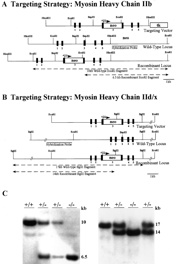
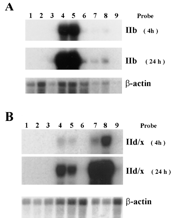
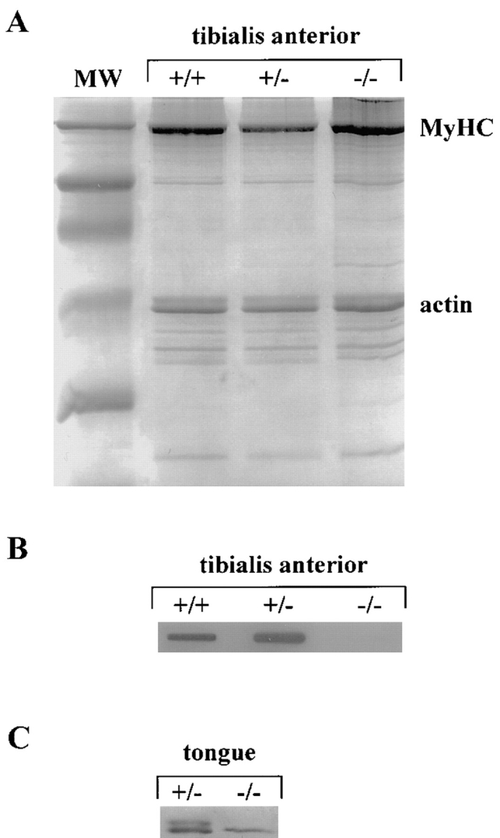
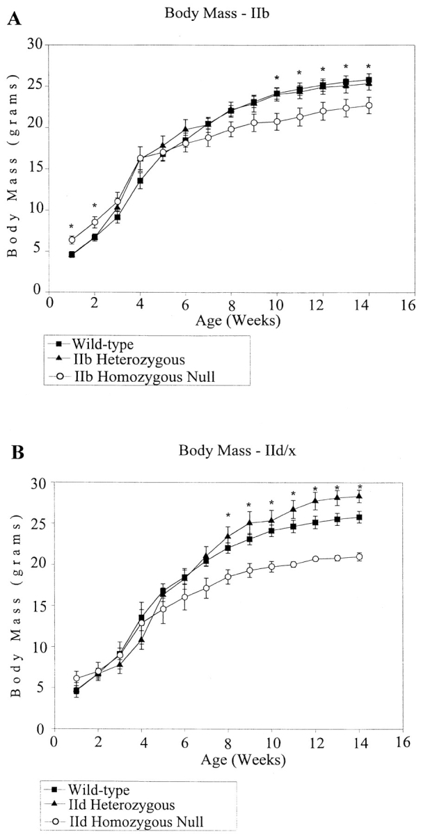
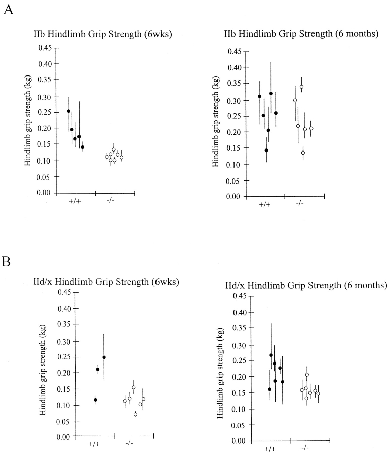
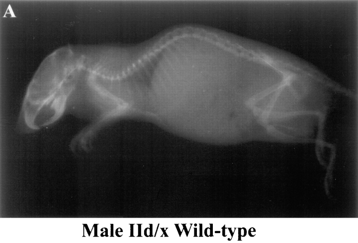
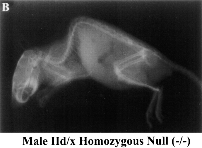
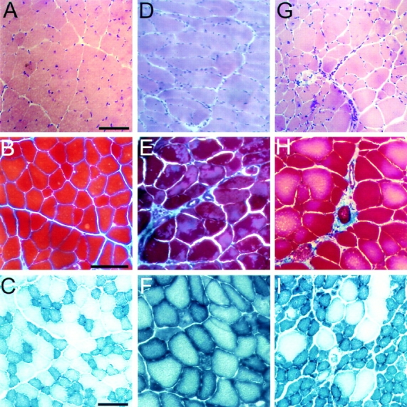
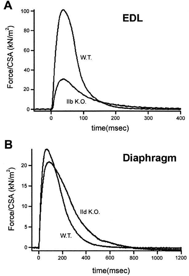
The three adult fast myosin heavy chains (MyHCs) constitute the vast majority of the myosin in adult skeletal musculature, and are >92% identical. We describe mice carrying null mutations in each of two predominant adult fast MyHC genes, IIb and IId/x. Both null strains exhibit growth and muscle defects, but the defects are different between the two strains and do not correlate with the abundance or distribution of each gene product. For example, despite the fact that MyHC-IIb accounts for >70% of the myosin in skeletal muscle and shows the broadest distribution of expression, the phenotypes of IIb null mutants are generally milder than in the MyHC-IId/x null strain. In addition, in a muscle which expresses both IIb and IId/x MyHC in wild-type mice, the histological defects are completely different for null expression of the two genes. Most striking is that while both null strains exhibit physiological defects in isolated muscles, the defects are distinct. Muscle from IIb null mice has significantly reduced ability to generate force while IId null mouse muscle generates normal amounts of force, but has altered kinetic properties. Many of the phenotypes demonstrated by these mice are typical in human muscle disease and should provide insight into their etiology.


Figures

Figure 1
Figure 1
The MyHC-IIb and IId/x targeting strategies and Southern blot analysis of F2 mice. Targeting vector, wild-type and recombinant loci are shown with relevant restriction sites. (A) A 6-kb HindIII fragment containing the first five exons of the MyHC-IIb gene was subcloned from the λ10a clone. The PGK-neomycin cassette was inserted into the third exon, downstream of the translational start site, and in the opposite orientation to the MyHC gene. The PGK-thymidine kinase gene for negative selection was placed 3′ of the construct as shown. Homologously recombined clones were assayed for by EcoRI Southern blot analysis using a probe 3′ of the construct, as indicated. (B) A 11-kb EcoRI fragment containing the first five exons of the MyHC-IId/x gene was subcloned from the λ19a clone. The PGK-neomycin cassette was inserted into a unique BspEI site, downstream of the translational start site, in the same orientation as the MyHC gene. Homologous recombination was assayed by BglII Southern Blot analysis using a probe 5′ of the targeting construct, as shown. (C) The first panel depicts a EcoRI southern blot analysis of F2 mice from the IIb targeting experiment. Wild-type, heterozygous and homozygous null animals are shown. The second panel depicts a BglII southern blot analysis of F2 mice from the IId/x targeting experiment.
Figure 2
Figure 2
RNA analysis. In both the IIb and IId/x animals, total RNA from heart (lanes 1– 3), lower leg (lanes 4– 6), and masseter (lanes 7–9) was examined for the expression of IIb and IId/x RNA, respectively. A β-actin probe was used to control for the amount of RNA. (A) The expression of MyHC-IIb was analyzed by RNase protection using a 120-nt fragment containing the 3′ UTR for the MyHC-IIb gene. The heart RNA is negative for IIb expression in wild-type (lane 1), heterozygous (lane 2), and homozygous null animals (lane 3). In skeletal muscle samples, wild-type (lanes 4 and 7) and heterozygous (lanes 5 and 8) but not homozygous animals (lanes 6 and 9) express of IIb RNA. (B) The expression of MyHC-IId/x RNA was analyzed using a 90-nt fragment containing the 3′ UTR for the gene. The heart RNA is negative for expression in wild-type (lane 1), heterozygous (lane 2), and homozygous null animals (lane 3). The lower leg and masseter samples show expression in wild-type (lanes 4 and 7), and heterozygous animals (lanes 5 and 8), but not in homozygous null samples (lanes 6 and 9).
Figure 3
Figure 3
MyHC-IIb protein expression. Myofibrils were prepared from tibialis anterior muscles of MyHC-IIb+/+, MyHC-IIb+/−, and MyHC-IIb−/− mice. (A) Myofibrils were separated by SDS-PAGE and stained with Coomassie blue. Lane 1, MW; lane 2, IIb+/+; lane 3, IIb+/−; lane 4, IIb−/−. (B) Myofibrils were blotted onto nitrocellulose and probed with an antibody against MyHC-IIb (BFF3). (C) High resolution gel electrophoresis of MyHC from tongue from +/+ and −/− IId mice.
Figure 4
Figure 4
Growth rates of MyHC-IIb and IId/x mice. Weight measurements for wild-type (▪), heterozygous (▴), and homozygous (○) male animals were taken once a week for a time period of 100 d. The homozygous null mice weigh significantly less than their +/+ or +/− littermates. Asterisks indicate time points at which a statistically significant difference (P < 0.05) exists between combined average +/+ and −/− body weights. For the IIb mice, n = 14, 4, and 4; for the IId/x mice, n = 14, 7, and 8 for wild-type, heterozygous, and homozygous mice, respectively.
Figure 5
Figure 5
Hindlimb grip strength measurements in MyHC-IIb and IId/x mice. Peak grip strengths were measured on the hindlimbs of mice. Five individual measurements were made for each mouse. Each point in the graph represents the average of the five measurements. There is a statistically significant difference (P < 0.01) between the combined average grip-strengths between −/− and +/+ mice at 6 wk of age, but at 6 mo, only the IId/x null mice exhibit sustained deficiencies in grip strength.
Figure 6
Figure 6
Kyphosis in MyHC-IId/x null mice. Male mice at 12 wk of age (wild-type) and 8 wk of age (MyHC-IId/x−/−) were anesthetized and x-rayed. (A) MyHC-IId/x wild-type +/+ male. (B) MyHC-IId/x−/− male.
Figure 6
Figure 6
Kyphosis in MyHC-IId/x null mice. Male mice at 12 wk of age (wild-type) and 8 wk of age (MyHC-IId/x−/−) were anesthetized and x-rayed. (A) MyHC-IId/x wild-type +/+ male. (B) MyHC-IId/x−/− male.
Figure 7
Figure 7
Histologic analysis of tibialis anterior muscle from MyHC-IId/x mice. Cross-sections of tibialis anterior muscle from 6-wk-old wild-type (A–C), MyHC-IIb−/− (D–F), and MyHC-IId/x−/− (G–I) mice were stained with hematoxylin and eosin (H&E) (A, D, and G), Masson trichrome (B, E, and H), and NADH-TR (C, F, and I). Note the general disarray (MyHC-IId/x−/−, G), change in fiber diameter in H&E and NADH-TR stains (F and I) and the interstitial fibrosis visualized with Masson Trichrome staining (E and H).
Figure 8
Figure 8
Representative examples of single twitches from whole EDL muscles (A) and small diaphragm strips (B) at 22°C. Muscle length and stimulation voltage (0.5-ms duration) was set to produce maximum twitch force. Muscle was stimulated at time zero. Mouse genotype is as indicated.

References

    1. Barany M. ATPase activity of myosin correlated with speed of muscle shortening. J Gen Physiol. 1967;50:197–218. - PMC - PubMed
    1. Berne, R.M., and M.N. Levy. 1996. Principles of Physiology. Mosby-Year Book, Inc., St. Louis, MO.
    1. Bouvagnet PF, Strehler EE, White GE, Strehler-Page MA, Nadal-Ginard B, Mahdavi V. Multiple positive and negative 5′ regulatory elements control the cell-type-specific expression of the embryonic skeletal myosin heavy-chain gene. Mol Cell Biol. 1987;7:4377–4389. - PMC - PubMed
    1. Bridges LR, Coulton GR, Howard G, Moss HJ, Mason R. The neuromuscular basis of hereditary kyphoscoliosis in the mouse. Muscle Nerve. 1992;15:172–179. - PubMed
    1. Carter GT, Abresch RT, Fowler WM, Jr, Johnson ER, Kilmer DD, McDonald CM. Profiles of neuromuscular diseases. Spinal muscular atrophy. Am J Phys Med Rehab. 1995;74:150–159. - PubMed

Publication types

Substances