. 1997 Dec 1;139(5):1255-69.
doi: 10.1083/jcb.139.5.1255.

Supervillin (p205): A novel membrane-associated, F-actin-binding protein in the villin/gelsolin superfamily

Affiliations

Supervillin (p205): A novel membrane-associated, F-actin-binding protein in the villin/gelsolin superfamily

K N Pestonjamasp et al. J Cell Biol. .

Abstract

Actin-binding membrane proteins are involved in both adhesive interactions and motile processes. We report here the purification and initial characterization of p205, a 205-kD protein from bovine neutrophil plasma membranes that binds to the sides of actin filaments in blot overlays. p205 is a tightly bound peripheral membrane protein that cosediments with endogenous actin in sucrose gradients and immunoprecipitates. Amino acid sequences were obtained from SDS-PAGE-purified p205 and used to generate antipeptide antibodies, immunolocalization data, and cDNA sequence information. The intracellular localization of p205 in MDBK cells is a function of cell density and adherence state. In subconfluent cells, p205 is found in punctate spots along the plasma membrane and in the cytoplasm and nucleus; in adherent cells, p205 concentrates with E-cadherin at sites of lateral cell-cell contact. Upon EGTA-mediated cell dissociation, p205 is internalized with E-cadherin and F-actin as a component of adherens junctions "rings." At later times, p205 is observed in cytoplasmic punctae. The high abundance of p205 in neutrophils and suspension-grown HeLa cells, which lack adherens junctions, further suggests that this protein may play multiple roles during cell growth, adhesion, and motility. Molecular cloning of p205 cDNA reveals a bipartite structure. The COOH terminus exhibits a striking similarity to villin and gelsolin, particularly in regions known to bind F-actin. The NH2 terminus is novel, but contains four potential nuclear targeting signals. Because p205 is now the largest known member of the villin/gelsolin superfamily, we propose the name, "supervillin." We suggest that supervillin may be involved in actin filament assembly at adherens junctions and that it may play additional roles in other cellular compartments.


Figures

Figure 1
Figure 1
Neutrophil plasma membranes are highly enriched in p205. Fractions highly enriched in plasma membranes (A) or secretory vesicles (B) were analyzed for the presence of p205 by F-actin blot overlay (C). Aliquots (100 μg protein) were fractionated on a 5% SDS–polyacrylamide gel, transferred to nitrocellulose, and then probed with 125I-labeled F-actin. Lane 1, plasma membranes; lane 2, secretory vesicles; and lane 3, cytosol. E/M, the position of ezrin and moesin. Other cytosolic actin-binding proteins also are detected by this method (lane 3).
Figure 1
Figure 1
Neutrophil plasma membranes are highly enriched in p205. Fractions highly enriched in plasma membranes (A) or secretory vesicles (B) were analyzed for the presence of p205 by F-actin blot overlay (C). Aliquots (100 μg protein) were fractionated on a 5% SDS–polyacrylamide gel, transferred to nitrocellulose, and then probed with 125I-labeled F-actin. Lane 1, plasma membranes; lane 2, secretory vesicles; and lane 3, cytosol. E/M, the position of ezrin and moesin. Other cytosolic actin-binding proteins also are detected by this method (lane 3).
Figure 2
Figure 2
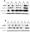
p205 is a peripheral component of the neutrophil plasma membrane skeleton. (A) Neutrophil plasma membranes were extracted with either buffer alone (lane 1), or buffer containing a final concentration of 2.5 mM MgATP (lane 2), 0.25 M KCl (lane 3), 1.0 M KCl (lane 4), 0.1 M sodium carbonate (lane 5), or 0.1 M NaOH (lane 6). For each extraction condition, high speed supernatants (S) and pellets (P) from 130-μg membranes were electrophoresed, blotted, and then probed with 125I-labeled F-actin and with specific antibodies. (B) Neutrophil plasma membranes (100 μg per treatment) were extracted with either buffer alone (lane 1), or buffer containing 1% Triton X-100 (lane 2), 1% Triton X-100, 50 mM NaCl (lane 3), 1% Triton X-100, 250 mM NaCl (lane 4), 3% octylglucoside, 250 mM NaCl (lane 5), or 0.1% SDS (lane 6) and processed as above. A positive control consisted of membranes extracted with 1% SDS at 70°C for 10 min (lane 7). The higher mobility F-actin–binding polypeptide present in B is consistently observed after detergent treatment; this band also reacts with an antibody against p205 sequences (see below), suggesting a close structural relationship with p205.
Figure 3
Figure 3
Purification of p205 by SDS-PAGE. (A) Neutrophil plasma membranes (lane 1) and Triton X-100–insoluble pellets (lanes 2–5) were separated on a 5% polyacrylamide gel and stained with silver (lanes 1 and 2), or electrotransfered to nitrocellulose and probed with either 125I-labeled F-actin (lane 3), or with antibodies against myosin II (lane 4), or nonerythroid spectrin/fodrin (lane 5). Loads represent 100 μg membranes or equivalent amounts of Triton X-100–insoluble pellets. The location of p205 is indicated (arrowheads). (B) Eight microsequences were obtained from proteolytic digests of SDS-PAGE–purified p205. Polyclonal rabbit antibodies were generated against synthetic peptides corresponding to two of these sequences (pepA and pepB). Residues at variance with the deduced amino acid sequence (Fig. 11 A) are underlined; a lysine deduced from the cleavage specificity of Endo-LysC is shown in parentheses.
Figure 3
Figure 3
Purification of p205 by SDS-PAGE. (A) Neutrophil plasma membranes (lane 1) and Triton X-100–insoluble pellets (lanes 2–5) were separated on a 5% polyacrylamide gel and stained with silver (lanes 1 and 2), or electrotransfered to nitrocellulose and probed with either 125I-labeled F-actin (lane 3), or with antibodies against myosin II (lane 4), or nonerythroid spectrin/fodrin (lane 5). Loads represent 100 μg membranes or equivalent amounts of Triton X-100–insoluble pellets. The location of p205 is indicated (arrowheads). (B) Eight microsequences were obtained from proteolytic digests of SDS-PAGE–purified p205. Polyclonal rabbit antibodies were generated against synthetic peptides corresponding to two of these sequences (pepA and pepB). Residues at variance with the deduced amino acid sequence (Fig. 11 A) are underlined; a lysine deduced from the cleavage specificity of Endo-LysC is shown in parentheses.
Figure 11
Figure 11
Strategy used for the cloning of sequences representing the full-length supervillin cDNA (A), and Northern blots with probes from the 5′ and 3′ ends of the predicted sequence. (A) A schematic of the supervillin cDNA showing the 5,376-bp coding region (gray box), the 5′ and 3′ untranslated regions, and the sequences used as probes for the Northern blots (hatched bars). 18 overlapping clones encoding the full-length supervillin cDNA (∼6.5 kb) were produced by PCR. Left arrows, 5′-RACE products; right arrows, 3′-RACE products, and no arrows, products obtained with two gene-specific primers. (B) Poly(A)+ RNA (5 μg) was separated on a 0.8% formaldehyde–agarose gel, blotted onto Duralon membranes and probed with the 32P-labeled probes indicated in A. Both the 659-bp probe 1 (lane 1) and the 465-bp probe 2 (lane 2) recognize a message of ∼7.2 kb. The smaller band of ∼1.8 kb observed in lane 2 may represent cross-hybridization with comigrating, residual ribosomal RNA (not shown).
Figure 11
Figure 11
Strategy used for the cloning of sequences representing the full-length supervillin cDNA (A), and Northern blots with probes from the 5′ and 3′ ends of the predicted sequence. (A) A schematic of the supervillin cDNA showing the 5,376-bp coding region (gray box), the 5′ and 3′ untranslated regions, and the sequences used as probes for the Northern blots (hatched bars). 18 overlapping clones encoding the full-length supervillin cDNA (∼6.5 kb) were produced by PCR. Left arrows, 5′-RACE products; right arrows, 3′-RACE products, and no arrows, products obtained with two gene-specific primers. (B) Poly(A)+ RNA (5 μg) was separated on a 0.8% formaldehyde–agarose gel, blotted onto Duralon membranes and probed with the 32P-labeled probes indicated in A. Both the 659-bp probe 1 (lane 1) and the 465-bp probe 2 (lane 2) recognize a message of ∼7.2 kb. The smaller band of ∼1.8 kb observed in lane 2 may represent cross-hybridization with comigrating, residual ribosomal RNA (not shown).
Figure 4
Figure 4
F-actin blot overlay shows that p205 is specifically immunoprecipitated from bovine neutrophil plasma membranes by antibodies against p205 peptides, pepA, and pepB. Proteins were immunoprecipitated from SDS-solubilized, Triton X-100–insoluble pellets by preimmune (Preimmune) or immune (Immune) sera from four different rabbits (lanes 1–4) that were immunized with either peptide A (lanes 1 and 2) or peptide B (lanes 3 and 4). Antibody specificity is indicated by the absence of p205 from immunoprecipitates generated either with preimmune sera or with immune sera plus the appropriate competing peptide (Immune + pepA/pepB). S, p205 in the initial RIPA supernatant.
Figure 5
Figure 5
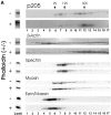
Treatment of membranes with phalloidin increases the sedimentability of p205, as well as actin. (A) Neutrophil plasma membranes treated without (−) or with (+) phalloidin were solubilized in TEB and fractionated on 20–55% sucrose gradients. The initial membrane extract (Load) and gradient fractions (lanes 1–17) were analyzed for the presence of cytoskeletal proteins as described in Materials and Methods. Positions of calibration standards (9S, 19S, and 30S) are indicated. (B) Phalloidin- induced shift in the sedimentability of p205. Average distribution of p205 in gradient fractions, expressed as a percent of the total F-actin binding at 205 kD on F-actin blot overlays (n = 3). Similar results were observed when blot strips were re-probed with anti-pepA antibody, indicating that a single 205-kD F-actin–binding polypeptide is present in the 13S, 26S, and 30S complexes.
Figure 5
Figure 5
Treatment of membranes with phalloidin increases the sedimentability of p205, as well as actin. (A) Neutrophil plasma membranes treated without (−) or with (+) phalloidin were solubilized in TEB and fractionated on 20–55% sucrose gradients. The initial membrane extract (Load) and gradient fractions (lanes 1–17) were analyzed for the presence of cytoskeletal proteins as described in Materials and Methods. Positions of calibration standards (9S, 19S, and 30S) are indicated. (B) Phalloidin- induced shift in the sedimentability of p205. Average distribution of p205 in gradient fractions, expressed as a percent of the total F-actin binding at 205 kD on F-actin blot overlays (n = 3). Similar results were observed when blot strips were re-probed with anti-pepA antibody, indicating that a single 205-kD F-actin–binding polypeptide is present in the 13S, 26S, and 30S complexes.
Figure 6
Figure 6
p205 cosediments with endogenous, phalloidin-stabilized actin (A), and actin coimmunoprecipitates with p205 (B). (A) Neutrophil plasma membranes (lane 1) were treated with either fluorescein-phalloidin (lanes 2 and 3) or unlabeled phalloidin (lane 4), solubilized in TEB, and then incubated with nonspecific IgG (lane 2) or antifluorescein IgG (lanes 3 and 4) bound to protein A–agarose. p205 was visualized by staining with affinity-purified pepA IgG and by F-actin blot overlays (not shown). (B) IgG and actin in immunoprecipitates generated with either nonspecific rabbit IgG (lane 1) or affinity-purified, anti-pepA antibody (lane 2) after three washes with RIPA buffer. The relative amounts of actin cited in the text were normalized by reference to the amounts of IgG visualized by labeling with radiolabeled secondary IgG.
Figure 6
Figure 6
p205 cosediments with endogenous, phalloidin-stabilized actin (A), and actin coimmunoprecipitates with p205 (B). (A) Neutrophil plasma membranes (lane 1) were treated with either fluorescein-phalloidin (lanes 2 and 3) or unlabeled phalloidin (lane 4), solubilized in TEB, and then incubated with nonspecific IgG (lane 2) or antifluorescein IgG (lanes 3 and 4) bound to protein A–agarose. p205 was visualized by staining with affinity-purified pepA IgG and by F-actin blot overlays (not shown). (B) IgG and actin in immunoprecipitates generated with either nonspecific rabbit IgG (lane 1) or affinity-purified, anti-pepA antibody (lane 2) after three washes with RIPA buffer. The relative amounts of actin cited in the text were normalized by reference to the amounts of IgG visualized by labeling with radiolabeled secondary IgG.
Figure 7
Figure 7
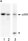
p205 is present in many cell types but is not the only ∼205-kD F-actin–binding protein. (A) Nitrocellulose blots of HeLa cells (lane 1), MDBK cells (lane 2), and bovine neutrophils (lane 3) stained with affinity-purified anti-pepA antibodies. Only the neutrophils contain a cross-reactive protein at ∼90 kD (arrowhead). This 90-kD protein cofractionates with pooled neutrophil granules (66), specialized vesicles involved in host defense, and is distinct from the ∼90-kD cytosolic actin-binding protein in Fig. 1 C, lane 3. (B) Blots of HeLa (lane 1), SHSY5Y neuroblastoma (lane 2), 3T3 (lane 3), MDBK (lane 4), NRK (lane 5), LLC-PK1 (lane 6), and COS-7 cells (lane 7) were stained in parallel with antibodies to pepA and F-actin. A higher molecular mass protein that binds F-actin, but not anti-pepA, is observed in lanes 3 and 5 (*).
Figure 7
Figure 7
p205 is present in many cell types but is not the only ∼205-kD F-actin–binding protein. (A) Nitrocellulose blots of HeLa cells (lane 1), MDBK cells (lane 2), and bovine neutrophils (lane 3) stained with affinity-purified anti-pepA antibodies. Only the neutrophils contain a cross-reactive protein at ∼90 kD (arrowhead). This 90-kD protein cofractionates with pooled neutrophil granules (66), specialized vesicles involved in host defense, and is distinct from the ∼90-kD cytosolic actin-binding protein in Fig. 1 C, lane 3. (B) Blots of HeLa (lane 1), SHSY5Y neuroblastoma (lane 2), 3T3 (lane 3), MDBK (lane 4), NRK (lane 5), LLC-PK1 (lane 6), and COS-7 cells (lane 7) were stained in parallel with antibodies to pepA and F-actin. A higher molecular mass protein that binds F-actin, but not anti-pepA, is observed in lanes 3 and 5 (*).
Figure 8
Figure 8
Distribution of p205 (A and C) and E-cadherin (B and D) in MDBK cells grown at low cell density. Cells were double labeled with affinity-purified, rabbit anti-pepA IgG and with monoclonal anticadherin antibodies, as described in Materials and Methods. Regions of p205 and cadherin colocalization at the plasma membrane (white arrows), and regions of anti-pepA staining alone (hollow arrows) are indicated. Bar, 10 μm.
Figure 9
Figure 9
Distribution of p205 (A and C) and E-cadherin (B and D) in MDBK cells grown to high cell density. Cells were double labeled with affinity-purified, rabbit anti-pepA IgG and with monoclonal anticadherin antibodies. Regions of p205 and cadherin colocalization at the plasma membrane (white arrows) and regions of anti-pepA staining alone (hollow arrows) are indicated. Bar, 10 μm.
Figure 10
Figure 10
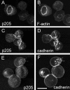
Colocalization of p205 (A, C, and E) with F-actin (B) and cadherin (D and F) in ringlike structures and cytoplasmic aggregates 10 min after EGTA treatment to disrupt cell adhesions. After 30 min (E and F), most of the cadherin is dissociated from p205, which is diffusely distributed in cytoplasmic puncta. Cells were double labeled with affinity-purified pepA antibodies and either fluorescein-phalloidin or anticadherin antibodies. Bar, 10 μm.
Figure 12
Figure 12
Predicted sequence and domain structure of bovine p205 (supervillin). (A) The amino acid sequence starting with the first methionine of the translated ORF includes all eight peptides obtained from purified p205 (double underline). The NH2-terminal half contains four putative nuclear targeting signals (gray boxes) the longest of which resembles the nucleoplasmin targeting signal (single underline). The COOH-terminal half of the molecule contains a potential tyrosine phosphorylation site (black). Amino acid positions are indicated by numbers in the left margin. These sequence data are available from GenBank/EMBL/DDBJ under accession number AF025996. (B) Schematic representation of the domain structure showing the NH2-terminal region with putative nuclear targeting regions (gray boxes) juxtaposed with potential protein kinase A phosphorylation sites (asterisks). The COOH-terminal domain (cross-hatched) shows extensive similarity to villin and gelsolin, with three regions of especially high homology that correspond to potential F-actin–binding sites (black boxes). The potential tyrosine phosphorylation site is indicated (·).
Figure 12
Figure 12
Predicted sequence and domain structure of bovine p205 (supervillin). (A) The amino acid sequence starting with the first methionine of the translated ORF includes all eight peptides obtained from purified p205 (double underline). The NH2-terminal half contains four putative nuclear targeting signals (gray boxes) the longest of which resembles the nucleoplasmin targeting signal (single underline). The COOH-terminal half of the molecule contains a potential tyrosine phosphorylation site (black). Amino acid positions are indicated by numbers in the left margin. These sequence data are available from GenBank/EMBL/DDBJ under accession number AF025996. (B) Schematic representation of the domain structure showing the NH2-terminal region with putative nuclear targeting regions (gray boxes) juxtaposed with potential protein kinase A phosphorylation sites (asterisks). The COOH-terminal domain (cross-hatched) shows extensive similarity to villin and gelsolin, with three regions of especially high homology that correspond to potential F-actin–binding sites (black boxes). The potential tyrosine phosphorylation site is indicated (·).
Figure 13
Figure 13
Detailed comparison of the COOH terminus of supervillin (p205) with the full-length protein sequences of mouse gelsolin, mouse villin, chicken villin, Dictyostelium protovillin, and a predicted C. elegans protein (These sequence data are available under GenBank/EMBL/DDBJ accession numbers P13020, M98454, P02640, P36418, and U88311, respectively). (A) Mouse villin, mouse gelsolin, and the COOH terminus of supervillin were aligned with PileUp, and the percentage of identical residues in every consecutive 30–amino acid segment of supervillin were plotted vs. the number of the last residue in the segment. The locations of the gelsolin and villin homology segments (S1–S6) and the villin headpiece domain (HP) are drawn to scale. (B) The regions of highest identity between supervillin and the other proteins in this family include portions of segments 2 and 5, which are present in both gelsolin and villin, and the villin headpiece domain, which is absent from gelsolin. Both segment 2 and the villin headpiece contain known actin-binding motifs, indicated by asterisks above the sequence (23, 38, 48). Villin headpiece amino acids implicated in binding to F-actin and in stabilization of headpiece structure (26, 56) are designated by ♦ and ⋄, respectively. Identical residues (black boxes) and conservative replacements (gray boxes), defined as matches scoring ⩾0.6 on the Dayhoff matrix (22), are highlighted.
Figure 13
Figure 13
Detailed comparison of the COOH terminus of supervillin (p205) with the full-length protein sequences of mouse gelsolin, mouse villin, chicken villin, Dictyostelium protovillin, and a predicted C. elegans protein (These sequence data are available under GenBank/EMBL/DDBJ accession numbers P13020, M98454, P02640, P36418, and U88311, respectively). (A) Mouse villin, mouse gelsolin, and the COOH terminus of supervillin were aligned with PileUp, and the percentage of identical residues in every consecutive 30–amino acid segment of supervillin were plotted vs. the number of the last residue in the segment. The locations of the gelsolin and villin homology segments (S1–S6) and the villin headpiece domain (HP) are drawn to scale. (B) The regions of highest identity between supervillin and the other proteins in this family include portions of segments 2 and 5, which are present in both gelsolin and villin, and the villin headpiece domain, which is absent from gelsolin. Both segment 2 and the villin headpiece contain known actin-binding motifs, indicated by asterisks above the sequence (23, 38, 48). Villin headpiece amino acids implicated in binding to F-actin and in stabilization of headpiece structure (26, 56) are designated by ♦ and ⋄, respectively. Identical residues (black boxes) and conservative replacements (gray boxes), defined as matches scoring ⩾0.6 on the Dayhoff matrix (22), are highlighted.

Similar articles

Cited by

References

    1. Aberle H, Schwartz H, Kemler R. Cadherin-catenin complex: protein interactions and their implications for cadherin function. J Cell Biochem. 1996;61:514–523. - PubMed
    1. Altschul SF, Gish W, Miller W, Myers EW, Lipman DJ. Basic local alignment search tool. J Mol Biol. 1990;215:403–410. - PubMed
    1. Anderson JM, Van Itallie CM. Tight junctions and the molecular basis for regulation of paracellular permeability. Amer J Physiol. 1995;269:467–475. - PubMed
    1. Atkinson PH. HeLa cell plasma membranes. Methods Cell Biol. 1973;7:157–188. - PubMed
    1. Ausubel, F.M., R. Brent, R.E. Kingston, D.D. Moore, J.G. Seidman, J.A. Smith, and K. Struhl. 1989. Current Protocols in Molecular Biology. Vol. 1. John Wiley & Sons, New York.

Publication types

MeSH terms

Associated data