Comparative Study
. 1997 Dec 1;139(5):1271-80.
doi: 10.1083/jcb.139.5.1271.

Stu2p: A microtubule-binding protein that is an essential component of the yeast spindle pole body

Affiliations
Comparative Study

Stu2p: A microtubule-binding protein that is an essential component of the yeast spindle pole body

P J Wang et al. J Cell Biol. .

Abstract

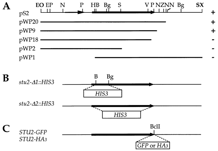
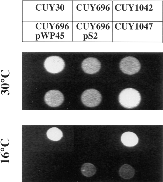
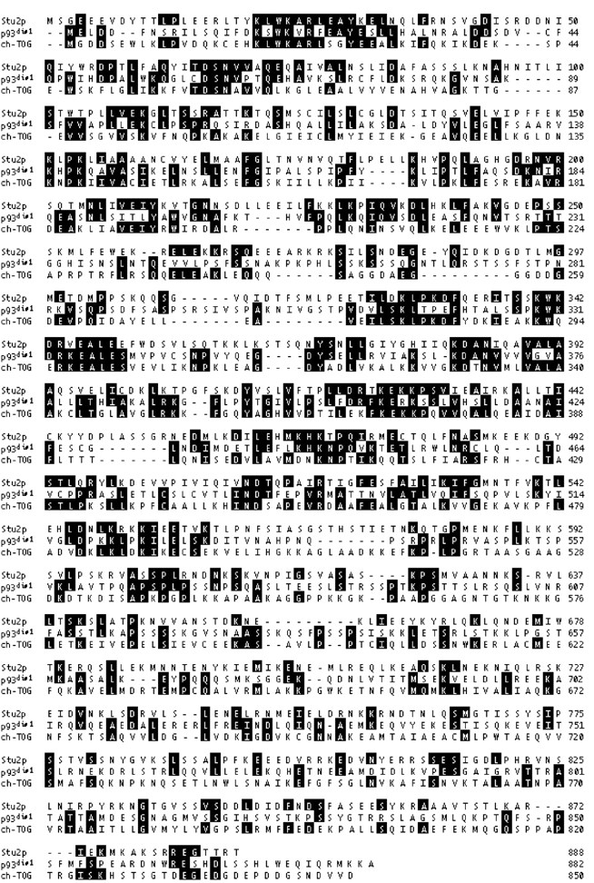
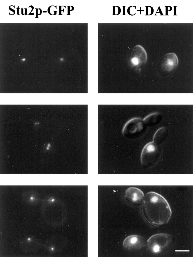
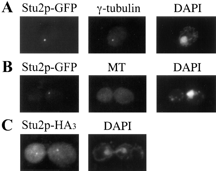
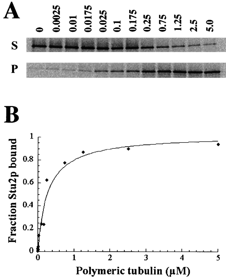
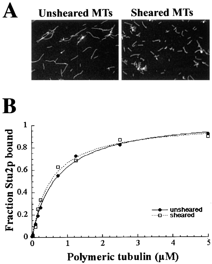
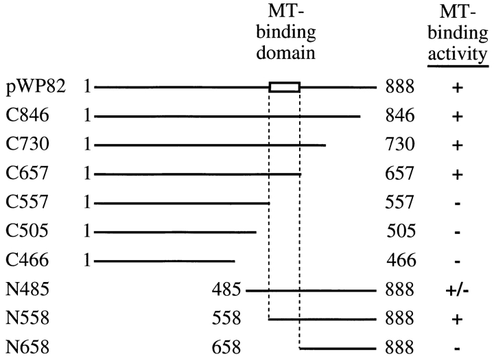
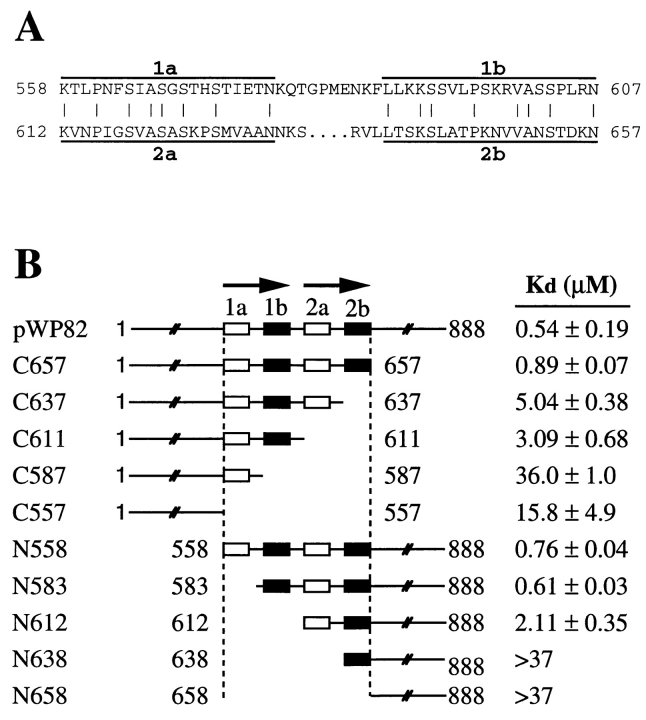
Previously we isolated tub2-423, a cold-sensitive allele of the Saccharomyces cerevisiae gene encoding beta-tubulin that confers a defect in mitotic spindle function. In an attempt to identify additional proteins that are important for spindle function, we screened for suppressors of the cold sensitivity of tub2-423 and obtained two alleles of a novel gene, STU2. STU2 is an essential gene and encodes a protein whose sequence is similar to proteins identified in a variety of organisms. Stu2p localizes primarily to the spindle pole body (SPB) and to a lesser extent along spindle microtubules. Localization to the SPB is not dependent on the presence of microtubules, indicating that Stu2p is an integral component of the SPB. Stu2p also binds microtubules in vitro. We have localized the microtubule-binding domain of Stu2p to a highly basic 100-amino acid region. This region contains two imperfect repeats; both repeats appear to contribute to microtubule binding to similar extents. These results suggest that Stu2p may play a role in the attachment, organization, and/or dynamics of microtubule ends at the SPB.


Figures

Figure 2
Figure 2
STU2 subclones and plasmid constructs. (A) The top bar represents the 6.7-kb genomic insert in pS2 that contains stu2-1. The stu2-1 open reading frame is indicated by the wide arrow. A smaller ORF, YLR406c, upstream of stu2-1 is indicated by the thin arrow. Subclones of the 6.7-kb insert were made by digestion with Exonuclease III and Nuclease S1. Right column indicates whether each construct could (+) or could not (−) suppress the cold sensitivity of tub2-423. (B) The stu21::HIS3 allele as created by replacing the 541-bp BamHI–BglII fragment with the 1.7-kb HIS3 gene. The stu22::HIS3 allele was made by replacing the entire STU2 ORF with HIS3. (C) A BclI restriction site was introduced just before the stop codon STU2. A 120-bp fragment encoding the HA3 or a 739-bp fragment encoding GFP was cloned into this site. Abbreviations of the restriction sites: B, BamHI; Bg, BglII; E, EcoRI; H, HindIII; N, NsiI; O, XhoI; P, PstI; S, SpeI; V, EcoRV; X, XbaI; Z, SphI. Sites in bold are in the vector.
Figure 1
Figure 1
Suppression of tub2-423 by stu2-1. Yeast cells were spotted on YPD plates and incubated at 30°C for 2 d and 16°C for 5 d. The relevant genotypes of strains are as follows: CUY30, TUB2 STU2; CUY696, tub2-423 STU2; CUY1042, tub2-423 stu2-1; CUY696/pWP45, tub2-423 STU2 (STU2 on YCp plasmid); CUY696/pS2, tub2-423 STU2 (stu2-1 on YCp plasmid); CUY1047, tub2-423/tub2-423 stu2-1/ STU2.
Figure 3
Figure 3
Alignments of the S. cerevisiae Stu2p, S. pombe p93dis1, and human ch-TOG. Note that only the amino-terminal half of ch-TOG is shown in this figure; the full length protein is 1,972 amino acids. The sequence of STU2 is available from GenBank/ EMBL/DDBJ under accession number U35247.
Figure 4
Figure 4
Localization of Stu2p–GFP fusion protein. GFP fluorescence was visualized in diploid cells, homozygous for the stu21::HIS3 disruption, that contain a plasmid carrying STU2-GFP (CUY1065). Cells were grown in the presence of 1 μg/ml DAPI for 2 h in SD media. Left column shows the fluorescence of Stu2p-GFP; right column shows both DNA staining with DAPI and differential interference contrast image of cells. Bar, 5 μm.
Figure 5
Figure 5
(A) Colocalization of Stu2p-GFP with γ-tubulin. CUY1060 cells were stained with anti-Tub4p antibody (middle) and DAPI (right). The left frame shows the fluorescence of Stu2p–GFP in the same cell. (B) CUY1060 cells were treated with nocodazole to depolymerize microtubules and stained with anti-tubulin antibody (middle) and DAPI (right). The left frame shows the fluorescence of Stu2p–GFP in the same cell. (C) Localization of Stu2p-HA3. CUY1066 cells were stained with anti- hemaglutinin antibody (left) and DAPI (right).
Figure 6
Figure 6
Measurement of the dissociation constant (Kd) for full length Stu2p. A constant amount of 35S-labeled in vitro translated Stu2p (∼10−11 M final concentration) was incubated with variable amounts of taxol-stabilized bovine brain microtubules. Microtubules were pelleted by centrifugation, and both bound Stu2p (in pellets) and unbound Stu2p (in supernatants) were subjected to SDS-PAGE analysis. Band intensities were quantitated using the phosphoimager software. (A) Autoradiographs of Stu2p in the supernatants (S) and in the corresponding pellets (P). Microtubule concentration ranging from 0 to 5 μM is shown above each lane. (B) The binding curve was generated from the above data. The Kd is equal to the concentration of polymerized tubulin required to cosediment 50% of the Stu2p in the reaction.
Figure 7
Figure 7
(A) Microtubules before (left) and after (right) shearing visualized by immunofluorescence. (B) The binding curves for Stu2p to both sheared and unsheared microtubules.
Figure 8
Figure 8
Identification of Stu2p microtubule-binding domain. Various truncated Stu2p polypeptides were translated in vitro and tested for microtubule-binding activity by the cosedimentation assay using a constant amount of microtubules (16.5 μM tubulin). The percentage of Stu2p that pelleted with microtubule is indicated by (+), >90%; (+/−), 70–90%; (−), <30%. The numbers adjacent to the endpoints represent the corresponding amino acid positions.
Figure 9
Figure 9
Dissection of the microtubule-binding domain of Stu2p. (A) Sequences of the two bipartite repeats in the microtubule-binding domain of Stu2p. The identical residues between these two repeats are indicated by vertical lines. (B) The Kd 's of Stu2p polypeptides were determined as in Fig. 6 and presented as the average of the measured values. The full-length Stu2p was assayed three times and standard error of the mean is shown. All the truncated polypeptides were assayed twice and the range is included. The top line represents the full-length Stu2p. The numbers adjacent to the endpoints represent the positions of the corresponding amino acids.

References

    1. Barnes G, Louie KA, Botstein D. Yeast proteins associated with microtubules in vitro and in vivo. Mol Biol Cell. 1992;3:29–47. - PMC - PubMed
    1. Biernat J, Gustke N, Drewes G, Mandelkow EM, Mandelkow E. Phosphorylation of Ser262 strongly reduces binding of tau to microtubules: distinction between PHF-like immunoreactivity and microtubule binding. Neuron. 1993;11:153–163. - PubMed
    1. Blose SH, Meltzer DI, Feramisco JR. 10-nm filaments are induced to collapse in living cells microinjected with monoclonal and polyclonal antibodies against tubulin. J Cell Biol. 1984;98:847–858. - PMC - PubMed
    1. Bramblett GT, Goedert M, Jakes R, Merrick SE, Trojanowski JQ, Lee VM. Abnormal tau phosphorylation at Ser396 in Alzheimer's disease recapitulates development and contributes to reduced microtubule binding. Neuron. 1993;10:1089–1099. - PubMed
    1. Byers, B. 1981. Cytology of the yeast life cycle. In The Molecular Biology of the Yeast Saccharomyces. J.N. Strathern, E.W. Jones, and J.R. Broach, editors. Cold Spring Harbor Laboratory, Cold Spring Harbor, New York. 59–96.

Publication types

MeSH terms

Substances

Associated data