. 1997 Dec 1;139(5):1307-15.
doi: 10.1083/jcb.139.5.1307.

Neurofilaments and orthograde transport are reduced in ventral root axons of transgenic mice that express human SOD1 with a G93A mutation

Affiliations

Neurofilaments and orthograde transport are reduced in ventral root axons of transgenic mice that express human SOD1 with a G93A mutation

B Zhang et al. J Cell Biol. .

Abstract

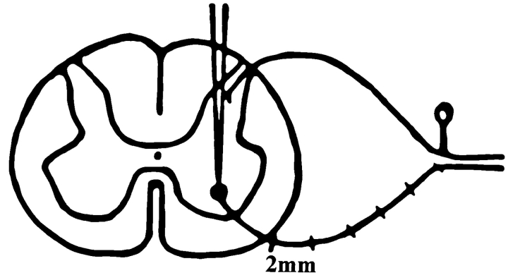
Mice engineered to express a transgene encoding a human Cu/Zn superoxide dismutase (SOD1) with a Gly93 --> Ala (G93A) mutation found in patients who succumb to familial amyotrophic lateral sclerosis (FALS) develop a rapidly progressive and fatal motor neuron disease (MND) similar to amyotrophic lateral sclerosis (ALS). Hallmark ALS lesions such as fragmentation of the Golgi apparatus and neurofilament (NF)-rich inclusions in surviving spinal cord motor neurons as well as the selective degeneration of this population of neurons were also observed in these animals. Since the mechanism whereby mutations in SOD1 lead to MND remains enigmatic, we asked whether NF inclusions in motor neurons compromise axonal transport during the onset and progression of MND in a line of mice that contained approximately 30% fewer copies of the transgene than the original G93A (Gurney et al., 1994). The onset of MND was delayed in these mice compared to the original G93A mice, but they developed the same neuropathologic abnormalities seen in the original G93A mice, albeit at a later time point with fewer vacuoles and more NF inclusions. Quantitative Western blot analyses showed a progressive decrease in the level of NF proteins in the L5 ventral roots of G93A mice and a concomitant reduction in axon caliber with the onset of motor weakness. By approximately 200 d, both fast and slow axonal transports were impaired in the ventral roots of these mice coincidental with the appearance of NF inclusions and vacuoles in the axons and perikarya of vulnerable motor neurons. This is the first demonstration of impaired axonal transport in a mouse model of ALS, and we infer that similar impairments occur in authentic ALS. Based on the temporal correlation of these impairments with the onset of motor weakness and the appearance of NF inclusions and vacuoles in vulnerable motor neurons, the latter lesions may be the proximal cause of motor neuron dysfunction and degeneration in the G93A mice and in FALS patients with SOD1 mutations.


Figures

Figure 1
Figure 1
Schematic diagram showing the microdissection of L5 ventral roots and the microinjection of [35S]methionine into the ventral horn of the mouse spinal cord in the experimental paradigm used here to analyze axonal transport.
Figure 2
Figure 2
Photomicrographs showing NF-rich inclusions in the perikarya and proximal axons of the spinal cord of the G93A mice. The NF inclusions contain NFL epitopes revealed by a rabbit anti-NFL polyclonal antiserum (A and B). Notably, the vacuolar pathology (arrowheads) in the rederived G93A mice in the terminal stages of MND (230 d of age in B) is less prominent than in the original line of G93A mice at a similar terminal stage (140 d of age in A). However, the inclusions (arrows) in the rederived G93A mice in the terminal stages of MND (230 d of age in B) are more abundant than in the original line of G93A mice at a similar terminal stage (140 d of age in A). The large arrow in B indicates an inclusion with light staining. In C and D, the RMdO9 mAb to poorly phosphorylated NFH stains the white matter of the spinal cord of the G93A mouse (D) more intensely than in the control mouse (C), and the small arrows in D indicate RMdO9-stained axonal inclusions containing poorly phosphorylated NFH. Bars: (B) 20 μm; (D) 40 μm.
Figure 3
Figure 3
Electron micrographs showing NF accumulations in a proximal axon of a spinal cord motor neuron and reduced NFs in an L5 ventral root of a 200-d-old G93A mouse. NFs are aligned in parallel in the normal axon (A). The NF inclusion is a mass of tightly packed disorganized NFs (B). This inclusion fills the axon and displaces cellular organelles in the proximal ventral root axons of the G93A mice. Normal NFs are shown in a cross-section of a ventral root axon from a control mouse (C), while there is a decreased density of NFs in the L5 ventral root axon of G93A mice (D). Bars: (B) 1 μm; (B, inset, and D) 0.5 μm.
Figure 4
Figure 4
Western blots showing a progressive decrease in the level of NF proteins in the L5 ventral roots of the G93A transgenic mice. The ventral roots of 24 G93A SOD1 and N1029 transgenic mice as well as age-matched control mice of four different ages (150, 180, 200, and 230 d, n = 3/age group) were harvested after lethal anesthesia as described. The relative levels of NFH are revealed by a rabbit anti-NFH polyclonal antiserum; those of NFM, by the mAb RMO189, and those of NFL, by the rabbit anti-NFL polyclonal antiserum. The levels of NF proteins and tubulin in the 150-d-old G93A mouse are comparable to that of the age-matched control mouse (A, CTR). However, there is a progressive decrease in NF proteins and tubulin in the ventral roots of the G93A mice at 180, 200, and 230 d of age compared with the age-matched CTR mice (B–D). Quantification of these data (E) shows that NFL and tubulin start to decrease in the G93A mice as early as 180 d, while a significant reduction in the levels of NFM, NFH, and tubulin occurs when the G93A mice reach 200 d of age. The ventral roots of the G93A mice lose 80– 90% of NF proteins when they are at the terminal stages of MND (230 d). vr1-5, Ventral root segments from 1 to 5; *P < 0.05; **P < 0.01.
Figure 5
Figure 5
EM photomicrographs show a drastic reduction in the axonal caliber of the L5 ventral roots of the G93A transgenic mice by 200 d of age. The ventral roots of the control mouse (A, C, and E) contain many large myelinated axons that are tightly packed and evenly distributed in the nerve. Note the relative abundance of small- and intermediate-sized axons (A, C, and E). There is no obvious difference in the ventral roots of the G93A transgenic mice (B) compared to control mice (A) at 150 d. In contrast, the ventral roots of the G93A mouse are mainly composed of axons of smaller caliber (D and F) compared to age-matched control mice (C and E). The abundant interaxonal space and the tangential or longitudinal orientation of axons probably reflects the collapse of these axons due to impaired transport. Bars: (A) 20 μm; (F) 10 μm.
Figure 6
Figure 6
SDS-PAGE shows a progressive retardation in slow transport in the L5 ventral root of the spinal cord in the G93A transgenic mice compared to the control (CTR) mice. 12 G93A and N1029 transgenic mice as well as age-matched control mice of two different ages (150 and 200 d, n = 3/age group) were killed 7 d after microinjection. Fluorographs show a decrease in the transport of a variety of cytoskeletal proteins such as NFH, NFM, NFL, tubulin, and actin in the 200-d-old G93A mice (B) but not in the 150-d-old G93A mice (A). The graphs in C and D illustrate quantitative measurements of individual proteins conveyed by slow axonal transport in pairs of age-matched G93A and CTR mice. The 150-d-old G93A mice fail to show any significant retardation of slow transport (C). However, the slow transport of several proteins is retarded in the 200-d-old G93A mice (D). The symbols to the right of each fluorograph in A and B are aligned with the respective proteins identified on the left, and they are used in C through E. Tub, Tubulin; Act, actin.
Figure 7
Figure 7
SDS-PAGE shows a progressive retardation in fast transport in the L5 ventral roots of the spinal cord in the G93A transgenic mice compared to the control (CTR) mice. 12 G93A and N1029 transgenic mice as well as age-matched control mice of two different ages (150 and 200 d, n = 3/age group) were killed 3 h after microinjection. Fluorographs show no change in the fast transport of several proteins (closed triangle, closed square, and closed circle) in the 150-d-old G93A mice (A and C), but the fast transport of some proteins is retarded in the 200-d-old G93A mice (B and D). The graphs in C and D illustrate quantitative measurements of individual proteins conveyed by fast axonal transport in pairs of age-matched G93A and CTR mice. The 150-d- old G93A mice fail to show any significant slowing of fast transport (C), but the fast transport of several proteins is retarded in the 200-d-old G93A mice (D). The symbols in A and B correspond to proteins analyzed in the graphs in C and D.

References

    1. Bannister JV, Bannister WH, Rotilio G. Aspects of the structure, function, and applications of superoxide desmutase. CRC Crit Rev Biochem. 1987;22:111–180. - PubMed
    1. Beyer W, Imlay J, Fridovich I. Superoxide dismutase. Prog Nucleic Acid Res Mol Biol. 1991;40:221–253. - PubMed
    1. Black MM, Lee VM-Y. Phosphorylation of neurofilament proteins in intact neurons: demonstration of phosphorylation in cell bodies and axons. J Neurosci. 1988;8:3296–3305. - PMC - PubMed
    1. Bruijn LI, Becher MW, Lee MK, Anderson KL, Jenkins NA, Copeland NG, Sisodia SS, Rothstein JD, Borchelt DR, Price DL, et al. ALS-linked SOD1 mutant G85R mediates damage to astrocytes and promotes rapidly progressive disease with SOD1-containing inclusions. Neuron. 1997;18:327–338. - PubMed
    1. Clark EA, Lee VM-Y. Dynamics of mammalian high-molecular-weight neurofilament subunit phosphorylation in cultured rat sympathetic neurons. J Neurosci Res. 1991;30:116–123. - PubMed

Publication types

MeSH terms