. 1997 Dec 1;186(11):1843-51.
doi: 10.1084/jem.186.11.1843.

Atypical disease after Bordetella pertussis respiratory infection of mice with targeted disruptions of interferon-gamma receptor or immunoglobulin mu chain genes

Affiliations

Atypical disease after Bordetella pertussis respiratory infection of mice with targeted disruptions of interferon-gamma receptor or immunoglobulin mu chain genes

B P Mahon et al. J Exp Med. .

Abstract

Using a murine respiratory challenge model we have previously demonstrated a role for Th1 cells in natural immunity against Bordetella pertussis, but could not rule out a role for antibody. Here we have demonstrated that B. pertussis respiratory infection of mice with targeted disruptions of the genes for the IFN-gamma receptor resulted in an atypical disseminated disease which was lethal in a proportion of animals, and was characterized by pyogranulomatous inflammation and postnecrotic scarring in the livers, mesenteric lymph nodes and kidneys. Viable virulent bacteria were detected in the blood and livers of diseased animals. An examination of the course of infection in the lung of IFN-gamma receptor-deficient, IL-4-deficient and wild-type mice demonstrated that lack of functional IFN-gamma or IL-4, cytokines that are considered to play major roles in regulating the development of Th1 and Th2 cells, respectively, did not affect the kinetics of bacterial elimination from the lung. In contrast, B cell-deficient mice developed a persistent infection and failed to clear the bacteria after aerosol inoculation. These findings demonstrate an absolute requirement for B cells or their products in the resolution of a primary infection with B. pertussis, but also define a critical role for IFN-gamma in containing bacteria to the mucosal site of infection.


Figures

Figure 1
Figure 1
Survival of IFN-γR−/− mice after B. pertussis challenge. In one experiment groups of 24 IFN-γR−/− (○) or 24 wild-type 129Sv/Ev (□) mice were aerosol challenged with B. pertussis which resulted in an inoculum of 6 × 10⁴ CFU/lung, determined from a sample group killed 2 h after challenge. In a separate experiment 6 IFN-γR−/− mice (•) received a higher dose challenge, which resulted in 1 × 10⁶ CFU/lung 2 h after challenge. Six wild-type 129Sv/Ev mice (▪) exposed to the higher dose of bacteria all survived the challenge (displayed offset for clarity).
Figure 2
Figure 2
Pathological findings in IFN-γR−/− mice after respiratory infection with B. pertussis. Mice were challenged with 2 × 10¹⁰ CFU/ml giving 6 × 10⁴ CFU/lung 2 h later. (A) Liver: aggregates of neutrophils and macrophages with reactive changes in adjoining hepatocytes. (B) Mesentery: macrophages and neutrophils surround a central area of necrosis (necrotic pyogranuloma). (C) Brain: neutrophils, macrophages and fibrin deposits in the leptomeninges. (D) Lung: granular basophilic debris (arrows) in the alveolar spaces with macrophage and neutrophil infiltration primarily in alveolar walls. (A–D, hematoxylin and eosin [H & E] staining) (E) Lung: B. pertussis antigen in the lumen of alveoli and a bronchiole (arrow). Anti–B. pertussis, haematoxylin counterstain. (F) Mesentry: intracytoplasmic deposits of B. pertussis antigen in macrophages in an area of pyogranulomatous inflammation. Anti–B. pertussis, haematoxylin counterstain. Results are representative of 15 mice from two challenge experiments. Similar pathological lesions were observed in tissue from all mice examined, except for the brain, where lesions were visible in only 3 out of 7 mice examined. Original magnifications: (A, C–F) ×400; (B) ×200.
Figure 2
Figure 2
Pathological findings in IFN-γR−/− mice after respiratory infection with B. pertussis. Mice were challenged with 2 × 10¹⁰ CFU/ml giving 6 × 10⁴ CFU/lung 2 h later. (A) Liver: aggregates of neutrophils and macrophages with reactive changes in adjoining hepatocytes. (B) Mesentery: macrophages and neutrophils surround a central area of necrosis (necrotic pyogranuloma). (C) Brain: neutrophils, macrophages and fibrin deposits in the leptomeninges. (D) Lung: granular basophilic debris (arrows) in the alveolar spaces with macrophage and neutrophil infiltration primarily in alveolar walls. (A–D, hematoxylin and eosin [H & E] staining) (E) Lung: B. pertussis antigen in the lumen of alveoli and a bronchiole (arrow). Anti–B. pertussis, haematoxylin counterstain. (F) Mesentry: intracytoplasmic deposits of B. pertussis antigen in macrophages in an area of pyogranulomatous inflammation. Anti–B. pertussis, haematoxylin counterstain. Results are representative of 15 mice from two challenge experiments. Similar pathological lesions were observed in tissue from all mice examined, except for the brain, where lesions were visible in only 3 out of 7 mice examined. Original magnifications: (A, C–F) ×400; (B) ×200.
Figure 3
Figure 3
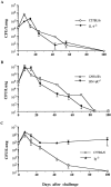
Course of B. pertussis respiratory infection in normal and gene knockout mice. IL-4−/− and C57BL/6 (A), IFN-γR−/− and 129Sv/Ev (B), and Ig−/− and C57BL/6 (C) mice were infected by aerosol with 2 × 10¹⁰ CFU/ml B. pertussis, giving an initial colonization of 2–8 × 10⁴ per lung. Groups of mice were killed at intervals after challenge and the number of viable bacteria estimated by performing colony counts on individual lung homogenates. Results are representative from two experiments and are presented as mean (±SE) CFU in the lungs estimated for three or four mice at each time point.
Figure 4
Figure 4
Subclass of B. pertussis–specific IgG in the serum 43 d after respiratory infection of wildtype and gene disrupted mice. Serum IgG was measured by B. pertussis–specific ELISA. Results are given as the geometric mean (±SE) ELISA titers from at least four mice determined in quadruplicate.
Figure 5
Figure 5
Cell mediated immune responses induced after B. pertussis infection of normal and gene disrupted mice. Proliferation (A), IL-2 (B), IFN-γ (C), and IL-5 (D) secretion by spleen cells from gene disrupted (IFN-γR−/−, IL-4−/−, and Ig−/−) and wild-type (129Sv/Ev and C57BL/6) mice examined 43 d after challenge. Spleen cells (2 × 10⁶/ml) were incubated with heat-killed whole B. pertussis, 1 × 10⁶ cells/ml (open bar), inactivated PT 5.0 μg/ml (solid bar), FHA (hatched bar) 5.0 μg/ml, or pertactin 5.0 μg/ml (horizontal shading). Results are the mean responses for 4 mice per group assayed individually in triplicate. Proliferative responses are expressed as CPM (±SE) after subtraction of background responses to medium alone, which ranged from 5,000–10,000 cpm.

References

    1. Cheers C, Gray DF. Macrophage behaviour during the complaisant phase of murine pertussis. Immunol. 1969;17:875–887. - PMC - PubMed
    1. Saukonen E, Cabellos C, Burroughs M, Prasad S, Tuomanen E. Integrin mediated localization of Bordetella pertussiswithin macrophages: role in pulmonary colonization. J Exp Med. 1991;173:1143–1149. - PMC - PubMed
    1. Masure HR. The adenylate cyclase toxin contributes to the survival of Bordetella pertussiswithin human macrophages. Microb Pathog. 1993;14:253–260. - PubMed
    1. Bromberg K, Tannis G, Steiner P. Detection of Bordetella pertussisassociated with the macrophages of children with human immunodeficiency virus. Infect Immun. 1991;59:4715–4719. - PMC - PubMed
    1. Ewanowich CA, Melton AR, Weiss AA, Sherburne RK, Peppler MS. Invasion of Hela 229 cells by virulent Bordetella pertussis. . Infect Immun. 1989;57:2698–2704. - PMC - PubMed

Publication types

MeSH terms