Comparative Study
. 1997 Dec 1;186(11):1897-910.
doi: 10.1084/jem.186.11.1897.

Qualitative regulation of B cell antigen receptor signaling by CD19: selective requirement for PI3-kinase activation, inositol-1,4,5-trisphosphate production and Ca2+ mobilization

Affiliations
Comparative Study

Qualitative regulation of B cell antigen receptor signaling by CD19: selective requirement for PI3-kinase activation, inositol-1,4,5-trisphosphate production and Ca2+ mobilization

A M Buhl et al. J Exp Med. .

Abstract

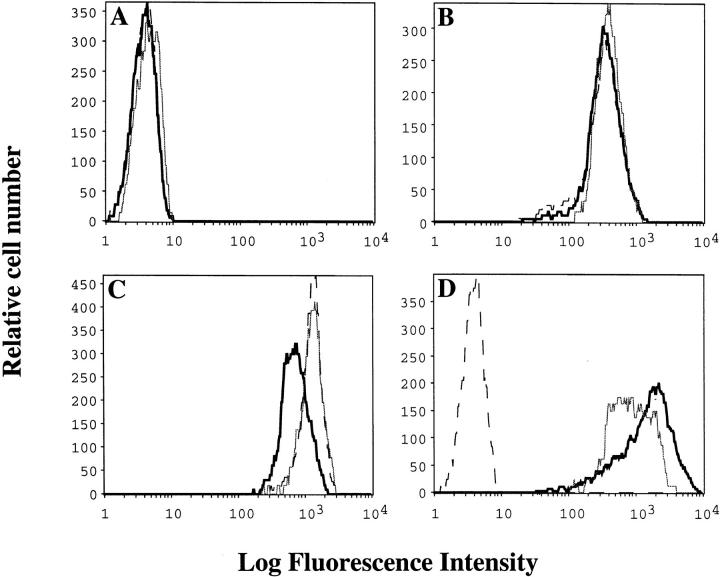
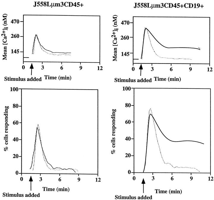
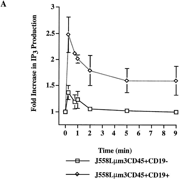
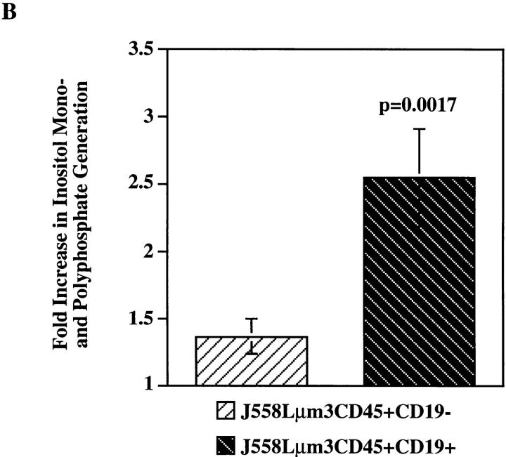
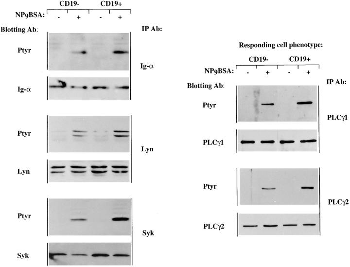
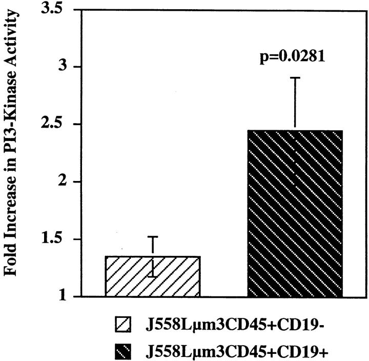
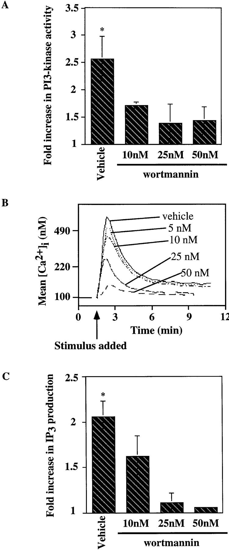
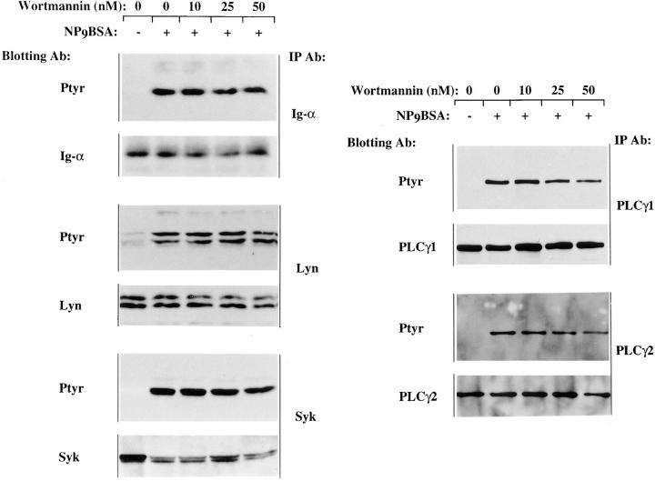
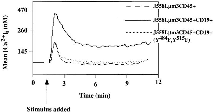
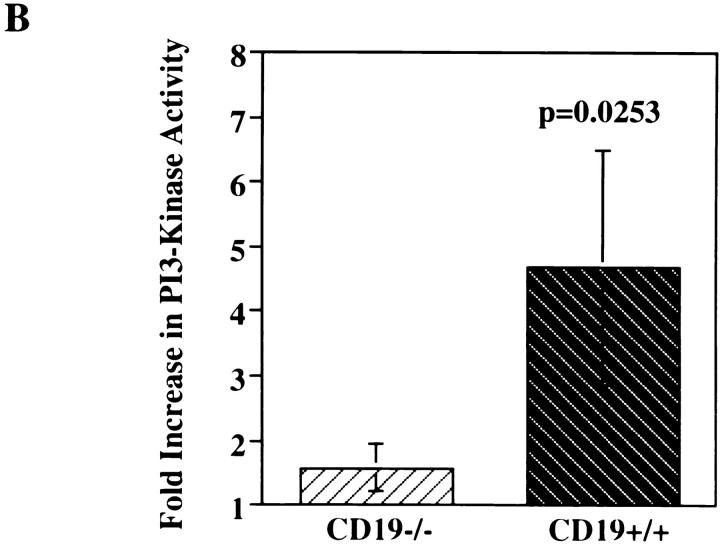
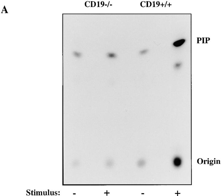
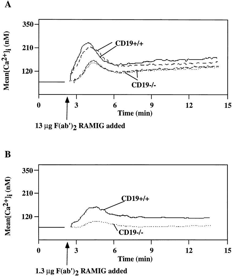
Genetic ablation of the B cell surface glycoprotein CD19 severely impairs the humoral immune response. This requirement is thought to reflect a critical role of CD19 in signal transduction that occurs upon antigen C3dg coligation of antigen receptors with CD19 containing type 2 complement receptors (CR2). Here we show that CD19 plays a key accessory role in B cell antigen receptor signaling independent of CR2 coligation and define molecular circuitry by which this function is mediated. While CD19 is not required for antigen-mediated activation of receptor proximal tyrosines kinases, it is critical for activation of phosphatidylinositol 3-kinase (PI3-kinase). PI3-Kinase activation is dependent on phosphorylation of CD19 Y484 and Y515. Antigen-induced CD19-dependent PI3-kinase activation is required for normal phosphoinositide hydrolysis and Ca2+ mobilization responses. Thus, CD19 functions as a B cell antigen receptor accessory molecule that modifies antigen receptor signaling in a qualitative manner.


Figures

Figure 1
Figure 1
Expression levels of surface markers on the plasmacytoma cell lines. Flow cytometric analysis was performed on the J558Lμm3CD45+ (dotted lines), J558Lμm3CD45+CD19+ (thick lines), and J558L μm3CD45+ CD19+ (Y484F,Y515F) (thin lines) cell lines. Shown is staining with secondary alone (A), biotinylated b-7-6 anti-IgM (B), biotinylated I32.5 anti-CD45 (C), or biotinylated HD37 anti-hCD19 (D) followed by PE-conjugated streptavidin.
Figure 2
Figure 2
The CD19-negative and -positive J558Lμm3CD45+ plasmacytoma variants mobilize [Ca2+]i to different extent after antigen stimulation. J558Lμm3CD45+CD19 and J558Lμm3CD45+CD19+ cells were loaded with indo-1 AM, and analysis of [Ca2+]i initiated before antigen stimulation (250 ng NP9BSA/ 106cells/ml). Mean [Ca2+]i (top) and cells responding (bottom) after antigen stimulation of J558Lμm3CD45+CD19 and J558Lμm3CD45+CD19+. The analysis was conducted under conditions of 60 nM (dotted line) or 1.3 mM (solid line) extracellular free calcium concentration buffered as calculated by the CalCalc program (74). [Ca2+]i was calculated according to Grynkiewicz et al. (75). Approximately 600 cells were analyzed per second. [Ca2+]i in resting cells is 100 nM.
Figure 3
Figure 3
Antigen-induced IP3 and total inositol phosphate release is greater in the CD19-positive than in CD19-negative J558Lμm3CD45+ cells. (A) Cells (10 × 106/ml) were stimulated with 2.5 μg NP9BSA for various times and the responses terminated by addition of 100% TCA (20% TCA final). After organic extraction the aqueous phase was assayed for IP3 content in a [3H]IP3 receptor binding inhibition assay (NEN-DuPont). The results are expressed as fold increase of IP3 release over basal (3 pmol IP3/2.5 × 106 cell equivalents). Shown is mean fold increase from three independent experiments ± standard error. (B) Cells were labeled with Myo- [2-3H(N)]-inositol (21.0 Ci/mmol; 2 μCi/ml) in inositol-free medium containing 5% FCS at 106 cells/ml for 18 h and the generation of inositol phosphates was measured after stimulation of cells for 25 min with 2.5 μg NP9BSA/107 cells/ml. The fold increase in inositol phosphate release was calculated; the two cell lines had comparable basal rates of inositol turnover as determined by the basal inositol phosphate release. Shown are the mean fold increases from three independent experiments ± standard error. Statistical significance was determined by JMP version 3.16 statistical software (SAS Institute, Cary, NC).
Figure 3
Figure 3
Antigen-induced IP3 and total inositol phosphate release is greater in the CD19-positive than in CD19-negative J558Lμm3CD45+ cells. (A) Cells (10 × 106/ml) were stimulated with 2.5 μg NP9BSA for various times and the responses terminated by addition of 100% TCA (20% TCA final). After organic extraction the aqueous phase was assayed for IP3 content in a [3H]IP3 receptor binding inhibition assay (NEN-DuPont). The results are expressed as fold increase of IP3 release over basal (3 pmol IP3/2.5 × 106 cell equivalents). Shown is mean fold increase from three independent experiments ± standard error. (B) Cells were labeled with Myo- [2-3H(N)]-inositol (21.0 Ci/mmol; 2 μCi/ml) in inositol-free medium containing 5% FCS at 106 cells/ml for 18 h and the generation of inositol phosphates was measured after stimulation of cells for 25 min with 2.5 μg NP9BSA/107 cells/ml. The fold increase in inositol phosphate release was calculated; the two cell lines had comparable basal rates of inositol turnover as determined by the basal inositol phosphate release. Shown are the mean fold increases from three independent experiments ± standard error. Statistical significance was determined by JMP version 3.16 statistical software (SAS Institute, Cary, NC).
Figure 4
Figure 4
Tyrosine phosphorylation of signaling molecules upon antigen stimulation of the CD19-negative and CD19-positive cell lines. J558Lμm3 CD45+ variants were stimulated for 1 min with 2.5 μg NP9BSA/107cells/ml, lysed, and intermediary signaling molecules immunoprecipitated with various antibodies and fractionated on 10% SDS-PAGE. After electrophoretic transfer to PVDF membrane, both anti-phosphotyrosine and anti-effector immunoblotting were performed. Data are representative of at least three independent experiments.
Figure 5
Figure 5
PI3-kinase activity is significantly increased only upon antigen stimulation of the CD19-positive J558Lμm3CD45+ cells. CD19-negative and CD19-positive J558Lμm3CD45+ cells were stimulated for 2 min with 1 μgNP9BSA/4 × 106/ml, lysed and PI3-kinase immunoprecipitated with anti-p85 antibody. Immunoprecipitates were washed and assayed for P-I3 kinase activity. Fold increase in PI3-kinase activity after stimulation was determined using a PhosphorImager. The data shown is the mean fold increase over basal in eight independent experiments ± standard error of the mean. Statistical significance was determined by JMP version 3.16 statistical software (SAS Institute, Cary, NC).
Figure 6
Figure 6
Wortmannin inhibits the antigen-induced increase in PI3-kinase activity, [Ca2+]i mobilization and IP3 generation in the J558Lμm3 CD45+CD19+ plasmacytoma. (A) Cells were preincubated with DMSO (vehicle) or wortmannin at various concentrations (in 0.2% final DMSO concentration) for 25 min at 25°C, prewarmed for 4 min at 37°C, and then stimulated for 2 min with 1 μg NP9BSA/4 × 106 cells/ml before analysis of PI3-kinase activation as described in Fig, 4. Shown is the mean fold increase in activity over basal from four independent experiments ± standard error of the mean. Asterisk indicates a statistical significance of P <0.05 determined by JMP 3.16 statistical software. (B) To study the effects of wortmannin on the calcium response, J558Lμm3CD45+CD19+ cells were Indo-1 AM loaded and preincubated with DMSO (vehicle) or wortmannin at various concentrations (resulting in 0.2% final DMSO) for 25 min at 25°C, prewarmed for 4 min at 37°C, and stimulated at arrow with 250 ng NP9BSA/106 cells/ml. Shown is the mean [Ca2+]i over time with analysis of ∼600 cells/s. [Ca2+]i in resting cells is 100 nM. (C) J558Lμm3CD45+CD19+ cells were preincubated with DMSO or increasing concentrations of wortmannin, prewarmed, and stimulated with 2.5 μg NP9BSA/107/ml for 1 min before IP3 production was measured by the IP3 receptor binding inhibition assay. Shown is the mean fold increase over basal in three independent experiments ± standard error of the mean. Asterisk indicates a statistical significance of P <0.05 determined by JMP 3.16 statistical software.
Figure 7
Figure 7
In the CD19-positive J558Lμm3CD45+ cells, antigen-induced tyrosine phosphorylation of PLCγ1 and PLCγ2 is slightly inhibited by wortmannin, while Lyn, Ig-α and Syk inductive tyrosine phosphorylation is insensitive to wortmannin. J558Lμm3CD45+CD19+ cells were preincubated with DMSO or increasing concentrations of wortmannin, prewarmed and stimulated for 1 min with NP9BSA (2.5 μg/107 cells/ml) and lysed. Immunoprecipitation, SDS-PAGE fractionation and electrophoretic transfer were performed. For each immunoprecipitation, both anti-phosphotyrosine and anti-effector immunoblots are shown. Results are representative of at least three independent experiments.
Figure 8
Figure 8
Mutant CD19 (CD19Y484F,Y515F) does not support antigen-induced [Ca2+]i mobilization. J558Lμm3CD45+CD19, J558Lμm3CD45+ CD19+ and J558Lμm3CD45+CD19+(Y484F,Y515F) cells were loaded with Indo-1 AM, and analysis of [Ca2+]i initiated before antigen stimulation (250 ng NP9BSA/106cells/ml). Shown is the mean [Ca2+]i over time with analysis of ∼600 cells/s. [Ca2+]i in resting cells is 100 nM.
Figure 9
Figure 9
Antigen induction of [Ca2+]i mobilization in splenic B cells from normal mice is sensitive to wortmannin. Splenic B cells (ρ ⩾1.066) were isolated from spleens of normal mice, and loaded with Indo-1 AM. The cells were preincubated with DMSO or increasing concentrations of wortmannin for 25 min at 25°C. They were then warmed to 37°C for 4 min and stimulated with 13.2 μg F(ab′)2RAMIG/106/ml (at arrow). Shown is the mean [Ca2+]i over time with analysis of ∼600 cells/s. [Ca2+]i in resting cells is 70 nM.
Figure 10
Figure 10
BCR-mediated stimulation of PI3-kinase activation is diminished in splenic B cells from CD19−/− mice compared with CD19+/+ littermates. Splenic B cells (ρ ⩾1.066) were purified from spleens of CD19−/− or CD19+/+ mice, prewarmed, stimulated with F(ab′)2 RAMIG (26.4 μg/5 × 106/ml) for 2 min, lysed, and then immunoprecipitated with an anti-p85 antibody. Immunoprecipitates were washed and assayed for PI3-kinase activity. Fold increase in PI3-kinase activity after stimulation was determined using a PhosphorImager. (A) Primary autoradiographic data from a representative experiment. (B) Mean fold increase over basal in three independent experiments ± standard error of the mean. Statistical significance was determined by JMP version 3.16 statistical software (SAS Institute).
Figure 10
Figure 10
BCR-mediated stimulation of PI3-kinase activation is diminished in splenic B cells from CD19−/− mice compared with CD19+/+ littermates. Splenic B cells (ρ ⩾1.066) were purified from spleens of CD19−/− or CD19+/+ mice, prewarmed, stimulated with F(ab′)2 RAMIG (26.4 μg/5 × 106/ml) for 2 min, lysed, and then immunoprecipitated with an anti-p85 antibody. Immunoprecipitates were washed and assayed for PI3-kinase activity. Fold increase in PI3-kinase activity after stimulation was determined using a PhosphorImager. (A) Primary autoradiographic data from a representative experiment. (B) Mean fold increase over basal in three independent experiments ± standard error of the mean. Statistical significance was determined by JMP version 3.16 statistical software (SAS Institute).
Figure 11
Figure 11
BCR-mediated [Ca2+]i mobilization is reduced in splenic B cells from CD19−/− mice compared with CD19+/+ littermates at both optimal and suboptimal doses of antibody. B cells (ρ ⩾1.066) were isolated from spleen of CD19−/− and CD19+/+ mice and loaded with Indo-1 AM. [Ca2+]i mobilization was monitored before and after stimulation of the cells with F(ab′)2 RAMIG. (A) Cells were stimulated with 13 μg/106 cells/ml; shown is an overlay of the [Ca2+]i mobilization pattern in an experiment where splenic B cells from two CD19−/− and two CD19+/+ mice were compared independently. This experiment was performed 10 times with similar results. (B) Cells were stimulated with a suboptimal dose of F(ab′)2 RAMIG (1.3 μg/106 cells/ml). Shown is the mean [Ca2+]i over time with analysis of ∼600 cells/s. [Ca2+]i in resting cells is 70 nM.

References

    1. Fearon DT, Carter RH. The CD19/CR2/ TAPA-1 complex of B lymphocytes: linking natural to acquired immunity. Ann Rev Immunol. 1995;13:127–149. - PubMed
    1. Carter RH, Doody GM, Bolen JB, Fearon DT. Membrane IgM-induced tyrosine phosphorylation of CD19 requires a CD19 domain that mediates association with components of the B cell antigen receptor complex. J Immunol. 1997;158:3062–3069. - PubMed
    1. Tedder TF, Isaacs CM. Isolation of cDNAs encoding the CD19 antigen of human and mouse B lymphocytes. A new member of the immunoglobulin superfamily. J Immunol. 1989;143:712–717. - PubMed
    1. Roifman CM, Ke S. CD19 is a substrate of the antigen receptor-associated protein tyrosine kinase in human B cells. Biochem Biophys Res Commun. 1993;194:222–225. - PubMed
    1. Uckun FM, Burkhardt AL, Jarvis L, Jun X, Stealey B, Dibirdik I, Myers DE, Tuel AL, Bolen JB. Signal transduction through the CD19 receptor during discrete developmental stages of human B-cell ontogeny. J Biol Chem. 1993;268:21172–21184. - PubMed

Publication types

MeSH terms