. 1997 Dec 1;186(11):1939-44.
doi: 10.1084/jem.186.11.1939.

T cell receptor signals enhance susceptibility to Fas-mediated apoptosis

Affiliations

T cell receptor signals enhance susceptibility to Fas-mediated apoptosis

B Wong et al. J Exp Med. .

Abstract

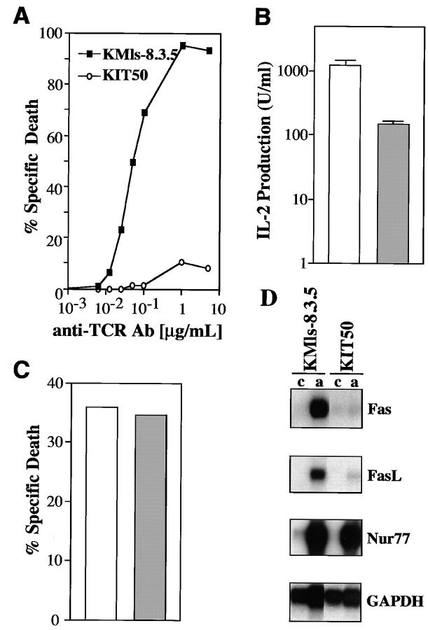
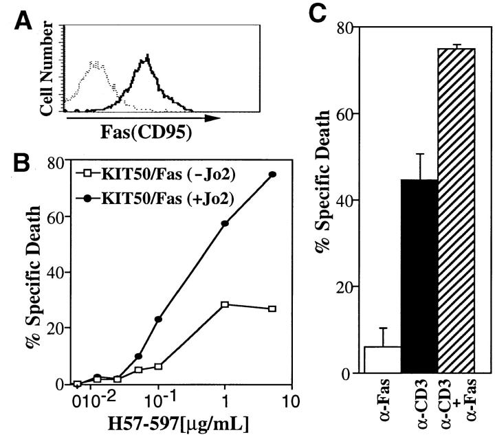
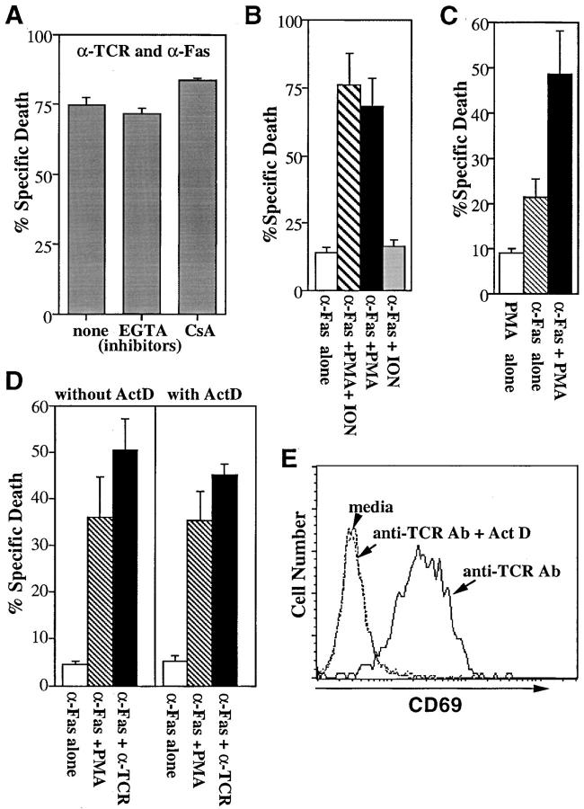
Fas(CD95) and its ligand (FasL) interaction plays a pivotal role in T cell receptor (TCR)-mediated apoptosis. However, the susceptibility of T cells to Fas-mediated apoptosis is tightly regulated during immune responses, a regulation which is thought to maintain the antigen-specificity of T cell apoptosis. Here we show that TCR stimulation enhances the induction of Fas-mediated apoptosis. In addition, using a mutant T cell hybridoma with impaired FasL expression, we show that the synergy provided by TCR stimulation can be mimicked by activators of PKC but not calcium influx. This effect cannot be inhibited by actinomycin D, suggesting that TCR stimulation leads to the alteration in preexisting signaling molecules to enhance Fas-mediated apoptosis. Our results therefore provide a mechanism of how Fas-FasL interactions lead to T cell death in an antigen-specific manner via repetitive antigen stimulation.


Figures

Figure 1
Figure 1
Isolation and characterization of a T cell hybridoma mutant with impaired Fas and FasL expression. (A) The T cell hybridoma mutant KIT50 is resistant to anti-TCR Ab (H57-597). Cells were incubated for 24 h on 96-well plates coated with increasing amounts of H57-597 and then assayed for apoptosis by PI uptake and FACS® analysis. Specific cell death was determined as described in Materials and Methods. (Squares) The parental cell line, KMls-8.3.5; (circles) the mutant cell line, KIT50. The representative results of at least 10 independent experiments are shown. (B) IL-2 production upon TCR stimulation. Cells were stimulated for 18 h on plates coated with anti-TCR Ab (10 μg/ml). Supernatants were collected and their activity was assayed using HT-2 cells as previously described (23). (White bar) The parental cell line, KMls-8.3.5; (hatched bar) the mutant cell line, KIT50. The representative results of three to seven independent experiments are shown. (C) Apoptotic cell death induced by dexamethasone. KMls-8.3.5 and KIT50 cells were incubated in the presence of 10 μM dexamethasone or in the media alone for 24 h and cell viability was tested by PI uptake. Spontaneous cell death in the media alone was <2–3%. (White bar) the parental cell line, KMls-8.3.5; (hatched bar) the mutant cell line, KIT50. The representative results of at least six independent experiments are shown. (D) Fas and FasL mRNA expression was impaired in KIT50. RNA was prepared from control or TCR-stimulated cells and then the expression of various genes was tested by Northern blot hybridization. (c) Control, unstimulated, (a) stimulated with anti-TCR Ab (10 μg/ml).
Figure 2
Figure 2
TCR-mediated signals enhance Fas-mediated apoptosis. (A) KIT50/Fas expresses relatively high levels of Fas on the cell surface. Surface Fas expression on KIT50-Fas cells were determined by FACS® analysis using biotinylated Jo2 Ab (PharMingen) and phycoerythrin (PE)- labeled streptavidin (Becton Dickinson). (Dotted line) PE-streptavidin alone; (solid line) biotinylated Jo2 plus PE-streptavidin. (B) TCR stimulation enhances Fas-mediated cell death. KIT50-Fas cells were cultured in 96-well plates coated with increasing amounts of H57-597 ± anti-Fas Ab (Jo2, 10 μg/ml). Cells were collected after 24 h and cell death was detected by PI uptake by FACS® analysis. The representative results of at least three independent experiments are shown. (Filled circles) KIT50/Fas stimulated with anti-Fas and anti-TCR Abs; (white squares) KIT50/Fas stimulated with anti-TCR Ab alone. (C) Synergistic effect of simultaneous Fas and TCR stimulation on activated lymph node T cells. ConA-IL-2–activated LNTC were incubated on plates coated with anti-CD3 (10 μg/ml) and/or anti-Fas Abs (10 μg/ml). Cell death was determined 48 h later by PI uptake. The representative results of three independent experiments are shown. Spontaneous cell death in the media alone was <5%. These experiments were done in the absence of cycloheximide. (White bar) anti-Fas Ab alone; (black bar) anti-CD3 Ab alone; (hatched bar) anti-Fas Ab plus anti-CD3 Ab.
Figure 3
Figure 3
A PKC dependent, calcium-independent pathway mediates synergy between TCR and Fas signaling. (A) EGTA or CsA does not block the synergy between TCR and Fas. KIT50-Fas cells were stimulated with plate-bound H57-597 (10 μg/ml) plus Jo2 (10 μg/ml) in the presence of media alone, EGTA or CsA. Cell death was measured 24 h later by PI uptake. The representative results of three independent experiments are shown. The similar treatment of EGTA or CsA completely blocked cell death induced by anti-TCR Ab alone (data not shown). The low level of apoptosis induced by anti-Fas Ab alone (2–10% specific cell death) was not affected by EGTA or CsA (data not shown). Spontaneous cell death in the media alone ± EGTA or CSA was <5%. (B) PMA can substitute for TCR signals to enhance Fas-mediated apoptosis. KIT50-Fas cells were cultured in 96-well plates coated with anti-Fas Ab (Jo2, 10 μg/ ml) in the presence of various stimuli. Cell death was assayed 24 h later by PI uptake. The representative results of at least three independent experiments are shown. Cells were stimulated with Jo2 in the presence of media alone (white bar), PMA plus ionomycin (hatched bar), PMA alone (black bar), or ionomycin alone (gray bar). In the absence of Jo2, PMA or ionomycin alone did not induce cell death and PMA plus ionomycin induced a low level (<15%) of apoptosis in these cells (data not shown). (C) PMA alone can enhance Fas-mediated cell death of another Fas-transfected T cell hybridoma, KCIT1-8.5/Fas. KCIT1-8.5/Fas cells (23) were stimulated with PMA alone (white bar), plate-bound 10 μg/ml anti-Fas Ab alone (hatched bar), or PMA plus plate-bound anti-Fas Ab (black bar). Cell death was assayed 24 h later by PI uptake. The representative results of three independent experiments are shown. (D) Actinomycin D does not inhibit the synergy mediated by PMA or anti-TCR Ab. KIT50/Fas cells were stimulated with various reagents in the presence or absence of actinomycin D. Cell death was determined 24 h later by PI uptake. The representative results of three independent experiments are shown. (White bars) plate-bound anti-Fas Ab alone, 10 μg/ml; (hatched bars) PMA plus plate-bound anti-Fas Ab; (black bars) plate-bound anti-TCR Ab plus anti-Fas Ab at 10 μg/ml each. In the absence of anti-Fas Ab, PMA alone did not induce significant cell death (<5%). Anti-TCR Ab without anti-Fas Ab induced ∼10% specific cell death (data not shown). (E) Actinomycin D inhibits CD69 induction by TCR stimulation. KIT50/Fas cells from D were stained by FITC-labeled anti-CD69 antibody and analyzed by FACS®.

References

    1. von Boehmer H. Positive selection of lymphocytes. Cell. 1994;76:219–228. - PubMed
    1. Nossal GJV. Negative selection of lymphocytes. Cell. 1994;76:229–239. - PubMed
    1. Nagata S, Golstein P. The Fas death factor. Science. 1995;267:1449–1456. - PubMed
    1. Lynch DH, Ramsdell F, Alderson MR. Fas and FasL in the homeostatic regulation of immune responses. Immunol Today. 1995;16:569–574. - PubMed
    1. van Parijs L, Abbas AK. Role of Fas-mediated cell death in the regulation of immune responses. Curr Opin Immunol. 1996;8:355–361. - PubMed

Publication types

MeSH terms

Substances