. 1997 Dec;110(6):749-62.
doi: 10.1085/jgp.110.6.749.

Caffeine-induced release of intracellular Ca2+ from Chinese hamster ovary cells expressing skeletal muscle ryanodine receptor. Effects on full-length and carboxyl-terminal portion of Ca2+ release channels

Affiliations

Caffeine-induced release of intracellular Ca2+ from Chinese hamster ovary cells expressing skeletal muscle ryanodine receptor. Effects on full-length and carboxyl-terminal portion of Ca2+ release channels

M B Bhat et al. J Gen Physiol. 1997 Dec.

Abstract

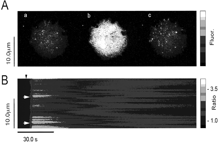
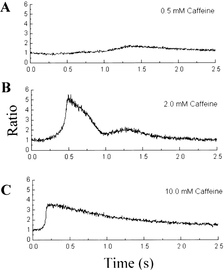
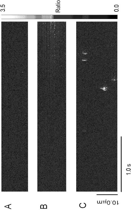
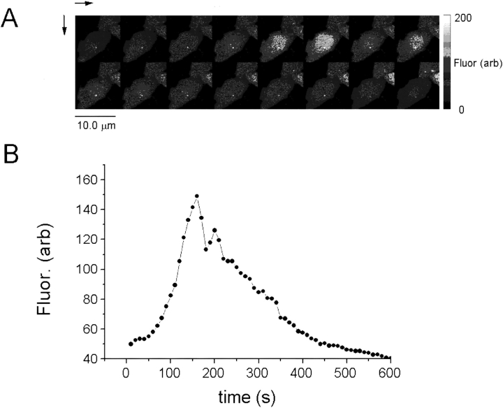
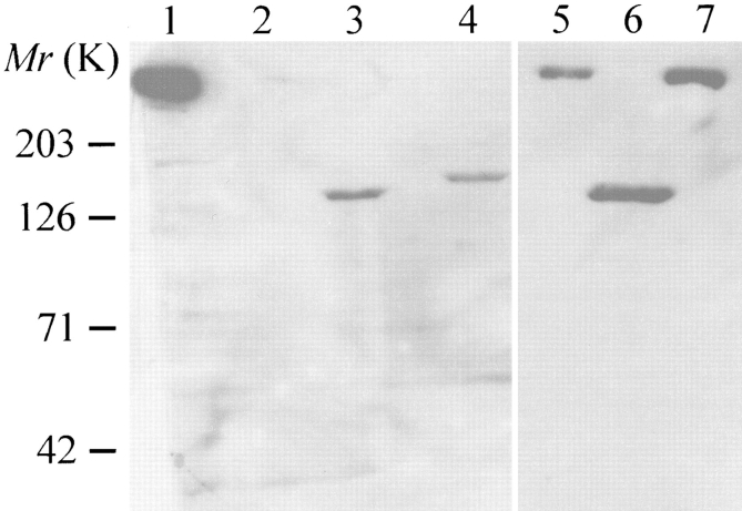
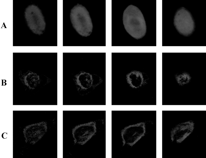
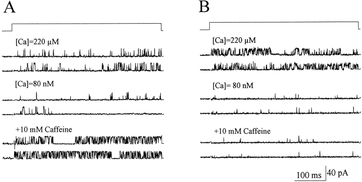
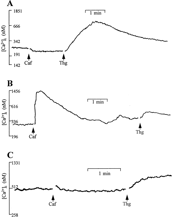
The ryanodine receptor (RyR)/Ca2+ release channel is an essential component of excitation-contraction coupling in striated muscle cells. To study the function and regulation of the Ca2+ release channel, we tested the effect of caffeine on the full-length and carboxyl-terminal portion of skeletal muscle RyR expressed in a Chinese hamster ovary (CHO) cell line. Caffeine induced openings of the full length RyR channels in a concentration-dependent manner, but it had no effect on the carboxyl-terminal RyR channels. CHO cells expressing the carboxyl-terminal RyR proteins displayed spontaneous changes of intracellular [Ca2+]. Unlike the native RyR channels in muscle cells, which display localized Ca2+ release events (i.e., "Ca2+ sparks" in cardiac muscle and "local release events" in skeletal muscle), CHO cells expressing the full length RyR proteins did not exhibit detectable spontaneous or caffeine-induced local Ca2+ release events. Our data suggest that the binding site for caffeine is likely to reside within the amino-terminal portion of RyR, and the localized Ca2+ release events observed in muscle cells may involve gating of a group of Ca2+ release channels and/or interaction of RyR with muscle-specific proteins.


Figures

Figure 5
Figure 5
Spatial and temporal patterns of caffeine-induced Ca2+ release in CHO cells expressing RyR. CHO cells expressing RyR-wt release Ca2+ upon exposure to caffeine. (A) Full frame confocal image of Fluo-3 fluorescence (Ca2+ activated) in CHO cells expressing the full length RyR: (a) control, (b) 0.5 s after addition of 10 mM caffeine, and (c) 5 min after washout of the caffeine. Increasing Ca2+-activated Fluo-3 fluorescence is indicated by the color bar on the right. Distance, in both x and y dimensions, is indicated by calibration bar indicating 10.0 μm. (B) Compressed line-scan image Fluo-3 fluorescence (proportional to [Ca2+]i) of a single CHO cell exposed to 10 mM caffeine. Calcium rises rapidly and cooperatively (see small initial “foot” in rise), and declines slowly.
Figure 6
Figure 6
Dose-dependent effect of caffeine on CHO cells expressing RyR-wt. Fluorescence ratio, derived from one selected pixel of line scan images such as those in Fig. 5 B, is plotted as a function of time. The pixel size was 0.2 μm. Results are from three different CHO cells. Doses of caffeine: (A) 0.5, (B) 2.0, and (C) 10 mM. The rate of rise in fluorescence ratio ([Ca2+]) is highly dose dependent, possibly indicating dose-dependent cooperativity of the opening of the Ca2+ release channels. The fact that the peak fluorescence ratio reached was greatest in 2.0 mM caffeine may be the result of variability in the amount of Ca2+ in the intracellular stores of different cells. The number of cells tested are n = 2 (0.1 mM), n = 5 (0.2 mM), n = 4 (0.5 mM), n = 4 (2 mM), n = 2 (5 mM), and n = 10 (10 mM).
Figure 7
Figure 7
Lack of Ca2+ sparks in CHO cells expressing RyR-wt. (A) Line-scan image of fluorescence ratio (Fluo-3) in c1148 cells under control conditions. No local calcium transients or sparks are evident. (B) Line-scan image of c1148 cells during the initial rise in [Ca2+]i in the presence of 0.5 mM caffeine. [Ca2+] rises very slowly from resting levels, but no sparks are evident. (C) Typical spontaneous Ca2+ sparks in a cardiac myocyte. All line-scan images were obtained and presented at exactly the same spatial and temporal resolution, with the same confocal microscope.
Figure 8
Figure 8
Spontaneous oscillation of intracellular Ca2+ in CHO cells expressing RyR-C. (A) Sequence of 16 full-frame images of Ca2+- activated Fluo-3 fluorescence taken at 20-s intervals. (B) Plot of Ca2+-activated Fluo-3 fluorescence as a function of time in the cell illustrated in A. The existence of spontaneous oscillations of [Ca2+]i may explain the apparent high resting [Ca2+]i in population measurements of [Ca2+]i in CHO cells expressing RyR-C.
Figure 1
Figure 1
Heterologous expression of skeletal muscle RyR in CHO cells. Western blot analysis of RyR proteins expressed in Chinese hamster ovary cells. Total cell lysates were run on a 3–12% gradient SDS polyacrylamide gel, and blotted with monoclonal antibody (RR2) against the rabbit skeletal muscle RyR (Bhat et al., 1997). Lane 1, SR vesicles from rabbit skeletal muscle; lane 2, untransfected CHO cells; lanes 3 and 6, COOH-terminal RyR proteins (∼130 kD) stably expressed in CHO cells (ryr-c); lane 4, CHO cells transfected with GFP-RyR-C; lane 5, CHO cells transfected with GFP-RyR; lane 7, full length RyR (∼560 kD) stably expressed in CHO cells (c1148).
Figure 2
Figure 2
Localization of RyR in intracellular membranes of CHO cells. Confocal measurement of green fluorescence in CHO cells transfected with (A) pEGFP-N1, (B) pcDNA3 (GFP-RyR), and (C) pcDNA3 (GFP-RyR-C). Cells expressing GFP alone exhibit a diffuse pattern of fluorescence throughout the cell (A), whereas cells expressing the GFP-RyR and GFP-RyR-C fusion proteins show localized fluorescence signal near the perinuclear region of the cells (B and C). The panels from left to right show representative sections through the cells at 0.5-μm intervals. The cells have an average thickness of ∼10 μm.
Figure 3
Figure 3
Effect of caffeine on single RyR-wt and RyR-C channels. Selected single channel currents at +50 mV from a continuous experiment with the RyR-wt (A) and RyR-C (B) channels incorporated into the lipid bilayer membrane. Under control conditions (220 μM [Ca2+]i), the RyR-wt had an average Po of 0.121 ± 0.044 (n = 19), and the RyR-C channel had an average Po of 0.201 ± 0.036 (n = 12). The addition of 10 mM EGTA lowered the free [Ca2+] to 0.08 μM and reduced the Po of RyR-wt to 0.013 ± 0.002 (n = 5), and Po of RyR-C to 0.008 ± 0.003 (n = 4). Subsequent addition of 10 mM caffeine increased the Po of RyR-wt to 0.198 ± 0.063 (n = 4). Po of the RyR-C channel remained unchanged after the addition of 10 mM caffeine (Po = 0.008 ± 0.002, n = 4).
Figure 4
Figure 4
Caffeine-induced release of Ca2+ in CHO cells. Changes in [Ca2+]i were measured in cells (106/ml) loaded with Fura-2 AM and maintained under continuous stirring in a cuvette. (A) Application of 10 mM caffeine (Caf) does not induce release of intracellular Ca2+ in untransfected CHO cells, whereas thapsigargin (Thg, 500 nM) causes a rise in [Ca2+], suggesting intact intracellular Ca2+ store in these cells. (B) In c1148 cells, an increase in [Ca2+]i from 0.192 ± 0.038 μM (n = 4) to 1.222 ± 0.257 μM (n = 3) is seen after stimulation with caffeine. (C) CHO cells expressing RyR-C do not respond to stimulation with caffeine. The resting intracellular [Ca2+] in these cells is 0.422 ± 0.071 μM (n = 7). All fluorescence traces are representative of at least three separate experiments. Gaps in the recordings are due to the opening of the compartment to make additions.

References

    1. Ahern GP, Junankar PR, Dulhunty AF. Subconductance states in single-channel activity of skeletal muscle ryano-dine receptors after removal of FKBP12. Biophys J. 1997;72:146–162. - PMC - PubMed
    1. Bhat MB, Ma J, Zhao JY, Hayek S, Freeman EC, Takeshima H. Deletion of amino acids 1641–2437 from the foot region of skeletal muscle ryanodine receptor alters the conduction properties of the Ca release channel. Biophys J. 1997a;73:1320–1328. - PMC - PubMed
    1. Bhat MB, Zhao JY, Takeshima H, Ma J. Functional calcium release channel formed by the carboxyl-terminal portion of ryanodine receptor. Biophys J. 1997b;73:1329–1336. - PMC - PubMed
    1. Blatter LA, Huser J, Rios E. Sarcoplasmic reticulum Ca2+ release flux underlying Ca2+sparks in cardiac muscle. Proc Natl Acad Sci USA. 1997;94:4176–4181. - PMC - PubMed
    1. Block BA, Imagawa T, Campbell KP, Franzini-Armstrong C. Structural evidence for a direct interaction between the molecular components of the transverse tubule/sarcoplasmic reticulum junction in skeletal muscle. J Cell Biol. 1988;107:2587–2600. - PMC - PubMed

Publication types

MeSH terms