. 1997 Oct;56(10):613-21.
doi: 10.1136/ard.56.10.613.

Long-term persistent accumulation of CD8+ T cells in synovial fluid of rheumatoid arthritis

Affiliations

Long-term persistent accumulation of CD8+ T cells in synovial fluid of rheumatoid arthritis

K Masuko-Hongo et al. Ann Rheum Dis. 1997 Oct.

Abstract

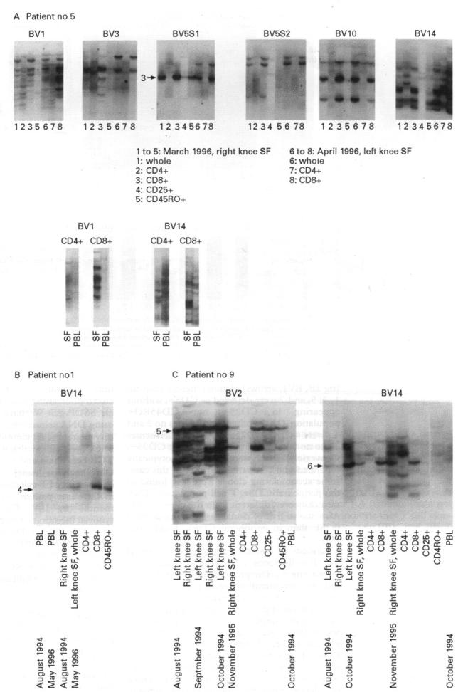
Objective: To characterise the type and kinetics of T cell clones in synovial lesions of patients with rheumatoid arthritis (RA).

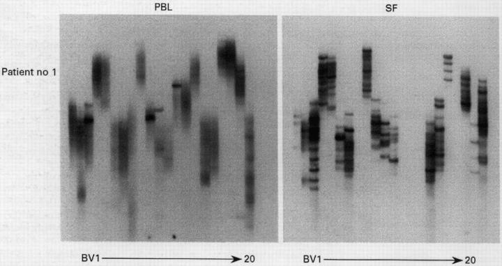
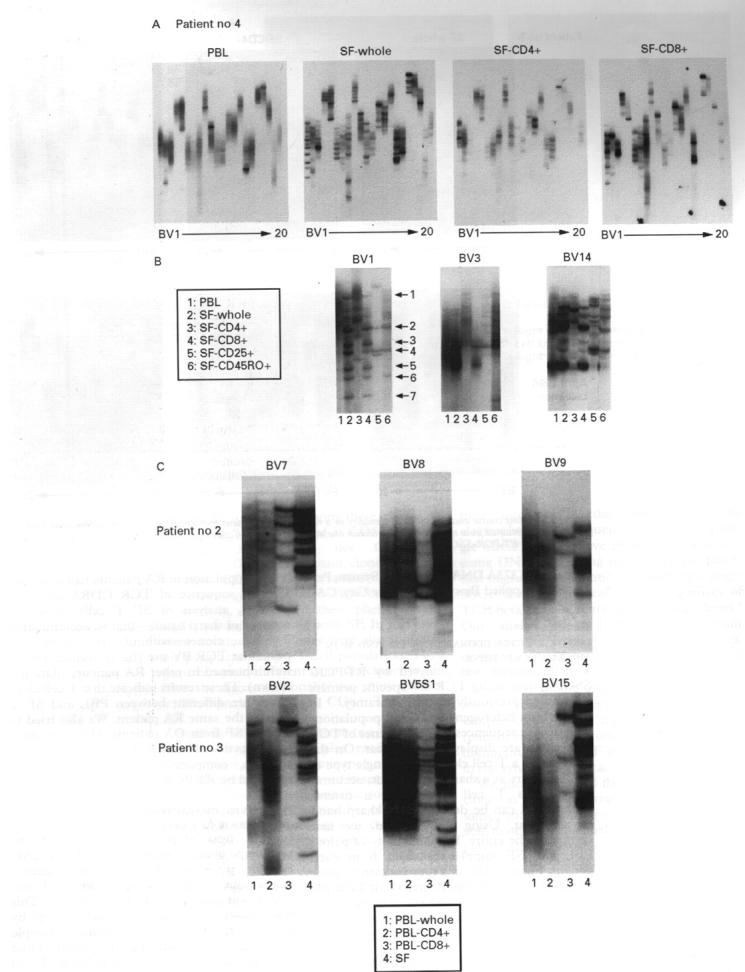
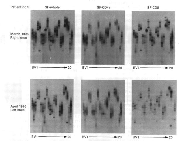
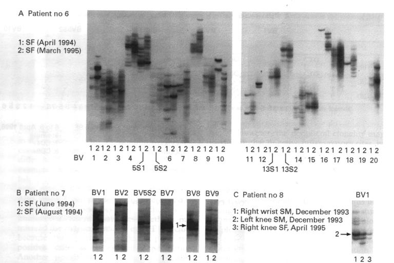
Methods: Mononuclear cells from serial samples of synovial fluid (SF) and peripheral blood from nine RA patients were separated phenotypically using antibody coated magnetic beads. After mRNA preparation, reverse transcription-polymerase chain reaction (RT-PCR) was performed to amplify V-D(N)-J (that is, the third complementarity determining, CDR3) regions of their T cell receptor beta chain genes. This was followed by single strand conformation polymorphism (SSCP) analysis to detect the clonotypes of accumulating T cells. Amino acid sequences of the dominant clones were also determined.

Results: Although peripheral T cells were heterogeneous, accumulation of oligoclonal T cells was detected in SF. The predominant accumulating clone was the CD8 subset, which was persistently present in serial samples obtained over almost one year of follow up. A proportion of these cells expressed CD25 or CD45RO, or both, suggesting they are 'memory' clones.

Conclusion: The persistent presence of CD8+ T cell clones in RA joints indicates that they may be involved in the perpetuation of the chronic inflammatory process in RA joints.


Figures

Figure 1
Figure 1
RT-PCR-SSCP analysis of T cells clonality of peripheral blood lymphocytes (PBL) and synovial fluid (SF) in a representative patient with rheumatoid arthritis. Results of final SSCP are displayed. Lanes: from left to right, BV 1 to 20 including BVSSI/BVSS2 and BV13SI/BV13S2 subfamilies.
Figure 2
Figure 2
Dominant clonal accumulations are demonstrated in CD8+ T cell subset of synovial T cells. (A) Results of analysis of T cell clonality of peripheral blood lymphocytes (PBL) and synovial fluid T cells (SFTC) of a representative patient with rheumatoid arthritis. SFTC were examined as a whole group, CD4+ and CD8+ populations, and analysed independently. (B) Amplified DNAs from each population were electrophoresised on the same SSCP gel to provide a comparison between the groups. (C) Results of T cell clonality analyses of PBL and SFTC, with a phenotypic separation of PBL.
Figure 3
Figure 3
A time course analysis of T cell clonality in a representative patient with rheumatoid arthritis. Synovial fluid samples were obtained from the same patient within one month from the left and right knee joints and analysed for T cell clonality using RT-PCR-SSCP.
Figure 4
Figure 4
Comparison of accumulating T cell clones in synovial fluid (SF) in two patients with rheumatoid arthritis at different time intervals. (A) and (B) SF T cell samples obtained at the indicated time intervals from two patients were individually analysed by RT-PCR-SSCP and finally electrophoresised on the same SSCP gel. (C) Two specimens of synovial membranes from the right wrist and left knee joint were obtained from another patient in 1993, followed by samples of synovial fluid from the right knee joint in 1995. Each sample was analysed independently and compared on the same SSCP gel. Arrows indicate dominantly accumulating clones.
Figure 5
Figure 5
Serial determination of accumulated T cell clones and their phenotype. (A) to (C) Serial samples of synovial fluid T cells and peripheral blood lymphocyte (PBL) were obtained from three RA patients and separated into T cell subsets. The detected clones were compared with each other by electrophoresising to the same SSCP gels. Arrows indicate dominantly accumulating clones. The amino acid sequences of the bands are shown in table 3.

References

    1. Int Immunol. 1994 Jun;6(6):897-907 - PubMed
    1. J Rheumatol. 1990 Feb;17(2):142-8 - PubMed
    1. Arthritis Rheum. 1994 Nov;37(11):1627-36 - PubMed
    1. J Clin Invest. 1994 Dec;94(6):2525-31 - PubMed
    1. J Immunol. 1995 Apr 1;154(7):3538-47 - PubMed

Publication types

MeSH terms

Substances