. 1997 Dec 1;11(23):3157-67.
doi: 10.1101/gad.11.23.3157.

Dual role of the Smad4/DPC4 tumor suppressor in TGFbeta-inducible transcriptional complexes

Affiliations

Dual role of the Smad4/DPC4 tumor suppressor in TGFbeta-inducible transcriptional complexes

F Liu et al. Genes Dev. .

Abstract

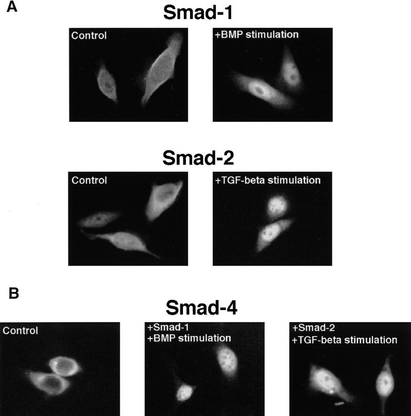
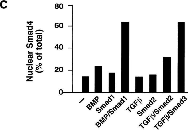
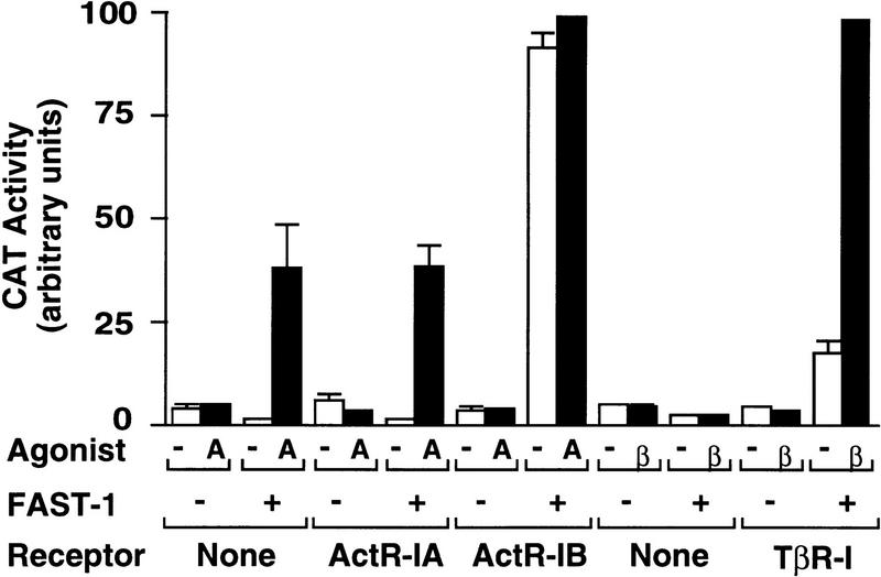
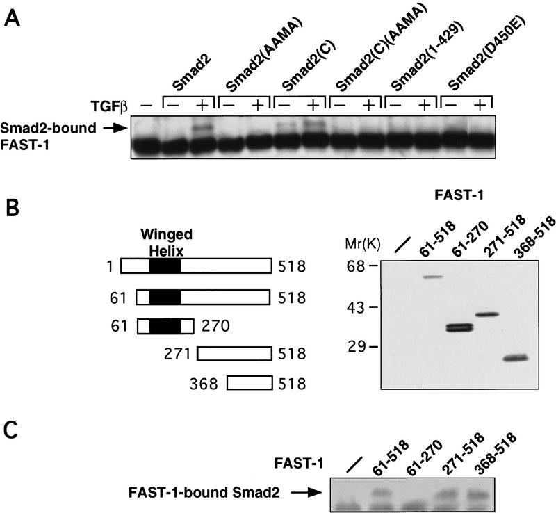
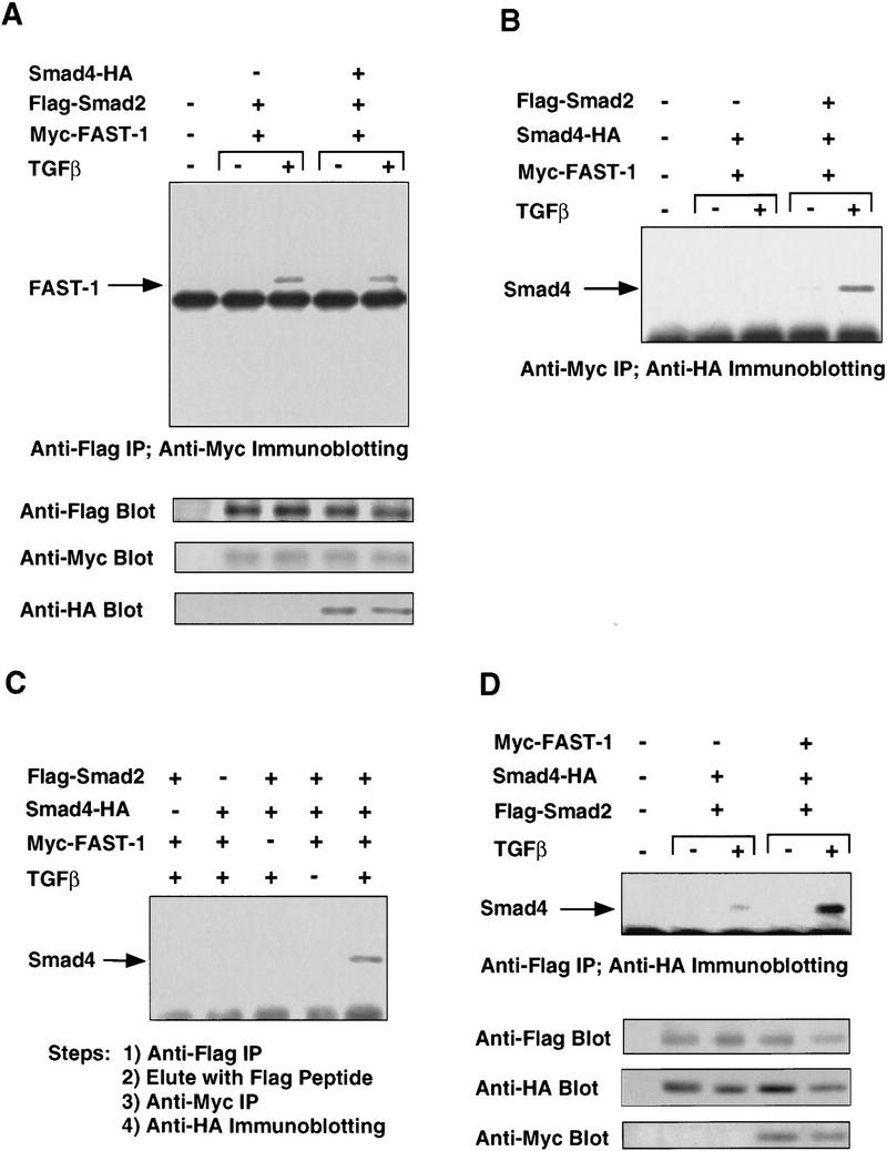
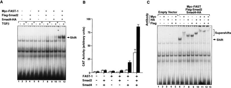
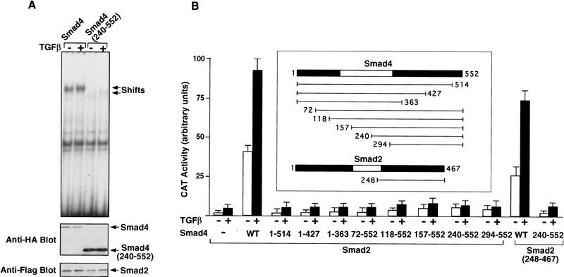
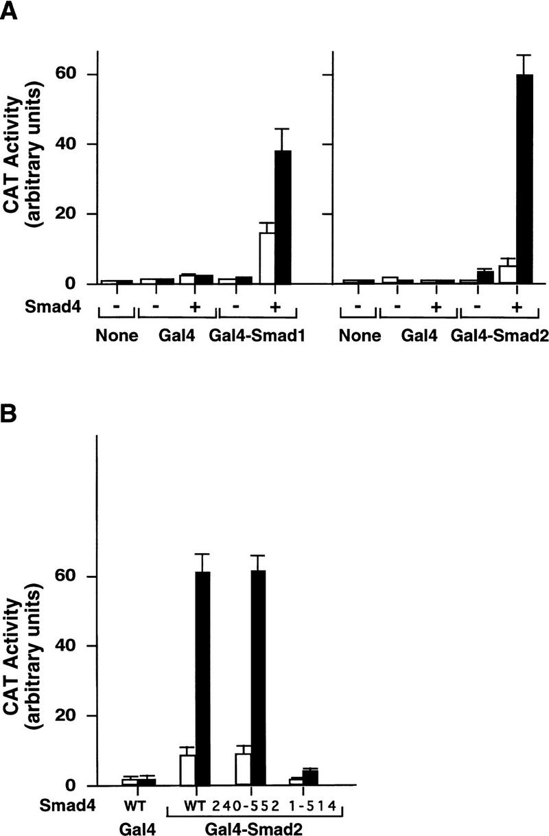
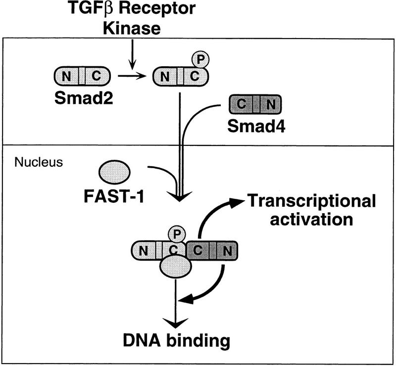
Upon ligand binding, the receptors of the TGFbeta family phosphorylate Smad proteins, which then move into the nucleus where they activate transcription. To carry out this function, the receptor-activated Smads 1 and 2 require association with the product of deleted in pancreatic carcinoma, locus 4 (DPC4), Smad4. We investigated the step at which Smad4 is required for transcriptional activation. Smad4 is not required for nuclear translocation of Smads 1 or 2, or for association of Smad2 with a DNA binding partner, the winged helix protein FAST-1. Receptor-activated Smad2 takes Smad4 into the nucleus where they form a complex with FAST-1 that requires these three components to activate transcription. Smad4 contributes two functions: Through its amino-terminal domain, Smad4 promotes binding of the Smad2/Smad4/FAST-1 complex to DNA; through its carboxy-terminal domain, Smad4 provides an activation function required for Smad1 or Smad2 to stimulate transcription. The dual function of Smad4 in transcriptional activation underscores its central role in TGFbeta signaling.


Figures

Figure 1
Figure 1
(A) Nuclear translocation of Smad1 and Smad2 in the absence of Smad4. SW480.7 cells were transfected with Flag–Smad1 or Flag–Smad2 and stimulated with BMP or TGFβ by cotransfection with activated BMP or TGFβ receptor plus treatment with BMP4 or TGFβ1. Immunofluorescence staining with anti–Flag antibody was then performed. After agonist stimulation, Flag–Smad1 staining was nuclear in up to 60% of the cells and Flag–Smad2 staining in up to 76% of the cells. (B) Smad4 requires a receptor-activated Smad for nuclear translocation in response to agonists. Amino-terminal Flag-tagged Smad4 was transfected alone or with the indicated SMADs and stimulated with BMP or TGFβ as in A. Shown are Flag immunostaining of cells transfected with Flag-Smad4 alone (control), with Smad1 and stimulated with BMP, or with Smad2 and stimulated with TGFβ. (C) A quantitation of Smad4 nuclear staining under various conditions in one representative experiment. BMP and TGFβ refers to BMP or TGFβ stimulation by cotransfection with activated BMP or TGFβ receptor and treatment with BMP4 or TGFβ1, respectively.
Figure 1
Figure 1
(A) Nuclear translocation of Smad1 and Smad2 in the absence of Smad4. SW480.7 cells were transfected with Flag–Smad1 or Flag–Smad2 and stimulated with BMP or TGFβ by cotransfection with activated BMP or TGFβ receptor plus treatment with BMP4 or TGFβ1. Immunofluorescence staining with anti–Flag antibody was then performed. After agonist stimulation, Flag–Smad1 staining was nuclear in up to 60% of the cells and Flag–Smad2 staining in up to 76% of the cells. (B) Smad4 requires a receptor-activated Smad for nuclear translocation in response to agonists. Amino-terminal Flag-tagged Smad4 was transfected alone or with the indicated SMADs and stimulated with BMP or TGFβ as in A. Shown are Flag immunostaining of cells transfected with Flag-Smad4 alone (control), with Smad1 and stimulated with BMP, or with Smad2 and stimulated with TGFβ. (C) A quantitation of Smad4 nuclear staining under various conditions in one representative experiment. BMP and TGFβ refers to BMP or TGFβ stimulation by cotransfection with activated BMP or TGFβ receptor and treatment with BMP4 or TGFβ1, respectively.
Figure 2
Figure 2
Reconstitution of the Mix2–FAST-1 transcriptional response in mammalian cells. R1B/L17 cells were cotransfected with the Mix2 reporter A3CAT and the indicated FAST-1 and receptor vectors, then treated with activin (A) or TGFβ (β). Reporter CAT activity was then determined. Data are the average ±s.d. of triplicates.
Figure 3
Figure 3
FAST-1 interaction with Smad2. (A) TGFβ-induced FAST-1 association with Smad2. R1B/L17 cells were cotransfected with Myc–FAST-1, the indicated Flag–Smad2 derivatives, and TβR-I for TGFβ stimulation. After incubation with or without TGFβ, cell lysates were immunoprecipitated with anti-Flag antibody and the precipitates subjected to anti-Myc immunoblotting. Smad2(AAMA) contains Ser to Ala mutations in the carboxy-terminal SSMS sequence (Macias-Silva et al. 1996; Kretzschmar et al. 1997a). Smad2(C) refers to the Smad2 C domain (amino acids 248–467) (Hata et al. 1997). Smad2(1–429) and Smad2(D450E) are tumor-derived inactive Smad2 mutants (Eppert et al. 1996). (B) FAST-1 truncation constructs and their expression (as Myc-tagged constructs) in R-1B/L17 cells, as determined by anti-Myc immunoblotting analysis of cell lysates. (Solid rectangles) The winged helix DNA-binding domain (Chen et al. 1996). (C) Smad2 interaction with the FAST-1 C domain. R1B/L17 cells were cotransfected with Flag–Smad2, TβR-I, and the indicated Myc–FAST-1 derivatives. Cells were treated with TGFβ and analyzed by immunoprecipitation with anti-Myc antibody followed by anti-Flag immunoblotting.
Figure 4
Figure 4
TGFβ-induced Smad2–Smad4–FAST-1 complex. (A) Smad2 interaction with FAST-1 does not require Smad4. SW480.7 cells were cotransfected with the indicated vectors and treated with TGFβ as indicated. Cell lysates were immunoprecipitated with anti-Flag antibody and the precipitates analyzed by anti-Myc immunoblotting. (B) Smad2-dependent interaction of Smad4 with FAST-1. SW480.7 cells were transfected with Myc–FAST-1, Flag–Smad2 and HA-tagged Smad4 vectors, and treated with TGFβ as indicated. Cell lysates were immunoprecipitated with anti-Myc antibody and the precipitates analyzed by anti-HA immunoblotting. (C) Agonist-induced Smad2–Smad4–FAST-1 ternary complex. COS cells were cotransfected with the indicated vectors, and TβR-I(T204D) for TGF-β stimulation. Cells lysates were immunoprecipitated with agarose-immobilized anti-Flag antibody. Beads were eluted with Flag peptide, and the eluate precipitated by anti-myc antibody followed by anti-HA immunoblotting. (D) FAST-1 enhances the Smad2–Smad4 interaction. COS cells were cotransfected with the indicated vectors, and TβR-I(T204D) for TGFβ stimulation. Cell lysates were precipitated with anti-Flag antibody and the precipitates analyzed by anti-HA immunoblotting.
Figure 5
Figure 5
Smad4 is essential for the formation of a TGFβ-inducible DNA-binding complex and transcriptional activation. (A) A TGFβ-inducible DNA-binding complex requiring FAST-1, Smad2, and Smad4. SW480.7 cells were cotransfected with Myc–FAST-1, Flag–Smad2, and Smad4–HA and treated with TGFβ as indicated. Nuclear extracts were used to perform gel mobility shift assays by use of the 50-bp ARE oligonucleotide as a probe. (B) Smad4 is essential for activation of the Mix2 reporter A3CAT. SW480.7 cells were cotransfected with the A3CAT reporter gene (Huang et al. 1995), FAST-1, Smad2, and Smad4, and treated with TGFβ (▪) or not (□), as indicated. CAT activity was then analyzed. (C) FAST-1, Smad2, and Smad4 bind together to DNA. SW480.7 cells were cotransfected with the indicated vectors and treated with TGFβ. Supershift assays were performed by use of nuclear extracts with the indicated antibodies individually or in various combinations.
Figure 6
Figure 6
(A) The amino-terminal region of Smad4 is required for DNA binding by the ternary complex. SW480.7 cells were cotransfected with Myc-FAST-1, Flag-Smad2, and either Smad4 or Smad4(240–552), both tagged with the HA epitope. Cells were treated with TGFβ as indicated. Gel shift experiment was performed with whole cell extracts. The expression level of Flag-Smad2, Smad4, and Smad4(240–552) was analyzed by immunoblotting with antibodies against the Flag or HA epitopes. (B) Both the N and the C domains of Smad4 are required to activate the A3CAT reporter gene. SW480.7 cells were cotransfected with A3CAT, FAST-1, the indicated full length or truncated Smad4 and Smad2 constructs. Cells were treated with TGFβ, and CAT activity was analyzed.
Figure 7
Figure 7
Ligand-induced transcriptional activity of GAL4–Smad1 and GAL4–Smad2 depends on the C domain of Smad4. SW480.7 cells were cotransfected with GAL4–Smad1, GAL4–Smad2, wild-type Smad4, Smad4(240–552), or Smad4(1–514) along with the GAL4 reporter gene G1E1BCAT (Lillie and Green 1989). Cells were incubated with 2 nm BMP4 (▪) in A (left) or without (□), or with 500 pm TGFβ in A (right) and B, as indicated, and CAT activity was then analyzed.
Figure 8
Figure 8
A model depicting the participation of Smad4 in a transcriptional complex with Smad2 and FAST-1, extending the previous model of Chen et al. (1996). Upon phosphorylation by the activated receptor, Smad2 translocates to the nucleus. Smad4 associates with Smad2 through their C domains but is not required for this translocation. In the nucleus, the Smad2/Smad4 complex associates with FAST-1. In this ternary complex, the N domain of Smad4 promotes binding to DNA, whereas the C domain of Smad4 is essential for activation of transcription.

References

    1. Baker J, Harland RM. A novel mesoderm inducer, mMadr-2, functions in the activin signal transduction pathway. Genes & Dev. 1996;10:1880–1889. - PubMed
    1. Barrett MT, Schutte M, Kern SE, Reid BJ. Allelic loss and mutational analysis of the DPC4 gene in esophageal adenocarcinoma. Cancer Res. 1996;56:4351–4353. - PubMed
    1. Cárcamo J, Weis FMB, Ventura F, Wieser R, Wrana JL, Attisano L, Massagué J. Type I receptors specify growth inhibitory and transcriptional responses to TGF-β and activin. Mol Cell Biol. 1994;14:3810–3821. - PMC - PubMed
    1. Chen X, Rubock MJ, Whitman M. A transcriptional partner of MAD proteins in TGF-β signaling. Nature. 1996;383:691–696. - PubMed
    1. Chen X, Weisberg E, Fridmacher V, Watanabe M, Naco G, Whitman M. Smad4 and FAST-1 in the assembly of activin-responsive factor. Nature. 1997;389:85–89. - PubMed

Publication types

MeSH terms

Substances

Associated data