. 1997 Dec 1;11(23):3218-31.
doi: 10.1101/gad.11.23.3218.

A novel cytosolic regulator, Pianissimo, is required for chemoattractant receptor and G protein-mediated activation of the 12 transmembrane domain adenylyl cyclase in Dictyostelium

Affiliations

A novel cytosolic regulator, Pianissimo, is required for chemoattractant receptor and G protein-mediated activation of the 12 transmembrane domain adenylyl cyclase in Dictyostelium

M Y Chen et al. Genes Dev. .

Abstract

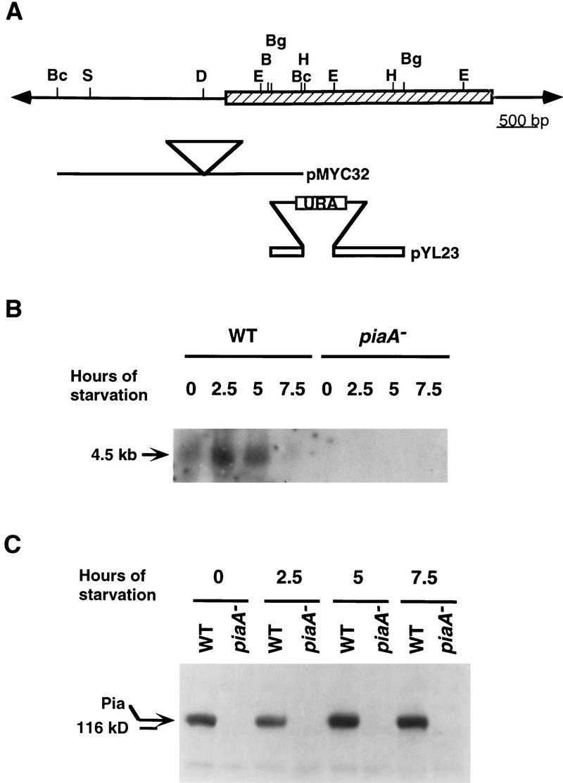
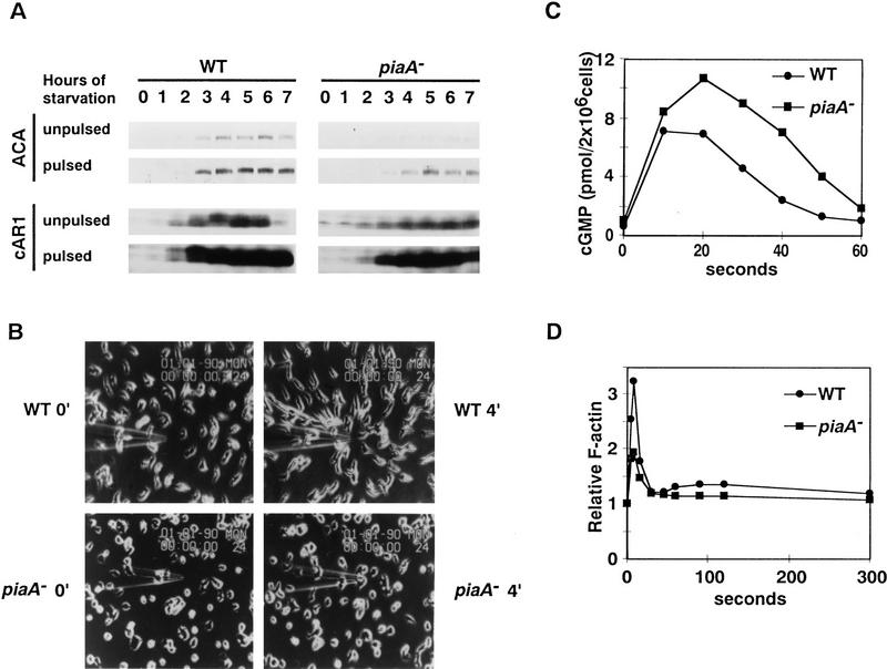
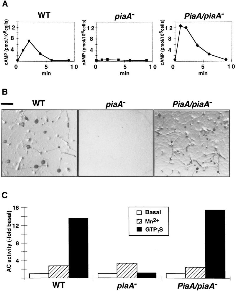
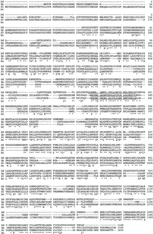
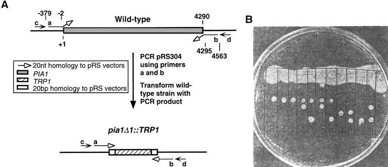
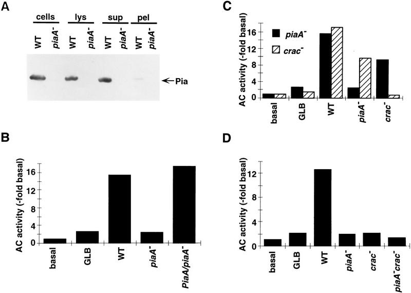
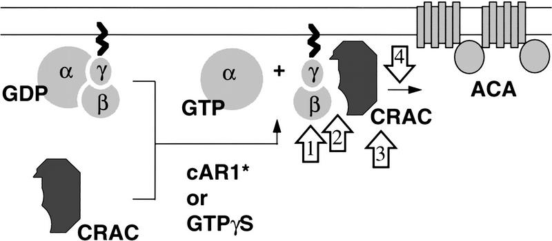
Genetic analysis was applied to identify novel genes involved in G protein-linked pathways controlling development. Using restriction enzyme-mediated integration (REMI), we have identified a new gene, Pianissimo (PiaA), involved in cAMP signaling in Dictyostelium discoideum. PiaA encodes a 130-kD cytosolic protein required for chemoattractant receptor and G protein-mediated activation of the 12 transmembrane domain adenylyl cyclase. In piaA- null mutants, neither chemoattractant stimulation of intact cells nor GTPgammaS treatment of lysates activates the enzyme; constitutive expression of PiaA reverses these defects. Cytosols of wild-type cells that contain Pia protein reconstitute the GTPgammaS stimulation of adenylyl cyclase activity in piaA- lysates, indicating that Pia is directly involved in the activation. Pia and CRAC, a previously identified cytosolic regulator, are both essential for activation of the enzyme as lysates of crac- piaA- double mutants require both proteins for reconstitution. Homologs of PiaA are found in Saccharomyces cerevisiae and Schizosaccaromyces pombe; disruption of the S. cerevisiae homolog results in lethality. We propose that homologs of Pia and similar modes of regulation of these ubiquitous G protein-linked pathways are likely to exist in higher eukaryotes.


Figures

Figure 1
Figure 1
The structure of the PiaA gene and its expression during growth and early development. (A) Structure of the PiaA locus. The hatched box represents the PiaA coding region. (Bc) BclI; (S) SphI; (D) DpnII; (E) EcoRI; (Bg) BglII; (B) BamHI; (H) HindIII. Solid lines and open bars represent genomic DNA and cDNA fragments, respectively. The plasmid pMYC32 was rescued from the original REMI mutant using BclI digestion (see Materials and Methods). The triangle represents the insertion of a REMI vector, pRHI30, at a DpnII site. Plasmid pYL23 is a cDNA construct used for gene targeting; when linearized by BglII digestion and transformed into wild-type cells, it disrupted the PiaA gene by homologous recombination, replacing 0.4 kb of the coding region (a HindIII–EcoRI fragment) with a vector carrying the URA selectable marker. (B) Wild-type (WT) and piaA cells were developed in suspension and samples were taken at times indicated. RNA was prepared and separated on a 1% agarose gel containing formaldehyde, blotted, and probed with a 2.4-kb 32P-labeled cDNA fragment. (C) Protein samples were prepared from the same cells as in B at times indicated, separated on a 7.5% SDS-PAGE gel, transferred onto a polyvinyldifluoride membrane, and probed with a rabbit polyclonal anti-Pia antibody.
Figure 2
Figure 2
Gene expression, chemotaxis, cGMP production, and actin polymerization in the piaA cells. (A) Wild-type and piaA cells were developed in suspension with or without addition of 100 nm cAMP pulses. Samples were taken at times indicated, separated on 10% SDS-PAGE gels, and transferred onto a polyvinyldifluoride membrane. Blots were each cut horizontally, and respective halves were probed with polyclonal antisera against ACA and cAR1, respectively. (B) Wild-type and piaA cells were developed for 5 hr with the addition of 100 nm cAMP at 6-min intervals. Cells were examined for chemotaxis to cAMP in a microneedle assay as described in Materials and Methods. At time 0 min, a microneedle filled with cAMP solution was positioned to stimulate the cells. The response of the cells at time 4 min is shown on the right. (C,D) Cells were developed as in B. cAMP-induced cGMP production (C) and actin polymerization (D) were assayed as described. Means of two to four experiments are shown.
Figure 3
Figure 3
Pia is required for receptor-mediated and GTPγS activation of ACA. (A) Chemoattractant receptor-mediated activation of adenylyl cyclase was assayed in 5-hr-developed wild-type, piaA and PiaA/piaA cells as described using 2′-deoxy-cAMP as the stimulus. (B) Wild-type, piaA, and PiaA/piaA cells were developed on non-nutrient agar plates and photographed at 48 hr. Bar, 1 mm. (C) Wild-type, piaA and PiaA/piaA cells were developed for 5 hr and assayed for adenylyl cyclase activity in the absence or presence of 5 mm MnSO4. GTPγS stimulation was determined in the presence of 40 μm GTPγS and 1 μm cAMP in the lysate. Means of 8–10 experiments are shown.
Figure 4
Figure 4
Amino acid sequences of Dictyostelium Pia and two yeast homologs. The alignment of three sequences is shown. Numbers on the right indicate amino acid positions. (Dd) Dictyostelium; (Sp) S. pombe; (Sc) S. cerevisiae; (Ho) homology between sequences. Residues identical in all three homologs are indicated by letters. Residues similar in all three homologs are indicated by an asterisk. Residues identical or similar in two sequences only are not marked. Segments identified by TMAP are underlined (see text for details).
Figure 5
Figure 5
Disruption of the S. cerevisiae PIA1 gene results in lethality. (A) A schematic diagram of the disruption of the PIA1 gene. Numbers indicate nucleotide positions. Primers a and b were both 60 nucleotides in length; each contained 40 nucleotides homologous to the locus, and 20 nucleotides homologous to pRS (indicated by open arrows). These two primers were used to amplify the TRP1 gene from pRS304 and the PCR product was transformed into a wild-type strain. The 40-bp region flanking TRP1 allowed homologous recombination at the PIA1 locus and deletion clones were first identified by PCR analysis (using primers a and d or primers b and c), then verified by Southern blot analysis (probed with oligonucleotide c). (B) The heterozygous pia1 deletion strain was sporulated and the tetrads were dissected by micromanipulation. The four spores from individual asci were aligned vertically and allowed to germinate on a YPD plate at 30°C. The picture was taken 5 days after dissection.
Figure 6
Figure 6
Reconstitution of GTPγS activation of ACA in mutant lysates. (A) Protein samples of whole cells (cells), filter lysates (lys), soluble (sup), and particulate (pel) fractions of lysates were separated on a 6% SDS-PAGE gel, blotted and probed with a rabbit antiserum directed against the carboxyl terminus of the Pia protein. Each lane was loaded with a sample equivalent to 4 × 106 cells. (B) Reconstitution of GTPγS activation of ACA in piaA lysate was performed, as described in Materials and Methods, on cells developed for 5.5 hr, using buffer (GLB) or supernatants prepared from different cell lines as indicated. Basal activity was assayed in the absence of GTPγS. Means of three to four experiments are shown. (C) Reconstitution of GTPγS activation of ACA in lysates prepared from piaA or crac cells. Buffer or supernatants from different cell lines were added as indicated. Shown are means of three to four experiments. (D) Reconstitution of GTPγS activation of ACA in lysates prepared from piaAcrac cells. Buffer or supernatants from different cell lines were added as indicated. Means of two to four experiments are shown.
Figure 7
Figure 7
A schematic model of activation of ACA. The double lines represent the plasma membrane. cAR1* represents the activated surface receptor. Upon receptor or GTPγS activation, CRAC translocates onto the membrane. The binding of CRAC to the membrane is a Gβ-dependent process. Open arrows indicate possible points of action of Pia. See Discussion for details.

References

    1. Altschul SF, Gish W, Miller W, Myers EW, Lipman DJ. Basic local alignment search tool. J Mol Biol. 1990;215:403–410. - PubMed
    1. Broek D, Samiy N, Fasano O, Fujiyama A, Tamanoi F, Northup J, Wigler M. Differential activation of yeast adenylate cyclase by wild-type and mutant RAS proteins. Cell. 1985;41:763–760. - PubMed
    1. Caterina MJ, Devreotes PN. Molecular insights into eukaryotic chemotaxis. FASEB J. 1991;5:3078–3085. - PubMed
    1. Chen J, DeVivo M, Dingus J, Harry A, Li J, Sui J, Carty DJ, Blank JL, Exton JH, Stoffel RH, Inglese J, Lefkowitz RJ, Logothetis DE, Hildebrandt JD, Iyengar R. A region of adenylyl cyclase 2 critical for regulation by G protein βγ subunits. Science. 1995;268:1166–1169. - PubMed
    1. Chen M-Y, Devreotes PN, Gundersen RE. Serine 113 is the site of receptor-mediated phosphorylation of the Dictyostelium G protein α-subunit Gα2. J Biol Chem. 1994;269:20925–20930. - PubMed

Publication types

MeSH terms

Associated data