. 1997 Dec 1;11(23):3254-64.
doi: 10.1101/gad.11.23.3254.

Hedgehog directly controls initiation and propagation of retinal differentiation in the Drosophila eye

Affiliations

Hedgehog directly controls initiation and propagation of retinal differentiation in the Drosophila eye

M Domínguez et al. Genes Dev. .

Abstract

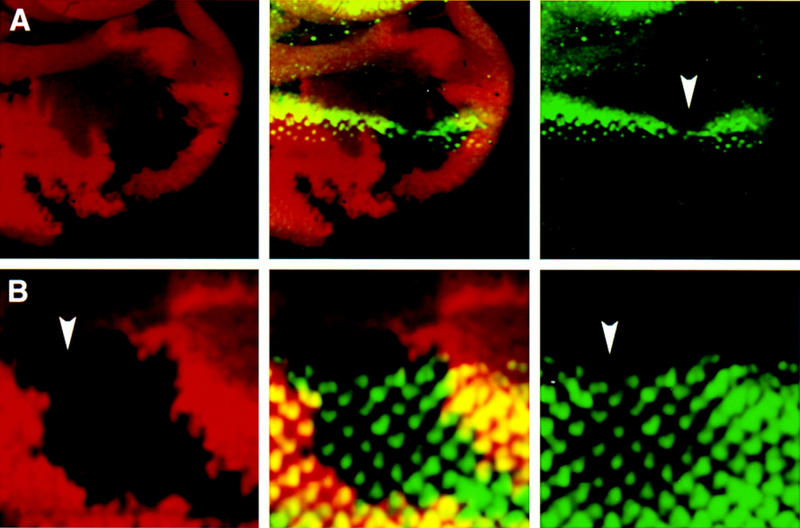
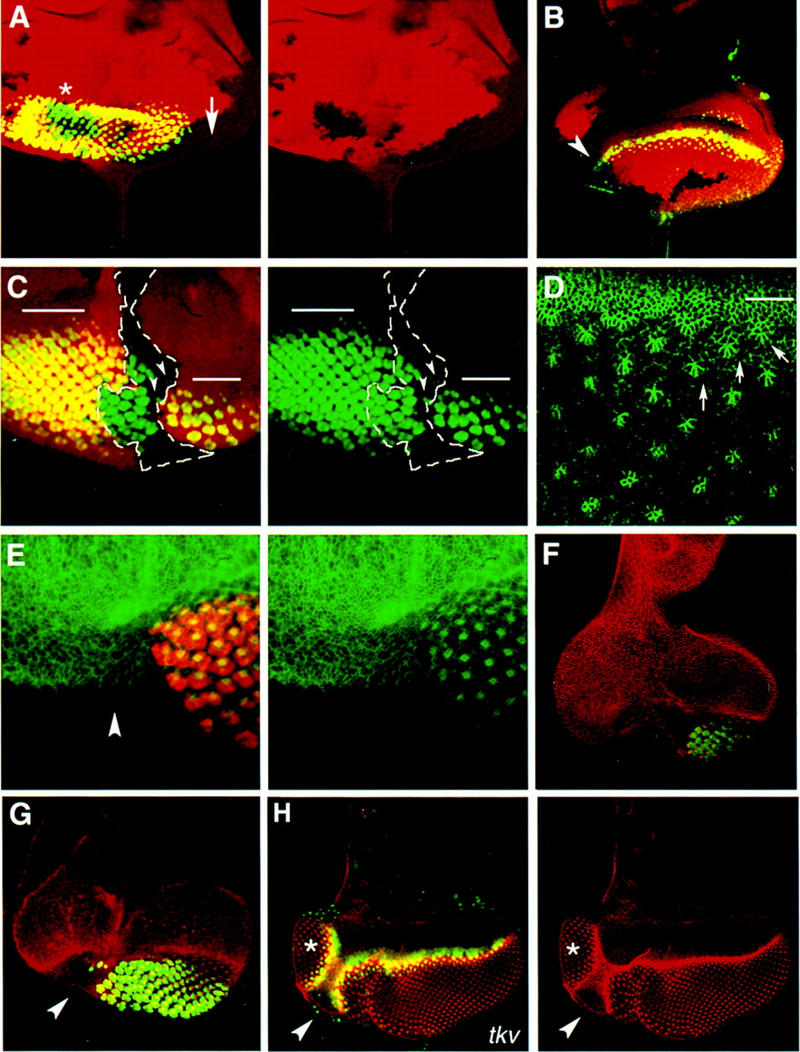
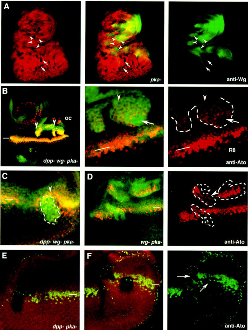
Patterning of the compound eye begins at the posterior edge of the eye imaginal disc and progresses anteriorly toward the disc margin. The advancing front of ommatidial differentiation is marked by the morphogenetic furrow (MF). Here we show by clonal analysis that Hedgehog (Hh), secreted from two distinct populations of cells has two distinct functions: It was well documented that Hh expression in the differentiating photoreceptor cells drives the morphogenetic furrow. Now we show that, in addition, Hh, secreted from cells at the posterior disc margin, is absolutely required for the initiation of patterning and predisposes ommatidial precursor cells to enter ommatidial assembly later. These two functions of Hh in eye patterning are similar to the biphasic requirement for Sonic Hh in patterning of the ventral neural tube in vertebrates. We show further that Hh induces ommatidial development in the absence of its secondary signals Wingless (Wg) and Dpp and that the primary function of Dpp in MF initiation is the repression of wg, which prevents ommatidial differentiation. Our results show that the regulatory relationships between Hh, Dpp, and Wg in the eye are similar to those found in other imaginal discs such as the leg disc despite obvious differences in their modes of development.


Figures

Figure 1
Figure 1
Effects of loss of hh function during MF propagation. In all images, eye discs are oriented dorsal to the right and anterior up. Third instar larval eye discs containing hh internal clones double stained with antibodies against β-galactosidase (red) and the proneural protein Ato (green in A) or the neuronal marker Elav (green in B). Single and superimposed confocal images are shown side by side. The homozygous mutant cells (hh/hh) are marked by the absence of lacZ staining. (A) The clone spans the MF and causes a reduction in the levels of Ato protein (arrowhead) in the MF. Note the slightly bent line of ato expression, suggesting that the MF has progressed more slowly in the region with limited Hh signal. (B) The disc contains two fused hh clones. The anterior clone (arrowhead) spans the MF at the time of dissection. The second clone is located posterior to the MF. Examination throughout the depth of the two clones shows that neuronal differentiation, assessed by Elav expression (green), has proceeded normally within the mutant tissue. On the basis of the rescue of mutant ommatidial units, we estimate that adequately high levels of Hh protein reach hh mutant cells across a distance of about three ommatidial clusters from the boundary with hh+ cells.
Figure 2
Figure 2
Role of hh in the initiation of the MF. (A–C and E–G) Mosaic eye discs carrying hh clones that include the disc margin. (A–C) hh clones are marked by the absence of lacZ (red) staining. (E–G) hh mutant tissue is marked indirectly by the absence of Elav (green). (A) The disc contains several hh clones. Single-channel image of the arm–lacZ staining to visualize the position of the clones is shown in the right panel. Neuronal differentiation, assessed by Elav expression, is normal in the internal hh clone (asterisk), whereas it is blocked in the marginal hh clone (arrow). The arrow points to the center of the marginal clone and also where marginal ommatidia are missing. (B) The expression of ato (green) is also affected in marginal hh clones. Note that only cells in close proximity to the wild-type border of the clone have detectable Ato protein levels (arrowhead). (C) A large hh clone (outlined in white) that runs along the center of the disc and spans half of the posterior eye margin. The single channel image of the Elav (green) is shown in the right panel. Neuronal differentiation is abolished in the area where the posterior margin is mutant for hh. In contrast, the hh+ margin initiated the MF, which progressed normally through an internal hh mutant area (note the presence of green ommatidia). Only a few mutant ommatidia (arrowheads) are rescued adjacent to the borders of the marginal part of the clone. The bars in the image show the approximate position of the MF. (D) A portion of a wild-type disc stained with a monoclonal antibody raised against Arm (see also Materials and Methods) to show changes in cell shape associated with cells in the MF (white line) and cells in the ommatidial clusters (some of them are marked with arrows). The image shows an apical focal plane. (E–G) Eye discs carrying marginal hh clones that span half of the posterior margin. hh cells marked by the absence of Elav staining (red in E, and green in F and G) do not constrict apically and fail to assemble into ommatidial clusters, as assessed by the Arm staining (green in E and red in F and G). The disc in E was dissected at the mid third instar stage to confirm that failure to initiate ommatidial differentiation is not an indirect effect of loss of hh in the ommatidial clusters. The Arm staining is in red, and the Elav in green. The clones in F and G were induced in a Minute background (see Materials and Methods). (H) Mosaic eye disc carrying a tkv clone also induced in a Minute background. The uniform Arm staining (red) and the absence of ato expression (green) in the region marked by the arrowhead indicates that this region lacks tkv. Adjacent to this area, a supernumerary eye field (asterisk) has formed in the ventral region of the eye disc (see Materials and Methods). The ectopic eye develops an equator as in the endogenous eye, indicating that loss of Dpp reception, but not loss of hh (cf. E–G with H), results in the reprogramming of positional information in the marginal cells to initiate an MF in an ectopic position.
Figure 3
Figure 3
Early pattern of hh expression in the second and early third instar eye disc. Anterior is up in all panels. (A) The first prominent accumulation of Ato protein (red, left panel) in the eye disc marks the onset of the MF, as assessed by Arm staining (green, right panel). The middle panel shows the combined image. (B–F) Confocal images of increasingly older discs stained with anti-Ato antibody (green) and β-galactosidase (red) to detect lacZ expression in the hhP30 (B–D), in the dpp-BS3.0 line (E) or in a wg–lacZ line (F). The expression of hh occurs in a line of cells along the posterior and dorsal margin of the eye disc prior to the initiation of the MF (B). This expression is elevated at the posterior most edge of the disc at the time of MF induction when the first Ato positive cells (green) are detectable (C). The right panel shows only hh–lacZ expression in the same disc. (D) An older eye disc, in which the MF has progressed three ommatidial rows (indicated by arrows). The expression of hh (arrow) has expanded anteriorly to the region of the presumptive dorsal head (dh) and is located posterior to the band of continuous Ato expression (arrowhead). The hh–lacZ-positive cells (red, arrow) are in between the single Ato-positive cells, which correspond to R8 precursor cells. Double staining with anti-Ato and anti-Elav showed no expression of Elav at this early stage (not shown). (E) dpp–lacZ expression was examined in relation to the early expression of ato. Note that the induction of the MF is not synchronous in the two eye discs of the same larva. The disc that shows no detectable accumulation of Ato protein also has weaker expression of dpp at the most posterior edge of the disc (arrow). The dpp–lacZ expression in this region becomes detectable at the time of ato induction, but dpp expression is still weaker and discontinuous (arrow) in this region compared with its expression in the lateral margins. (F) The disc is of the same age as that shown in D. wg–lacZ expression is confined to the anterior part of the disc. Note that the prominent wg–lacZ expression in the presumptive dorsal head region (dh) overlaps at this stage with hh–lacZ expression. (G–H) Young eye discs of hhP30 (G) and ptcAT96 (H) larvae stained with X-gal (blue). Note that the stripe of ptc–lacZ is broader than the stripe of hh–lacZ consistent with the notion that secreted Hh can induce gene expression in cells farther from the hh-expressing cells. The discrepancy between the time and place of expression of β-galactosidase in the hhP30 line shown here and the previous reported hh RNA distribution is most likely attributed to the different age of the eye discs used in these two studies. Whereas Ma et al. (1993) analyzed hh RNA distribution only in the third instar disc, we see hh–lacZ expression at the disc margin already in second instar disc. We used a cell marker (anti-Ato) and a morphological marker (anti-Arm) to assess directly the age of the eye discs. Abbreviations: an, antennal disc; dh, presumptive dorsal head.
Figure 4
Figure 4
Constitutive activation of the Hh signaling pathway, in the absence of wg and dpp activities, activates the expression of the proneural gene ato and causes ectopic MFs. (A) A third instar larval eye disc stained with anti-wg (green) and carrying multiple pkaDCO–B3 clones marked by the absence of arm–lacZ (red). The arrows point to eye internal single pkaDCO–B3 clones that do not activate wg expression. The arrowheads point to clones where wg expression is activated in response to removal of pka. (B,C) Eye discs carrying dppH61 wg CX4pkaDCO–B3, which are marked by the increased accumulation of Ci occurring in the pka mutant cells (Johnson et al. 1995; M. Domínguez and E. Hafen unpubl.). The dppd12 pkaDCO–B3 clones are marked by the absence of arm–lacZ staining. (B) A large dppH61 wg CX4pkaDCO–B3 triple mutant clone (arrowheads) located in the anterior part of the eye disc. ato expression is induced autonomously in the mutant cells. A detail of the posterior part of the clone is shown in the middle panel. The right panel shows a single image of ato expression in the posterior part of the clone (outlined in white) and ato expression in the endogenous MF (indicated with the bar). In the MF (white line), the uniform levels of Ato protein evolve into discrete single Ato-positive cells, the R8 photoreceptor cells. Note that most cells in the clone have uniform levels of Ato (arrowhead) as those found in the endogenous MF. Some of the mutant cells (arrow) have initiated ommatidial development, as inferred by the presence of regularly spaced Ato-positive cells. (C) A dppH61 wg CX4pkaDCO–B3 clone located posterior to the MF at the time of dissection. The clone caused an acceleration of the endogenous MF (arrowhead). (D) Aneye disc carrying three wg CX4pkaDCO–B3 mutant clones. Note the ectopic activation of ato expression even in the clones located far from the endogenous MF. The arrowhead points to some mutant cells where ectopic expression of ato is in separated singular cells. (E,F) Two eye discs carrying dppd12 pkaDCO–B3 mutant clones. Note that ato expression (green) is not induced in these clones, which are marked by the absence of arm–lacZ staining (red). The clones in F span the endogenous MF and have caused a reduction in the levels of expression of ato (arrowheads). (oc) Occellar region. The position of the endogenous MF is indicated by white bars.

References

    1. Basler K, Struhl G. Compartment boundaries and the control of Drosophila limb pattern by hedgehog protein. Nature. 1994;368:208–214. - PubMed
    1. Blackman RK, Sanicola M, Raftery LA, Gillevet T, Gelbart WM. An extensive 3′ cis-regulatory region directs the imaginal disk expression of decapentaplegic, a member of the TGF-beta family in Drosophila. Development. 1991;111:657–666. - PubMed
    1. Bonini NM, Choi KW. Early decisions in Drosophila eye morphogenesis. Curr Opin Genet Dev. 1995;5:507–515. - PubMed
    1. Bokor P, DiNardo S. The roles of hedgehog, wingless, and lines in patterning of the dorsal epidermis in Drosophila. Development. 1996;122:1083–1092. - PubMed
    1. Brook WJ, Cohen SM. Antagonistic interactions between Wingless and Decapentaplegic responsible for dorsal-ventral pattern in the Drosophila leg. Science. 1996;273:1373–1377. - PubMed

Publication types

MeSH terms

LinkOut - more resources