. 1997 Dec 15;17(24):9573-82.
doi: 10.1523/JNEUROSCI.17-24-09573.1997.

Nerve growth factor modulates synaptic transmission between sympathetic neurons and cardiac myocytes

Affiliations

Nerve growth factor modulates synaptic transmission between sympathetic neurons and cardiac myocytes

S T Lockhart et al. J Neurosci. .

Abstract

Regulation of heart rate by the sympathetic nervous system involves the release of norepinephrine (NE) from nerve terminals onto heart tissue, resulting in an elevation in beat rate. Nerve growth factor (NGF) is a neurotrophin produced by the heart that supports the survival and differentiation of sympathetic neurons. Here we report that NGF also functions as a modulator of sympathetic synaptic transmission. We determined the effect of NGF on the strength of synaptic transmission in co-cultures of neonatal rat cardiac myocytes and sympathetic neurons from the superior cervical ganglion (SCG). Synaptic transmission was assayed functionally, as an increase in the beat rate of a cardiac myocyte during stimulation of a connected neuron. Application of NGF produced a pronounced, reversible enhancement of synaptic strength. We found that TrkA, the receptor tyrosine kinase that mediates many NGF responses, is expressed primarily by neurons in these cultures, suggesting a presynaptic mechanism for the effects of NGF. A presynaptic model is further supported by the finding that NGF did not alter the response of myocytes to application of NE. In addition to the acute modulatory effects of NGF, we found that the concentration of NGF in the growth medium affects the level of synaptic transmission in cultures of sympathetic neurons and cardiac myocytes. These results indicate that in addition to its role as a survival factor, NGF plays both acute and long-term roles in the regulation of developing sympathetic synapses in the cardiac system.


Figures

Fig. 1.
Fig. 1.
Sympathetic neuron–myocyte co-culture. In 3-d-old co-cultures of neonatal rat cardiac myocytes and sympathetic neurons isolated from the superior cervical ganglion, neurons extend processes that form visible connections with spontaneously beating myocytes. Functional assays of synaptic transmission were performed by obtaining a whole-cell recording on the neuron, stimulating the neuron to fire action potentials, and measuring the change in beat rate of the connected myocyte.
Fig. 2.
Fig. 2.
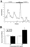
NGF modulates synaptic transmission between sympathetic neurons and beating myocytes. A, NGF reversibly potentiates myocyte response to stimulation of a connected neuron. A whole-cell recording was obtained from a neuron visibly connected to a beating myocyte. The neuron was stimulated at 2.5 Hz for a 3 min period (STIM; thin black bars), leading to a small increase in myocyte beat rate under control conditions. After superfusion of 50 ng/ml NGF into the bath solution (10 min) (the break in the timeline indicates that myocyte beat rate was not counted during this perfusion period), the myocyte response to neuronal stimulation was more than doubled. The response to stimulation returned to control levels after washout of the NGF (10 min). B, Magnitude and concentration dependence of synaptic potentiation in NGF. The average myocyte beat rate was measured for a 3 min baseline period before neuronal stimulation and again during the 3 min stimulation. Open bars show the stimulation-evoked elevation in beat rate under control conditions. NGF (10 or 50 ng/ml) was then superfused into the bath, and another 3 min baseline was counted. The neuron was stimulated, and the myocyte beat rate was counted for an additional 3 min period. The black bars show the stimulus-induced elevation in myocyte beat rate in the presence of NGF. 50 ng/ml NGF: significantly different from control, p < 0.002, paired Student’st test, n = 15; 10 ng/ml NGF: not significantly different from control, p < 0.74,n = 7; error bars represent SEM.
Fig. 3.
Fig. 3.
Dose dependence of neuronal survival in NGF. Duplicate wells containing neurons either alone (open bars) or in co-cultures with myocytes (black bars) were grown at varying concentrations of NGF. After 63 hr, a strip of neurons was counted across the tissue culture well for each condition. The average number of neurons remaining in the well was computed and plotted as a function of NGF concentration. In three independent experiments, 5 ng/ml NGF was sufficient to support neuronal survival. Error bars represent SDs.
Fig. 4.
Fig. 4.
Expression of TrkA in neuron–myocyte co-cultures.A, trkA mRNA in sympathetic neurons, cardiac myocytes, and neuron–myocyte co-cultures by RT-PCR. mRNA extracted from 3-d-old cultures was reverse-transcribed and PCR-amplified using primers specific for trkA. The resulting PCR products were analyzed by agarose gel electrophoresis and ethidium bromide staining. B, Expression of TrkA protein in sympathetic neurons and cardiac myocytes grown in co-culture was analyzed by immunocytochemistry. Neurons and myocytes were grown for 3 d in co-culture, fixed, and stained with the rtrkA antibody. Left panel, Phase-contrast photograph of neuron–myocyte co-culture after fixation. Right panel, TrkA staining. The arrow indicates a myocyte.
Fig. 5.
Fig. 5.
Effect of NGF on the NE-induced change in myocyte beat rate. A glass electrode with a tip diameter of 1 μm was micromanipulated close to a beating myocyte in a neuron–myocyte co-culture. A baseline myocyte beat rate was counted, and then a picospritzer was used to puff 10−5mNE at 20 psi for 75 msec. The myocyte beat rate was counted for 3 min after delivery of the NE, and the change in beat rate was calculated (Control). NGF (50 ng/ml) was superfused into the dish, another baseline was counted for the same myocyte, and again NE was puffed onto the myocyte. The increase in beat rate was calculated for NE application in the presence of NGF. NGF not different from control, p < 0.7; paired Student’st test; n = 13.
Fig. 6.
Fig. 6.
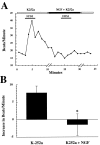
Effects of K252a. A, The kinase inhibitor K252a blocks potentiation and leads to inhibition of synaptic transmission by NGF. In the presence of 200 nmK252a alone, a myocyte showed a normal small elevation in beats per minute in response to stimulation of a connected neuron. However, in the presence of K252a, NGF did not potentiate, and led to a decrease in, the myocyte response to neuronal stimulation. The breaks in thex-axis indicate that myocyte beat rate was not counted during these perfusion periods (10 min for initial perfusion of NGF, 10 min for washout). B, In K252a, NGF reduces synaptic transmission. Averaged, pooled data. On average, in the presence of K252a, NGF induces a significant decrease in the responses of myocytes to neuronal stimulation (p < 0.05; paired Student’s t test; n = 5). Error bars represent SEM.
Fig. 7.
Fig. 7.
Effect of NGF concentration in growth medium on response of myocytes to neuronal stimulation. Response of myocytes to neuronal stimulation in control medium was greater in cultures grown in 50 ng/ml NGF than in cultures grown in 5 ng/ml. Neuron–myocyte co-cultures were grown for 3 d in 5 or 50 ng/ml NGF. Note that 5 ng/ml was sufficient to support full survival of sympathetic neurons in these cultures (Fig. 3). Neuron–myocyte pairs that had a visible process connecting the neuron and the myocyte were analyzed. After complete washout of growth medium, myocyte beat rate was counted before and during neuronal stimulation, and the change in beat rate during neuronal stimulation was calculated. Grown in 50 ng/ml NGF significantly different from grown in 5 ng/ml NGF: unpaired Student’st test, p < 0.03;n = 135 grown in 50 ng/ml NGF andn = 46 grown in 5 ng/ml NGF.

Similar articles

Cited by

References

    1. Albers KM, Wright DE, Davis BM. Overexpression of nerve growth factor in epidermis of transgenic mice causes hypertrophy of the peripheral nervous system. J Neurosci. 1994;14:1422–1432. - PMC - PubMed
    1. Armour JA, Ardell JL. Neurocardiology. Oxford UP; New York: 1994.
    1. Barrett GL, Bartlett PF. The p75 nerve growth factor receptor mediates survival or death depending on the stage of sensory neuron development. Proc Natl Acad Sci USA. 1994;91:6501–6505. - PMC - PubMed
    1. Birren SJ, Lo LC, Anderson DJ. Sympathetic neurons undergo a developmental switch in trophic dependence. Development. 1993;119:597–610. - PubMed
    1. Buckley KM, Landis SC. Morphological studies of synapses and varicosities in dissociated cell cultures of sympathetic neurons. J Neurocytol. 1983;12:67–92. - PubMed

Publication types

MeSH terms

LinkOut - more resources