. 1997 Dec 15;17(24):9642-55.
doi: 10.1523/JNEUROSCI.17-24-09642.1997.

The origin, location, and projections of the embryonic abdominal motorneurons of Drosophila

Affiliations

The origin, location, and projections of the embryonic abdominal motorneurons of Drosophila

M Landgraf et al. J Neurosci. .

Abstract

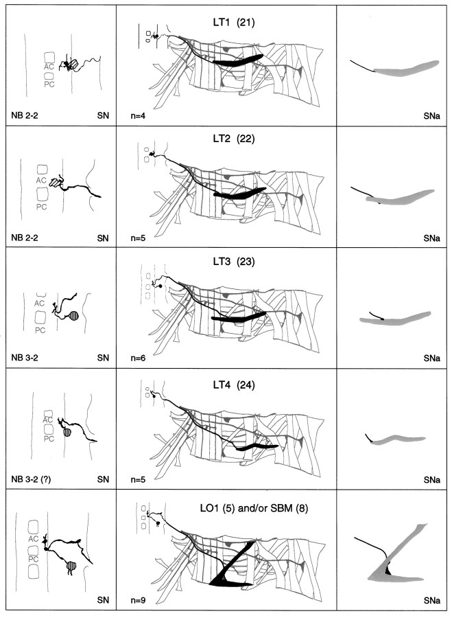
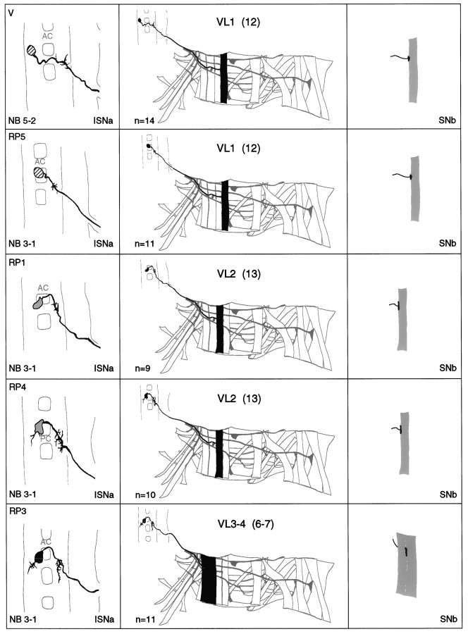
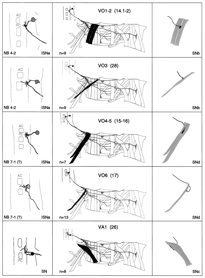
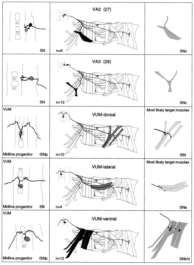
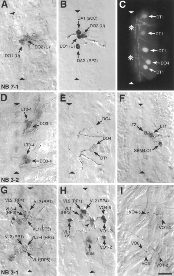
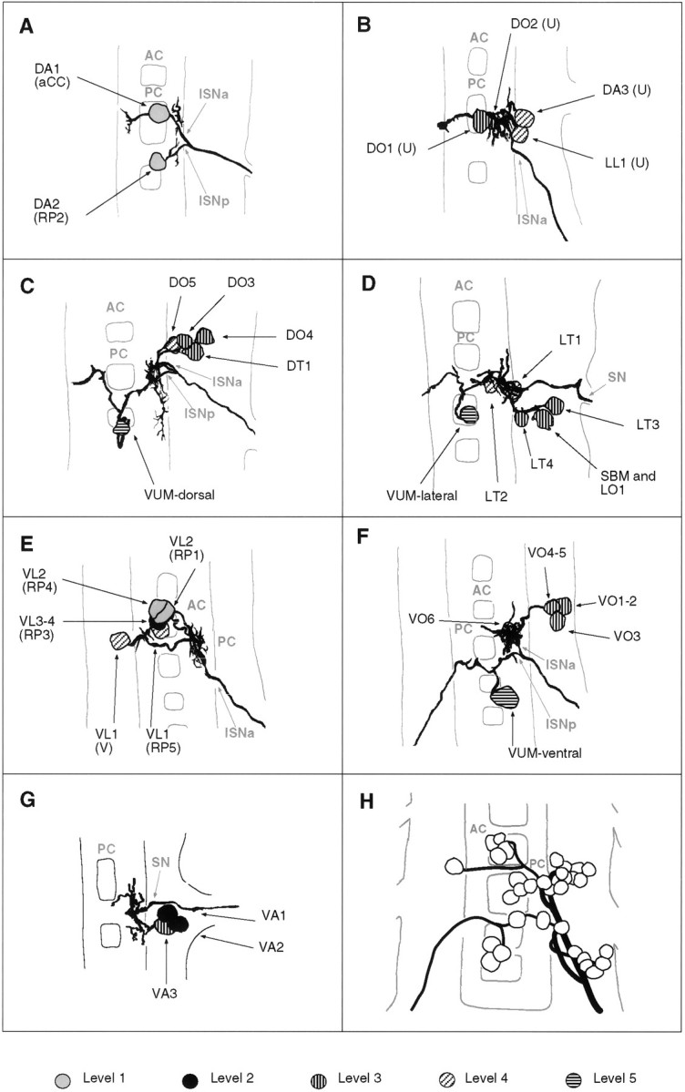
We have used a retrograde labeling technique to identify motorneurons for each of the 30 body wall muscles of an abdominal hemisegment in the late stage 16 Drosophila embryo. Each motorneuron has a characteristic cell body position, dendritic arborization, and axonal projection. In addition, we have determined the neuroblasts of origin for most of the motorneurons we describe. Some organizational principles for the neuromuscular system have become apparent: (1) There is no obvious topographic relationship between the cell body positions of motorneurons and the position or orientation of the muscles they innervate; (2) motorneurons that innervate muscles of similar position and orientation are often clustered and have overlapping dendritic trees; (3) morphologically similar motorneurons are generally derived from a common neuroblast and innervate operationally related muscles; and (4) neuroblasts can give rise to more than one morphological type of motorneuron.


Figures

Fig. 1.
Fig. 1.
The abdominal larval body wall muscles and peripheral nerve branches. A, Tracings of the larval body wall muscles and peripheral nerve branches in a single abdominal hemisegment (A2–A7) in a late stage 16 embryo as viewed from internal (top) and external (bottom). NMJs form at late stage 16, and each muscle is innervated at a characteristic position. Anterior is up, and the ventral midline is left. Muscle nomenclature according to Bate (1993): D, dorsal acute;DO, dorsal oblique; DT, dorsal transverse; LL, lateral longitudinal; LT, lateral transverse; LO, lateral oblique;SBM, segment border muscle; VL, ventral longitudinal; VO, ventral oblique; VA, ventral acute; VT, ventral transverse. Nerve branch nomenclature according to Thomas et al. (1984): ISN, intersegmental nerve; SN, segmental nerve;TN, transverse nerve. B, Diagrammatic transverse section of the ventral nerve cord to illustrate the relative dorsoventral positions of motorneuron cell bodies. We have arbitrarily divided the dorsoventral axis of the ventral nerve cord into five levels from dorsal (Level 1) to the neuropile (Level 2) to ventral (Level 5). We have given a characteristic shading to each of these five levels. To include information about dorsoventral position in the description of the motorneurons we have shaded motorneuron cell bodies according to this code (Figs. 3, 5). C, Diagrammatic dorsal view of the ventral nerve cord (seen from interior of embryo) to illustrate the organization of nerve roots. AC, Anterior commissure;PC, posterior commissure; ISNa, anterior root of the intersegmental nerve; ISNp, posterior root of the intersegmental nerve; SN, segmental nerve root;TN, transverse nerve root. Anterior isup.
Fig. 2.
Fig. 2.
Photoconverted DiI preparations of retrogradely labeled abdominal motorneurons and their target muscles. From dorsal to ventral: (A) U motorneuron innervating muscle DO1; (B) the motorneuron that innervates muscle LT1. C, The motorneurons that innervate muscle VT1 were labeled in two adjacent segments. Arrows point to the somata and broad arrows to drops of DiI, which were deposited at the NMJs. Dorsal is right and anterior isup. The ventral midline is indicated bytriangles. Scale bar (shown in C):A, 20 mm; B–C, 10 mm.
Fig. 3.
Fig. 3.
a–g. The embryonic motorneurons, their central and peripheral projections, and their target muscles. Theleft column shows individual but representative tracings of single motorneurons (therefore, dimensions of nerve cords and motorneurons may vary slightly between panels), indicating the position of the cell body and dendritic arborizations. Motorneuron names (where they exist) are given at the top left, and the neuroblast of origin (where known) is given at the bottom left (uncertainties are indicated by question marks). The nerve root through which the axon exits the CNS is given at the bottom right (AC, anterior commissure;PC, posterior commissure; ISNa, anterior root of the intersegmental nerve; ISNp, posterior root of the intersegmental nerve; SN, segmental nerve). Thecenter column shows the peripheral projection of the axon and the target muscle(s), which are named according to the nomenclatures of Bate (1993), and in parentheses,Crossley (1978). The right column shows the NMJ in more detail and indicates the peripheral nerve branch through which the motor axon projects (bottom right). We have been unable to show conclusively that muscles VA1 and VA2 are innervated by distinct motorneurons, but on the basis of the number of motorneurons projecting through the SN according to Sink and Whitington (1991a), we propose that they are. To indicate that we are uncertain about which of the dorsal and lateral muscles are innervated by VUM neurons, we have differentially highlighted those muscles that we think are the most likely targets, in agreement with Sink and Whitington (1991a).n = sample size for each labeled neuron.
Fig. 3.
Fig. 3.
a–g. The embryonic motorneurons, their central and peripheral projections, and their target muscles. Theleft column shows individual but representative tracings of single motorneurons (therefore, dimensions of nerve cords and motorneurons may vary slightly between panels), indicating the position of the cell body and dendritic arborizations. Motorneuron names (where they exist) are given at the top left, and the neuroblast of origin (where known) is given at the bottom left (uncertainties are indicated by question marks). The nerve root through which the axon exits the CNS is given at the bottom right (AC, anterior commissure;PC, posterior commissure; ISNa, anterior root of the intersegmental nerve; ISNp, posterior root of the intersegmental nerve; SN, segmental nerve). Thecenter column shows the peripheral projection of the axon and the target muscle(s), which are named according to the nomenclatures of Bate (1993), and in parentheses,Crossley (1978). The right column shows the NMJ in more detail and indicates the peripheral nerve branch through which the motor axon projects (bottom right). We have been unable to show conclusively that muscles VA1 and VA2 are innervated by distinct motorneurons, but on the basis of the number of motorneurons projecting through the SN according to Sink and Whitington (1991a), we propose that they are. To indicate that we are uncertain about which of the dorsal and lateral muscles are innervated by VUM neurons, we have differentially highlighted those muscles that we think are the most likely targets, in agreement with Sink and Whitington (1991a).n = sample size for each labeled neuron.
Fig. 4.
Fig. 4.
Examples of photoconverted preparations of clones from individually labeled NBs and of retrogradely labeled motorneurons. Photoconverted DiI preparations of clones from individual, identified, labeled NBs (A, D, G), of motorneurons (B, E, F, H), and of NMJs (I), and a fluorescent image of DiI-labeled motorneurons (C) are shown. The images are collages of several focal planes, which were assembled with Photoshop; the dorsal focal plane is uppermost.A, The two medial U motorneurons and the fpCC interneuron derived from NB 7-1. The other motorneurons derived from NB 7-1 (the two mediolateral U neurons and the motorneurons that most likely innervate muscles VO4-6) are not visible in this focal plane.B, The motorneurons that innervate the four dorsal muscles DA1-2 (aCC and RP2) and DO1-2 (the two medial U neurons) were retrogradely labeled in one abdominal segment. C, A fluorescent image of the motorneuron innervating muscle DT1, which was labeled in four adjacent abdominal segments. Note the characteristic cell body position and dendritic projection. Theasterisks indicate T-shaped sensory axons that were labeled by chance in two of the segments. The motorneuron that innervates muscle DO4 was labeled in one segment. D, Motorneurons derived from two NB 3-2 clones. The anterior clone is thoracic (T3); the posterior clone is abdominal (A2). NB 3-2 gives rise to two morphological types of motorneurons: first, motorneurons that project through the ISN and innervate muscles DT1 (not visible in this focal plane), DO3-4, and probably also DO5 (not visible in this focal plane); second, motorneurons that project through SNa and innervate muscle LT3 and probably also LT4. E, A photoconverted preparation similar to the one shown in C. F, Three motorneurons that project through SNa are shown. The motorneurons that innervate muscles LT1-2 are morphologically and clonally distinct from the ones innervating muscles LO1 and SBM. G, Two NB 3-1 clones, of which the anterior clone is thoracic (T3) and the posterior clone is abdominal (A1). NB 3-1 gives rise to the four morphologically similar RP1, -3, -4, and -5 motorneurons that innervate the ventral longitudinal muscles VL1-4.H, Most of the motorneurons that project through SNb and SNd were retrogradely labeled in a single abdominal segment to illustrate their relative cell body positions. In addition, the motorneuron that innervates muscles VO1-2 was labeled in the next posterior segment. Note that the V-neuron is morphologically distinct from the RP neurons but that their dendritic arbors are overlapping (arrowhead) (see text). The VUM neuron lies in the same segment as the muscles that it innervates, whereas the RP and V-neurons as well as the motorneurons that innervate muscles VO1-2 and VO4-5 lie in the next anterior segment. I, The endplates of two VUM neurons on the ventral oblique muscles VO1-6 in two adjacent abdominal segments (the VUMs were retrogradely labeled on the contralateral side). Anterior is up. The ventral midline is indicated by triangles and is on theleft in I. Scale bar, 10 μm.
Fig. 5.
Fig. 5.
Tracings of groups of motorneurons that innervate muscles that are related in operation by position and orientation. A–G, Composites of tracings from several preparations (A, 2; B, 3;C, 4; D, 3; E, 2;F, 3; G, 2). A, The aCC and RP2 motorneurons that innervate muscles DA1-2. B, The four U neurons that innervate the dorsal and lateral muscles DO1-2, DA3, and LL1 (note that this cluster of four neurons may be subdivided further by morphology and target muscles; see text for details).C, The motorneurons innervating the dorsal oblique muscles DO3-5 and muscle DT1. D, The motorneurons that innervate the lateral muscles LT1-4, LO1, and SBM. Note that muscles LO1 and SBM might be innervated by two distinct motorneurons, one of which is shown here. E, The RP1, -3, -4, -5, and the V-neurons innervate the ventral longitudinal muscles. F, Motorneurons innervating the ventral oblique muscles. G, Motorneurons innervating the ventral acute muscles. Note that there is uncertainty as to whether muscles VA1 and VA2 are innervated by two distinct motorneurons (Fig. 3). H, All embryonic motorneurons that innervate the larval body wall muscles of abdominal segment A7 at stage 16 (adapted from Sink and Whitington, 1991a). For details of the individual dendritic arborizations, refer to Figure 3. Anterior is up.

References

    1. Bate CM. Embryogenesis of an insect nervous system I. A map of the thoracic and abdominal neuroblasts in Locusta migratoria. J Embryol Exp Morphol. 1976;35:107–123. - PubMed
    1. Bate M. The embryonic development of larval muscles in Drosophila. Development. 1990;110:791–804. - PubMed
    1. Bate M. The mesoderm and its derivatives. In: Bate M, Martinez Arias A, editors. The development of Drosophila melanogaster. Cold Spring Harbor Laboratory; Cold Spring Harbor, NY: 1993. pp. 1013–1090.
    1. Bodmer R, Jan YN. Morphological differentiation of the embryonic peripheral neurons in Drosophila. Roux’s Arch Dev Biol. 1987;196:69–77. - PubMed
    1. Bossing T, Technau GM. The fate of the CNS midline progenitors in Drosophila as revealed by a new method for single cell labelling. Development. 1994;120:1895–1906. - PubMed

Publication types

MeSH terms

LinkOut - more resources