. 1997 Dec 15;17(24):9675-85.
doi: 10.1523/JNEUROSCI.17-24-09675.1997.

Lobular patterns of cerebellar activation in verbal working-memory and finger-tapping tasks as revealed by functional MRI

Affiliations

Lobular patterns of cerebellar activation in verbal working-memory and finger-tapping tasks as revealed by functional MRI

J E Desmond et al. J Neurosci. .

Abstract

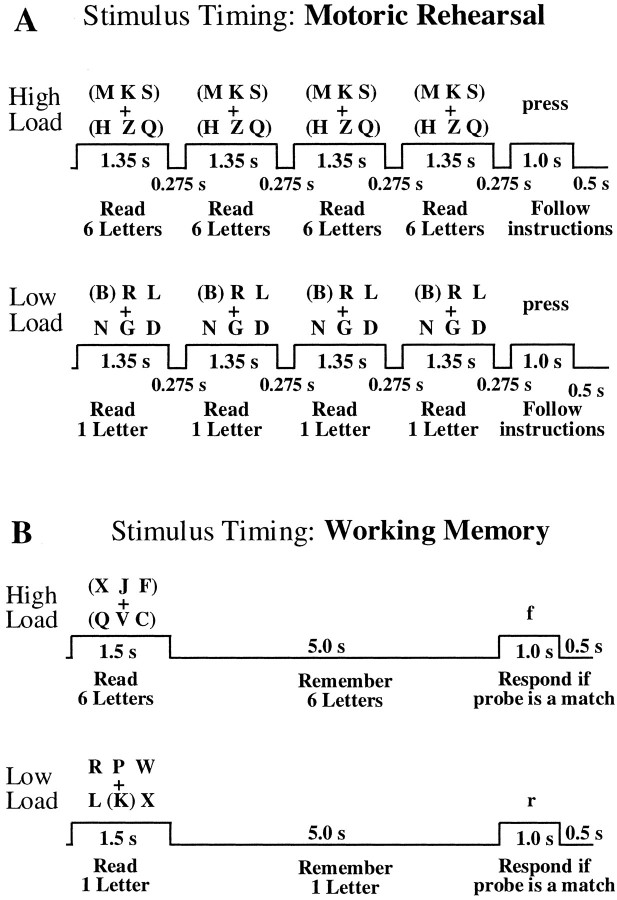
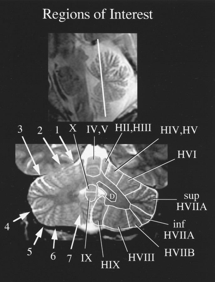
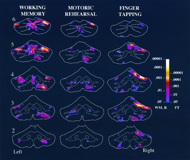
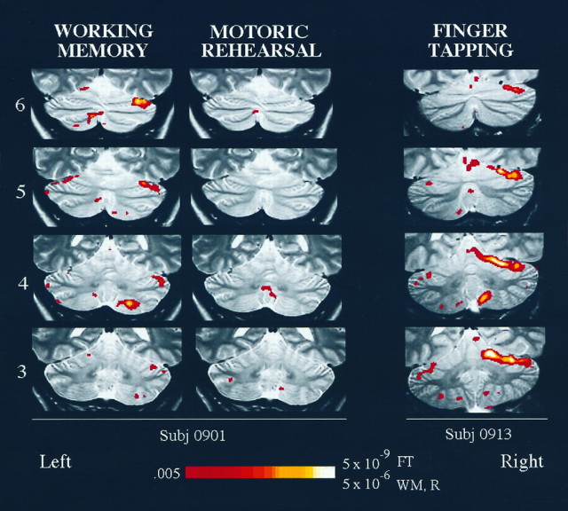
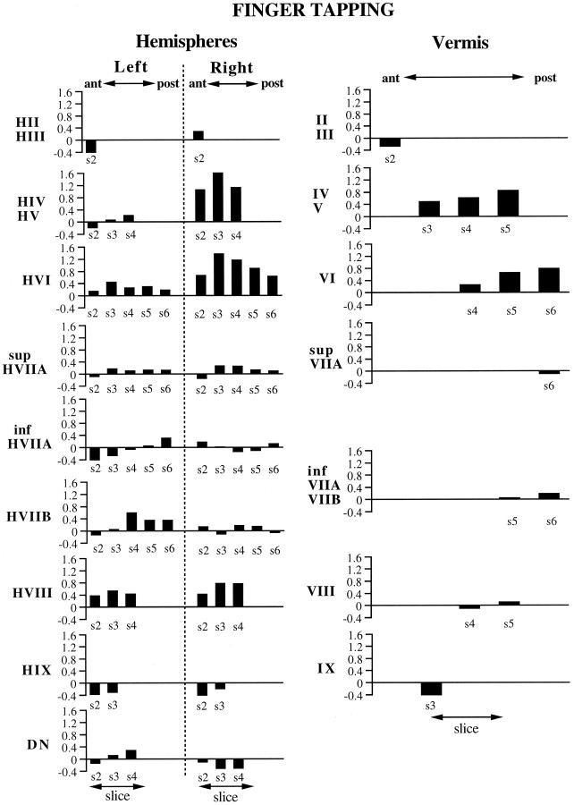
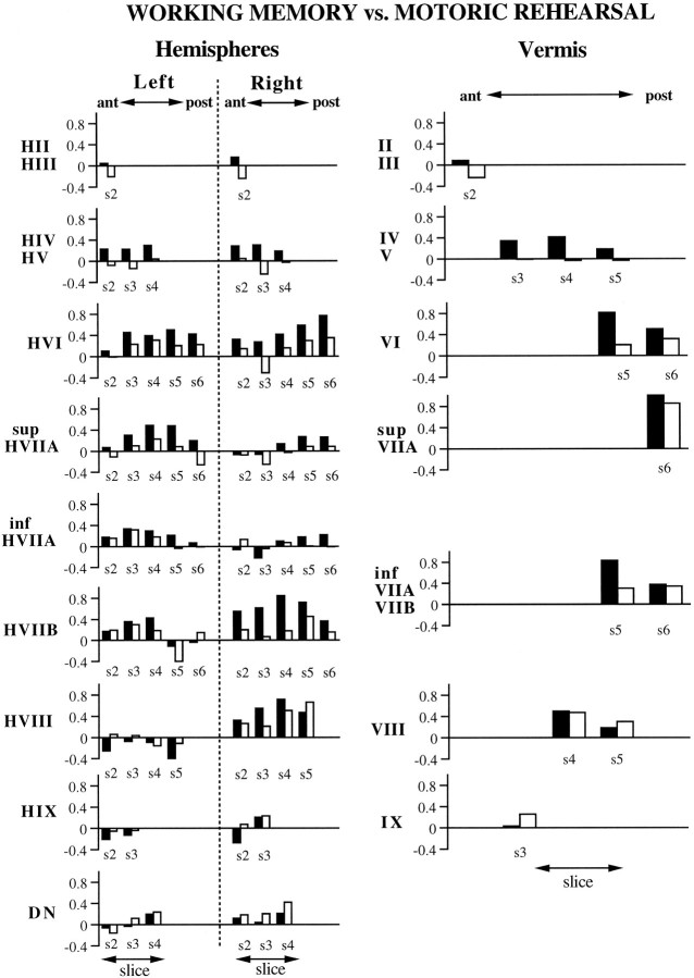
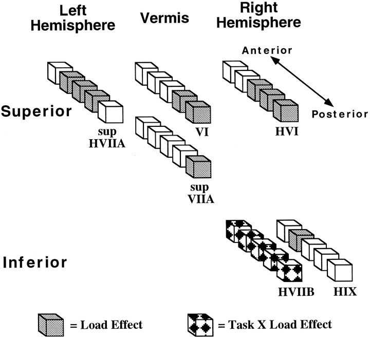
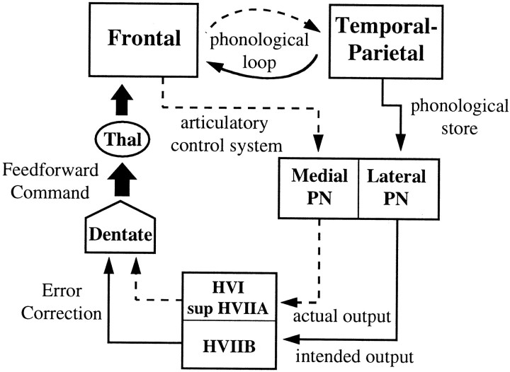
The lobular distributions of functional activation of the cerebellum during verbal working-memory and finger movement tasks were investigated using functional magnetic resonance imaging (fMRI). Relative to a rest control, finger tapping of the right hand produced ipsilateral-increased activation in HIV/HV [Roman numeral designations based on Larsell's () nomenclature] and HVI and weaker activation in HVIII that was stronger on the ipsilateral side. For a working-memory task, subjects were asked to remember six (high load) or one (low load) visually presented letters across a brief delay. To assess the motoric aspects of rehearsal in the absence of working memory, we asked the subjects to repeatedly read subvocally six or one letters at a rate that approximated the internally generated rehearsal of working memory (motoric rehearsal task). For both tasks, bilateral regions of the superior cerebellar hemispheres (left superior HVIIA and right HVI) and portions of posterior vermis (VI and superior VIIA) exhibited increased activation during high relative to low load conditions. In contrast, the right inferior cerebellar hemisphere (HVIIB) exhibited this load effect only during the working-memory task. We hypothesize that HVI and superior HVIIA activation represents input from the articulatory control system of working memory from the frontal lobes and that HVIIB activation is derived from the phonological store in temporal and parietal regions. From these inputs, the cerebellum could compute the discrepancy between actual and intended phonological rehearsal and use this information to update a feedforward command to the frontal lobes, thereby facilitating the phonological loop.


Figures

Fig. 1.
Fig. 1.
Timing diagrams for the motoric rehearsal (A) and working-memory (B) tasks used in this study.
Fig. 2.
Fig. 2.
Midline sagittal section illustrating the locations of the six planes acquired during the fMRI experiments.
Fig. 3.
Fig. 3.
An example of ROI identification on a T2-weighted oblique coronal section. The location of the section is illustrated on the sagittal localizer image (top). Fissures used to identify lobular boundaries are indicated on the left side of the oblique coronal section (bottom), and numbered arrows refer to the following:1, preculminate fissure; 2, primary fissure; 3, superior posterior fissure;4, horizontal fissure; 5, inferior posterior fissure; 6, inferior anterior fissure; and7, secondary fissure. ROIs for this section are depicted on the right side, and Roman numeral designations are based on Larsell’s (Larsell and Jansen, 1972) nomenclature (the ROI labeled D represents the deep nuclei, probably the emboliform as well as the dentate nucleus);sup, superior; inf, inferior.
Fig. 4.
Fig. 4.
Averaged fMRI activation in slices 2–6 for the working-memory, motoric rehearsal, and finger-tapping tasks. Sections represent oblique coronal slices taken parallel to the dorsal surface of the brain stem, as illustrated in Figure 2, and slice numbers appear on the left. Slice 1, which was the most anterior slice, exhibited almost no activation and was therefore omitted from the figure. The maps for working memory and motoric rehearsal were averaged across eight subjects, whereas finger tapping was averaged across five subjects. Regions depicted incolor represent areas that exhibited increased activation in high relative to low load conditions (in working memory and motoric rehearsal) or during finger tapping relative to rest. Decreases in activation during high load or finger tapping relative to their contrasting conditions were negligible and so are not depicted in this figure or in Figure 5. The color scale on theright represents the significance levels (one-tailedp values) of averaged Z scores and is scaled differently for finger tapping than for working memory and motoric rehearsal. The right side of the brain is depicted on theright. WM, Working memory;R, motoric rehearsal; FT, finger tapping.
Fig. 5.
Fig. 5.
Functional activation maps for individual subjects, overlaid on T2-weighted anatomy images. Activation obtained from the same subject for working-memory and motoric rehearsal tasks is illustrated on the left. The finger-tapping activation for a different subject is on the right. Slice numbers (3–6; see Fig. 2) appear on the left. The color scale at the bottom represents the significance levels (one-tailed p values) ofZ scores and is scaled differently for finger tapping than for working memory and motoric rehearsal. Color voxels represent regions that exhibited increased activation during high versus low load for the working-memory and motoric rehearsal tasks or during finger tapping versus rest for the finger-tapping task. The right side of the brain is depicted on theright. WM, Working memory;R, motoric rehearsal; FT, finger tapping.
Fig. 6.
Fig. 6.
ROI results for the finger-tapping task, representing the activations from five subjects. They-axis in each graph represents the averageZ score values obtained from the ROI. The name of the ROI appears next to the y-axis, with ROIs from the cerebellar hemispheres depicted on the left and ROIs from the vermis on the right. Thex-axis denotes the anterior–posterior dimension of the ROI, which corresponds to the slice number (denoteds2s6; see Fig. 2). The absence of a slice number on the x-axis means that the ROI did not appear on that slice.
Fig. 7.
Fig. 7.
ROI results for the working-memory (black bars) and motoric rehearsal (white bars) tasks, representing the activations from eight subjects. The organization of the figure is the same as that described for Figure 6.
Fig. 8.
Fig. 8.
Results of ANOVA on cerebellar lobular activation during working-memory and motoric rehearsal tasks. Eachcube within each linear set of cubesrepresents a slice position ranging from the most anterior (slice 2) to the most posterior (slice 6) location. The figure summarizes the regions that exhibited a significant effect of load (gray cubes, with high greater than low load) or a task-×-load interaction (patterned cubes, with high greater than low load for the working-memory but not for the motoric rehearsal task). Because a significant load-×-slice interaction was present for left superior HVIIA, right HVI, and right HIX, only the slices at which the load difference was significant are shaded. For the remaining lobules, no interaction with slice was observed, so all slices on which the lobule is found are shaded.
Fig. 9.
Fig. 9.
Model of cerebrocerebellar circuit proposed to be involved in verbal working memory. In addition to the phonological loop between frontal lobe structures (such as Broca’s area, comprising the articulatory control system and represented by adashed line) and temporal-parietal structures (such as the supramarginal gyrus, comprising the phonological store and represented by a solid line), a parallel path from these structures enters the cerebellar cortex via the pontine nuclei. Discrepancies between the actual and intended phonological output are computed and used to update a feedforward articulatory rehearsal command to the frontal cortex via dentatothalamic projections.PN, Pontine nuclei; Thal, thalamus.

References

    1. Adrian ED. Afferent areas in the cerebellum connected with the limbs. Brain. 1943;66:289–315.
    1. Allen G, Buxton RB, Wong EC, Courchesne E. Attention activation of the cerebellum independent of motor involvement. Science. 1997;275:1940–1943. - PubMed
    1. Awh E, Jonides J, Smith EE, Schumacher EH, Koeppe RA, Katz S. Dissociation of storage and rehearsal in verbal working memory: evidence from positron emission tomography. Psychol Sci. 1996;7:25–31.
    1. Baddeley A. Working memory. Science. 1992;255:556–559. - PubMed
    1. Brodal A. Neurological anatomy in relation to clinical medicine, Ed 3. Oxford UP; New York: 1981.

Publication types