Comparative Study
. 1997 Dec 15;17(24):9746-50.
doi: 10.1523/JNEUROSCI.17-24-09746.1997.

Effects of sleep on wake-induced c-fos expression

Affiliations
Comparative Study

Effects of sleep on wake-induced c-fos expression

R Basheer et al. J Neurosci. .

Abstract

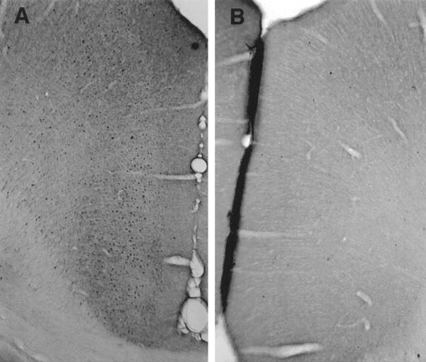
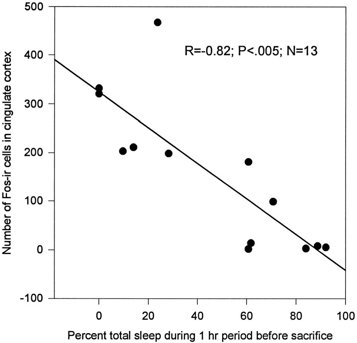
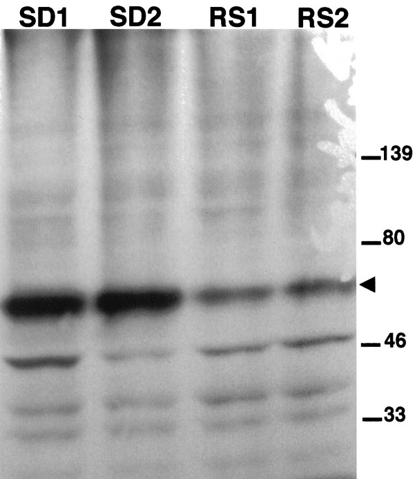
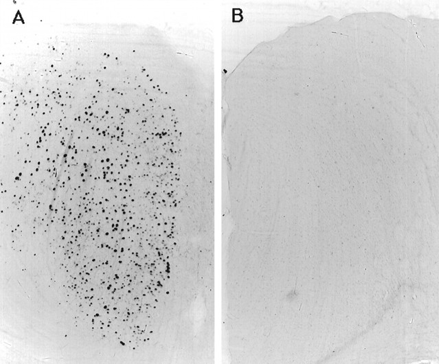
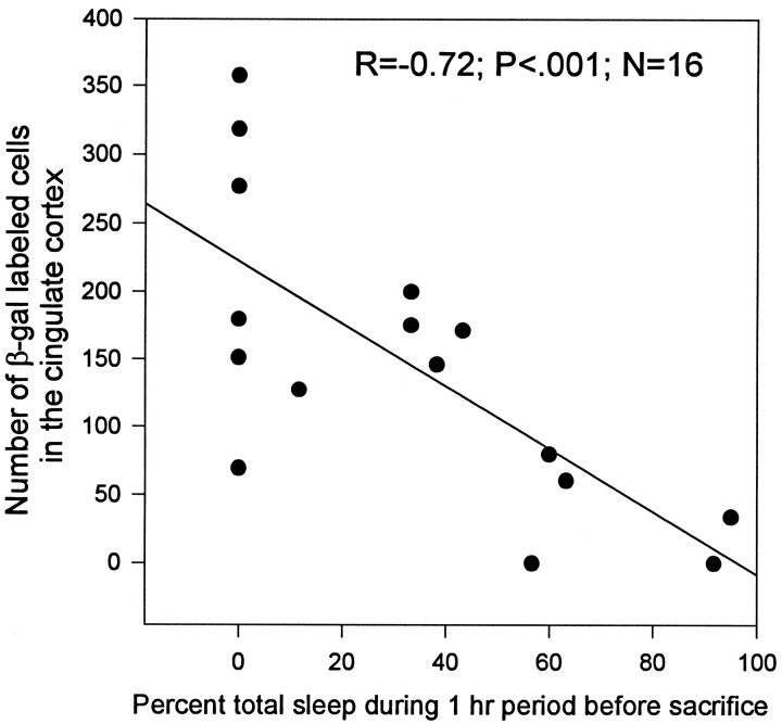
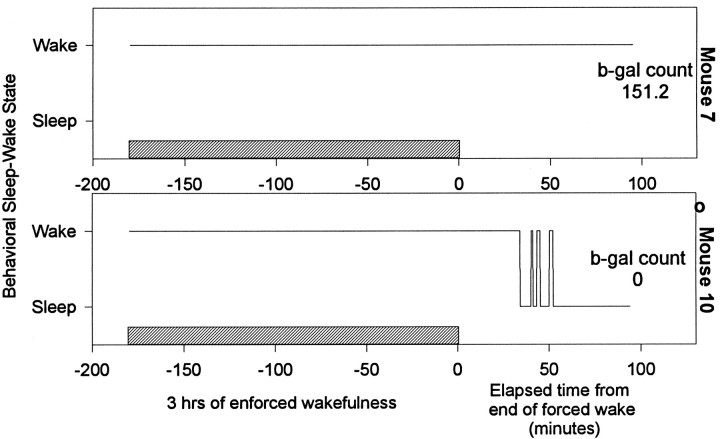
We investigated the effects of sleep on wake-induced c-fos expression in the cerebral cortex of rats and c-fos-lacZ transgenic mice. In the cortex of rats, the levels of c-Fos, detected both by immunocytochemistry and Western blot, remained high during 6 or 12 hr of enforced wakefulness but declined rapidly (within 1 hr) with increasing time of recovery sleep. Similarly, in the transgenic mice in which lacZ expression is driven from the c-fos promoter, beta-galactosidase activity was high after enforced wakefulness and declined with increasing amounts of sleep. These results suggest that the decrease in c-Fos protein in cortical neurons during sleep may be attributable to cessation of c-fos expression, activation of a process that degrades the wake-induced c-Fos, or both.


Figures

Fig. 1.
Fig. 1.
Immunohistochemical detection of c-Fos in cingulate cortex. Rats were kept awake by gentle handling for 12 hr (7:00 A.M.–7:00 P.M.) and then killed (n = 6) or allowed 1 hr of sleep (n = 7). c-Fos-labeled cells in cingulate cortex were detected using immunohisochemistry after prolonged wakefulness (A) and after 1 hr of sleep (B).
Fig. 2.
Fig. 2.
Enforced wakefulness and recovery from sleep-induced changes in the number of c-Fos-positive neurons in cortex. The number of c-Fos-positive cells was counted in cingulate cortex of rats that were kept awake for 12 hr or were allowed sleep after enforced wakefulness. Percent of sleep was calculated as total sleep time/60 min × 100. c-Fos levels declined in those animals who slept when they were permitted (n = 7) compared with those animals not permitted to sleep (n = 6). The number of c-Fos-positive cells declined with increasing percentage of sleep (n = 13; r = −0.82;p < 0.005).
Fig. 3.
Fig. 3.
Western blot analysis of nuclear proteins. To determine the size of c-Fos protein, Western blot analysis was performed using tissue extracts of cingulate cortex from rats after 6 hr of enforced wakefulness (SD1, SD2), and followed by 1 hr of sleep (RS1, RS2) before death. c-Fos protein (molecular weight, 62 kDa; arrowhead) decreased in rats that were allowed sleep before death.
Fig. 4.
Fig. 4.
Detection of β-galactosidase activity in cingulate cortex. The presence of β-gal activity in the cingulate cortex of c-fos-lacZ transgenic mice was detected using x-gal after 3 hr of enforced wakefulness (A) and then followed by 1 hr of sleep (B).
Fig. 5.
Fig. 5.
Sleep-induced decrease in β-galactosidase activity. The number of cells showing β-gal activity was correlated with the percent of sleep during the 60 min period before death of c-fos-lacZ transgenic mice. β-Galactosidase activity decreased with increasing amount of sleep (n = 16;r = −0.72; p < 0.001).
Fig. 6.
Fig. 6.
The wake–sleep profile of two representative mice. Both mice were kept awake for 3 hr and then left undisturbed for 1 hr. Mouse 7 did not sleep even when permitted to do so, and high levels of β-galactosidase staining were found in the cingulate cortex. Mouse 10 was asleep for 45 min before death, and it showed no β-galactosidase stained cells. The hatched barrepresents 3 hr period of enforced wakefulness.

References

    1. Aston-Jones G, Bloom FE. Activity of norepinephrine-containing locus coeruleus neurons in behaving rats anticipates fluctuations in the sleep-waking cycle. J Neurosci. 1981;1:876–886. - PMC - PubMed
    1. Cirelli C, Tononi G. Changes in protein phosphorylation patterns in the brain during the sleep-waking cycle. Soc Neurosci Abstr. 1996;22:688.
    1. Cirelli C, Pompeiano M, Tononi G. Fos-like immunoreactivity in the rat brain in spontaneous wakefulness and sleep. Arch Ital Biol. 1993a;131:327–330. - PubMed
    1. Cirelli C, Pompeiano M, Tononi G. Fos expression in the rat brain after variable periods of sleep deprivation. Sleep Res. 1993b;22:595. - PubMed
    1. Cirelli C, Pompeiano M, Tononi G. Sleep deprivation and c-fos expression in the rat brain. J Sleep Res. 1995;4:92–106. - PubMed

Publication types

MeSH terms

Substances

LinkOut - more resources