. 1997 Dec 9;94(25):13437-41.
doi: 10.1073/pnas.94.25.13437.

Regulation of ribonuclease III processing by double-helical sequence antideterminants

Affiliations

Regulation of ribonuclease III processing by double-helical sequence antideterminants

K Zhang et al. Proc Natl Acad Sci U S A. .

Abstract

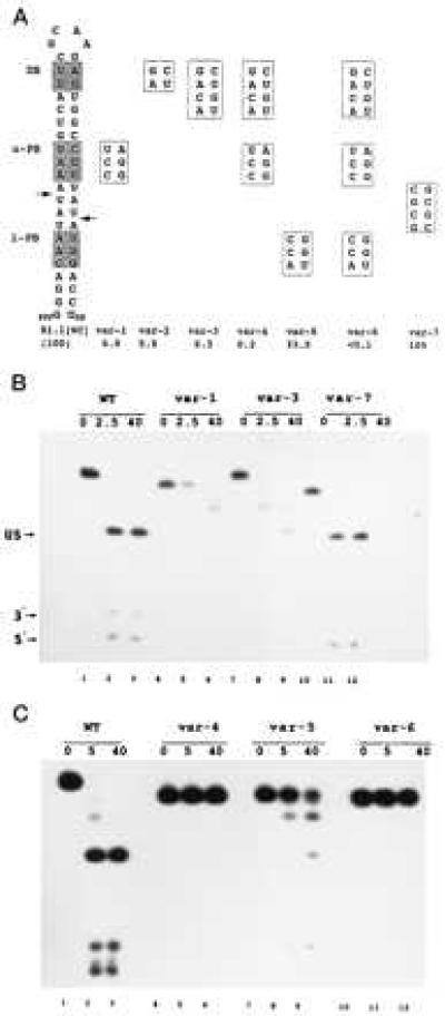
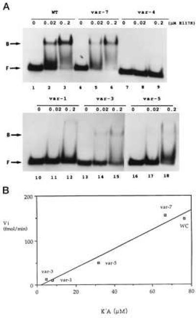
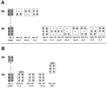
The double helix is a ubiquitous feature of RNA molecules and provides a target for nucleases involved in RNA maturation and decay. Escherichia coli ribonuclease III participates in maturation and decay pathways by site-specifically cleaving double-helical structures in cellular and viral RNAs. The site of cleavage can determine RNA functional activity and half-life and is specified in part by local tertiary structure elements such as internal loops. The involvement of base pair sequence in determining cleavage sites is unclear, because RNase III can efficiently degrade polymeric double-stranded RNAs of low sequence complexity. An alignment of RNase III substrates revealed an exclusion of specific Watson-Crick bp sequences at defined positions relative to the cleavage site. Inclusion of these "disfavored" sequences in a model substrate strongly inhibited cleavage in vitro by interfering with RNase III binding. Substrate cleavage also was inhibited by a 3-bp sequence from the selenocysteine-accepting tRNASec, which acts as an antideterminant of EF-Tu binding to tRNASec. The inhibitory bp sequences, together with local tertiary structure, can confer site specificity to cleavage of cellular and viral substrates without constraining the degradative action of RNase III on polymeric double-stranded RNA. Base pair antideterminants also may protect double-helical elements in other RNA molecules with essential functions.


Figures

Figure 1
Figure 1
Position-specific exclusion of sequence in RNase III substrates. (A) Substrate alignment analysis. Ten substrates were aligned whose cleavage sites were accurately determined, either by direct RNA sequence analysis or by primer extension of RNA cleaved in vitro by purified RNase III: 16S rRNA precursor (14), 23S rRNA precursor (15), T7 R0.3 (16), R0.5 (16), R1.1 (16) and R1.3 (16) substrates; lambda N leader (17), lambda sib (18) lambda cIII (19), and the RNase III operon transcript 5′-leader (20, 21). Each substrate provided two sequences for alignment, reflecting a two-fold symmetry about the cleavage site (11, 12). The cleavage site is indicated by vertical arrowhead between −1 and +1. The data represent the number of times (n = 20) each W-C bp occurs at positions +1 to −12. Position −12 represented the duplex limit for most substrates. P values from a Chi squared analysis also are provided. The significance of the disfavored A and U at positions −1 and −2, respectively, and the preferred C at −2 are not known, but may reflect specific sequence requirements for a structured asymmetric internal loop in a number of the substrates (22). The preference for CG at −6 is not known. (B) The “disfavored” bp, displayed in a dsRNA structure. The proximal box (PB) and distal box (DB) are included within an 11-bp helix. S = C or G, with S′ complementary to S. N, N′ indicate complementary nucleotides; while n, n′ indicate less strict complementarity. Arrowheads indicate the (blocked) cleavage sites. (C) Absence of conservation (degeneracy) of RNase III substrate sequence. H = A, G, U, with D′ (A, C, U) complementary to H; B = C, G, U, with V′ (G, C, U) complementary to B; W, W′ = A, U. (D) Secondary structure of the T7 R1.1 RNase III substrate, showing the proximal and distal boxes and the single cleavage site (arrowhead).
Figure 2
Figure 2
Bp-sequence-dependent inhibition of substrate cleavage. (A) Structure of R1.1[WC] RNA and the relevant sequences of seven variants. Cleavage of R1.1[WC] RNA occurs at the two indicated sites (arrows), determined by oligonucleotide sequence analysis of cleavage products (not shown). DB, distal box; u-PB, upper proximal box; l-PB, lower proximal box. Numbers given below the R1.1[WC] variants are cleavage rates relative to the R1.1[WC] RNA rate (100%, or ≈150 fmol product/min). The reported values are the average of at least three experiments, with standard deviations ≤13%. (B) Cleavage time course pattern for internally 32P-labeled R1.1[WC] RNA (WT, lanes 1–3) and variant 1 (lanes 4–6), variant 3 (lanes 7–9), and variant 7 (lanes 10–12). Time points (minutes) are indicated. (C) Same as experiment in B, but examining variant 4 (lanes 4–6), variant 5 (lanes 7–9), and variant 6 (lanes 10–12). Lanes 1–3 are for R1.1[WC] RNA. The cleavage products are the upper stem (US, 28 nucleotides); a 5′-end-containing fragment (5′, 10 nucleotides), and a 3′-end-containing fragment (3′, 8 nucleotides). The slighter slower rate of cleavage for R1.1[WC] RNA in the experiment in the lower panel reflects typical variation from experiment to experiment. The differing gel mobilities of the uncleaved RNAs reflect conformational differences in 7M urea, also seen elsewhere (23). For several of the variants, the two species with mobilities between the substrate and the 28-nt upper stem product represent products of single-site cleavage.
Figure 3
Figure 3
W-C bp substitution inhibits RNase III binding. (A) Gel shift assay of R1.1[WC] RNA variant binding to RNase III. 5′-32P-labeled RNA (104 dpm; 1.4 fmol) was combined with 0, 0.02, or 0.2 μM of the [E117K] RNase III mutant, then electrophoresed in a nondenaturing gel. (Upper) Lanes 1–3, R1.1[WC] RNA (WT); lanes 2–4, variant 7; and lanes 7–9, variant 4. (Lower) Lanes 1–3, variant 1; lanes 4–6, variant 3; and lanes 7–9, variant 5. (B) Correlation between initial cleavage rate (Vi) and binding affinity (1/KD). WC refers to R1.1[WC] RNA. The Vi values are expressed as fmol product formed per minute (37°). The measured KD values (nM ± SD) are the average of at least three experiments, and are: R1.1[WC] RNA, 13.1 ± 9.3; var-7, 15.0 ± 4.0; var-5, 31.5 ± 16.5; var-1, 126 ± 30; and var-3, 213 ± 117. Given the similarity in cleavage rates (Fig. 2A), the binding affinity of variant 2 was assumed to be similar to that for variant 3. The weak binding affinities of variants 4 and 6 prevented KD determination.
Figure 4
Figure 4
Inhibition of cleavage by single or double bp substitutions and by the tRNASec AD. (A) Relative cleavage rates of internally 32P-labeled R1.1[WC] RNA variants exhibiting single or double W-C bp substitution in the distal or proximal box. The proximal box (PB) and distal box (DB) sequences of R1.1[WC] RNA are shown on the left. (B) Inhibition by the tRNASec AD and sequence variants. The var-1 RNA value is from Fig. 2. (A and B) The numbers below the variant RNAs are cleavage rates, relative to that of R1.1[WC] RNA (100%), and represent the average of three experiments, with SDs averaging ≤22% of the values.

References

    1. Belasco J G, Brawerman G, editors. Control of Messenger RNA Stability. San Diego: Academic; 1993.
    1. Nicholson A W. Prog Nucleic Acids Res Mol Biol. 1996;52:1–65. - PubMed
    1. Meegan J M, Marcus P I. Science. 1989;244:1089–1091. - PubMed
    1. Robertson H D, Webster R E, Zinder N D. J Biol Chem. 1968;243:82–91. - PubMed
    1. Court D. In: Control of Messenger RNA Stability. Belasco J G, Brawerman G, editors. San Diego: Academic; 1993. pp. 71–116.

Publication types

MeSH terms

LinkOut - more resources