Comparative Study
. 1997 Dec 9;94(25):13452-7.
doi: 10.1073/pnas.94.25.13452.

Structure of the recombinant full-length hamster prion protein PrP(29-231): the N terminus is highly flexible

Affiliations
Comparative Study

Structure of the recombinant full-length hamster prion protein PrP(29-231): the N terminus is highly flexible

D G Donne et al. Proc Natl Acad Sci U S A. .

Abstract

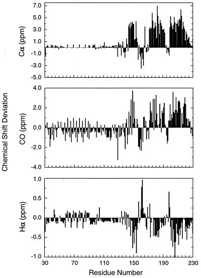
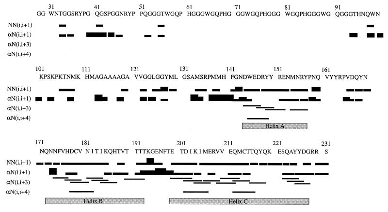
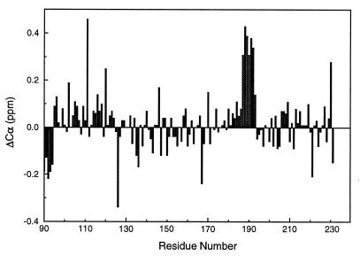
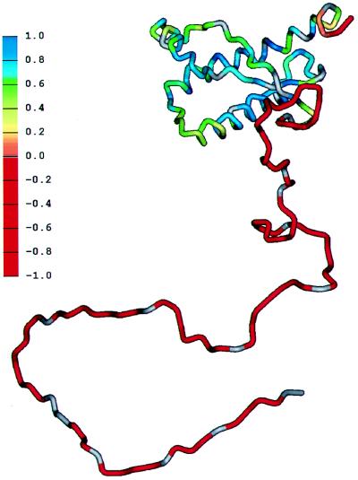
The prion diseases seem to be caused by a conformational change of the prion protein (PrP) from the benign cellular form PrPC to the infectious scrapie form PrPSc; thus, detailed information about PrP structure may provide essential insights into the mechanism by which these diseases develop. In this study, the secondary structure of the recombinant Syrian hamster PrP of residues 29-231 [PrP(29-231)] is investigated by multidimensional heteronuclear NMR. Chemical shift index analysis and nuclear Overhauser effect data show that PrP(29-231) contains three helices and possibly one short beta-strand. Most striking is the random-coil nature of chemical shifts for residues 30-124 in the full-length PrP. Although the secondary structure elements are similar to those found in mouse PrP fragment PrP(121-231), the secondary structure boundaries of PrP(29-231) are different from those in mouse PrP(121-231) but similar to those found in the structure of Syrian hamster PrP(90-231). Comparison of resonance assignments of PrP(29-231) and PrP(90-231) indicates that there may be transient interactions between the additional residues and the structured core. Backbone dynamics studies done by using the heteronuclear [1H]-15N nuclear Overhauser effect indicate that almost half of PrP(29-231), residues 29-124, is highly flexible. This plastic region could feature in the conversion of PrPC to PrPSc by template-assisted formation of beta-structure.


Figures

Figure 1
Figure 1
Summary of Cα, Hα, and 13CO chemical shift deviations from random-coil values (38, 39) used to identify the secondary structure of PrP(29–231). The chemical shift deviations in ppm are plotted against residue numbers in PrP(29–231). Note the repeating nature of shift values in the octarepeat region.
Figure 2
Figure 2
Sequential and medium-range NOEs used to verify the secondary structure elements derived from CSI analysis. NOEs between amide protons of consecutive residues and between Hα and the amide proton of subsequent residues are represented by bars connecting the residues. For NN(i, i+1) and αN(i, i+1) NOEs, the thickness of the bar qualitatively represents the relative intensity (weak, medium, or strong) of the NOE. The NOEs for four octarepeats are not shown because they were assigned only to residue types in the repeat region because of overlap. The three unambiguously identified α-helices also are shown.
Figure 3
Figure 3
Difference in the Cα chemical shift between values measured for PrP(29–231) and PrP(90–231) plotted against residue number.
Figure 4
Figure 4
Summary of heteronuclear [1H]-15N NOE data used to assess the backbone flexibility of the PrP(29–231) and PrP(90–231). (A) The mean values of heteronuclear NOE from two sets of data are plotted against residue number in the full-length SHa PrP(29–231). The error bars represent the difference between the two data sets. The NOEs in four of the octarepeats (residues 59–91) are approximate values estimated by dividing the degenerate peak volumes by four. (B) Heteronuclear NOE values for PrP(90–231). All spectroscopic parameters and processing procedures are the same as those of PrP(29–231).
Figure 5
Figure 5
Schematic diagram showing the flexibility of the polypeptide chain for PrP(29–231). The structure of the portion of the protein representing residues 90–231 was taken from the coordinates of PrP(90–231) (10). The remainder of the sequence was hand-built for illustration purposes only. The color scale shows the heteronuclear [1H]-15N NOE data from Fig. 4A, from red for the lowest (most negative) values, where the polypeptide is most flexible, to blue for the highest (most positive) values in the most structured and rigid regions of the protein.

Comment in

References

    1. Gajdusek D C. Science. 1977;197:943–960. - PubMed
    1. Prusiner S B. Science. 1997;278:245–251. - PubMed
    1. Prusiner S B. Science. 1982;216:136–144. - PubMed
    1. Prusiner S B. Science. 1991;252:1515–1522. - PubMed
    1. Pan K-M, Baldwin M, Nguyen J, Gasset M, Serban A, Groth D, Mehlhorn I, Huang Z, Fletterick R J, Cohen F E, Prusiner S B. Proc Natl Acad Sci USA. 1993;90:10962–10966. - PMC - PubMed

Publication types

MeSH terms