. 1997 Dec 9;94(25):13594-9.
doi: 10.1073/pnas.94.25.13594.

Aldehyde dehydrogenase class 3 expression: identification of a cornea-preferred gene promoter in transgenic mice

Affiliations

Aldehyde dehydrogenase class 3 expression: identification of a cornea-preferred gene promoter in transgenic mice

W T Kays et al. Proc Natl Acad Sci U S A. .

Abstract

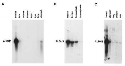
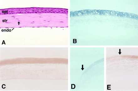
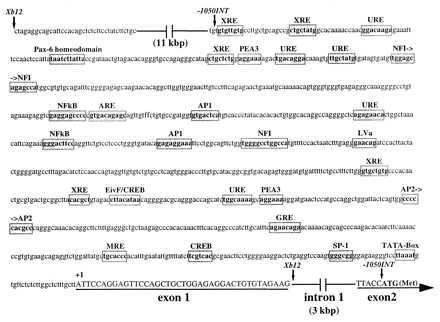
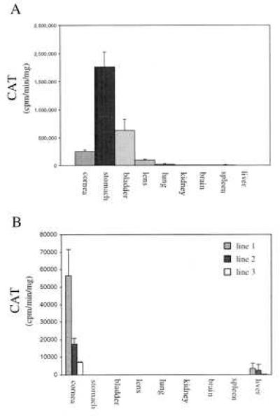
Aldehyde dehydrogenase class 3 (ALDH3) constitutes 20-40% of the total water-soluble proteins in the mammalian cornea. Here, we show by Northern blot analysis that ALDH3 expression in the mouse is at least 500-fold higher in the cornea than in any other tissue examined, with very low levels of expression detected in the stomach, urinary bladder, ocular lens, and lung. Histochemical localization reveals that this exceptional level of expression in the mouse cornea occurs in the anterior epithelial cells and that little ALDH3 is present in the keratocytes or corneal endothelial cells. A 13-kbp mouse ALDH3 promoter fragment containing >12 kbp of the 5' flanking sequence, the 40-bp untranslated first exon, and 29 bp of intron 1 directed cat reporter gene expression to tissues that express the endogenous ALDH3 gene, except that transgene promoter activity was higher in the stomach and bladder than in the cornea. By contrast, when driven by a 4.4-kbp mouse ALDH3 promoter fragment [1,050-bp 5' flanking region, exon 1, intron 1 (3.4 kbp), and 7 bp of exon 2] expression of the cat reporter gene was confined to the corneal epithelial cells, except for very low levels in the liver, effectively reproducing the corneal expression pattern of the endogenous ALDH3 gene. These results indicate that tissue-specific expression of ALDH3 is determined by positive and negative elements in the 5' flanking region of the gene and suggests putative silencers located in intron 1. We demonstrate regulatory sequences capable of directing cornea-specific gene expression, affording the opportunity for genetic engineering in this transparent tissue.


Figures

Figure 1
Figure 1
Northern blot analysis of ALDH3 expression in mouse, bovine, and human tissues. (A) Hybridization of a mouse ALDH3 cDNA probe to a Northern blot of total RNA from adult mouse cornea (2 μg), lens (20 μg), adrenal gland (20 μg), urinary bladder (20 μg), brain (20 μg), liver (20 μg), lung (20 μg), and stomach (20 μg) revealed ALDH3 transcript levels in the cornea to be at least 500-fold higher than those observed in any other tissue examined. (B) Significant levels of ALDH3 transcript also were detected in total RNA from bovine cornea (10 μg) and human corneal epithelium (10 μg) but not in human corneal endothelium (5 μg). (C) A Northern blot analysis in which the autoradiogram purposely has been overdeveloped to demonstrate hybridization of a mouse ALDH3 cDNA probe to total RNA from mouse cornea (2 μg), stomach (20 μg), urinary bladder (20 μg), lung (20 μg), and lens (20 μg), indicating very low but detectable levels of ALDH3 expression in these tissues.
Figure 2
Figure 2
Histochemical examination of ALDH3 and cat reporter gene expression in the mouse cornea. (A) The structure of the adult mouse cornea is seen in a hematoxylin/eosin-stained section. The stratified corneal epithelium (epi) overlies a stromal layer (str) maintained by numerous corneal keratocytes (arrow). The one-cell-thick endothelium (endo) forms the posterior extent of the cornea. (B) Histochemical localization demonstrates ALDH3 activity primarily in the cells of the central epithelium of the normal mouse cornea. (C) Histochemical localization of CAT activity in the cornea of a −1050INT transgenic mouse demonstrates the ability of the mouse ALDH3 promoter fragment to target reporter gene expression to the corneal epithelium, faithfully reproducing the epithelial-specific pattern demonstrated for the endogenous mouse ALDH3 gene. In the peripheral cornea, localization of both ALDH3 (D) and CAT (E) activities diminishes near the limbal border (arrows), with little activity detected in limbal epithelial cells themselves.
Figure 3
Figure 3
Structure of the 5′ end of the mouse ALDH3 gene. A 40-bp untranslated exon (exon 1) was identified ≈3 kbp upstream of the translation initiation ATG codon in exon 2. A sequence analysis, employing Wisconsin Sequence Analysis Package software (Genetics Computer Group, Madison, WI), identified putative binding sites for a number of transcription factors within the initial 1,050 bp of sequence 5′ to the +1 transcription initiation site. Of particular interest are the antioxidant responsive element (ARE) and UV-responsive elements (URE), which suggest a potential to up-regulate expression in response to oxidative and UV-induced stress in the cornea. The promoter fragment utilized to generate transgenic mice is delineated by arrows (↓). Exon sequence is indicated by underlined uppercase characters.
Figure 4
Figure 4
ALDH3 promoter-driven reporter gene expression in the tissues of adult transgenic mice. (A) In Xb12/cat transgenic animals (n = 4), significant expression of the reporter gene, expressed as CAT activity, is seen in stomach, bladder, cornea, lens, lung, and spleen (trace) although not at relative levels consistent with expression of the endogenous ALDH3 gene (see Fig. 1 A and C). (B) By contrast, transgene expression in −1050INT/cat transgenic mice is restricted primarily to the corneas of mice from three independent transgenic lines (n = 4 animals/transgenic line). Very low levels of transgene expression are seen in the livers of animals from each transgenic line, with CAT activity ranging from 5 to 12% of corneal activity in the respective transgenic line.

References

    1. Overbeek P A, Chepelinsky A B, Khillan J S, Piatigorsky J, Westphal H. Proc Natl Acad Sci USA. 1985;82:7815–7819. - PMC - PubMed
    1. Cvekl A, Piatigorsky J. Bioessays. 1996;18:621–630. - PubMed
    1. Liou G I, Geng L, al-Ubaidi M R, Matragoon S, Hanten G, Baehr W, Overbeek P A. J Biol Chem. 1990;265:8373–8376. - PubMed
    1. Lem J, Applebury M L, Falk J D, Flannery J G, Simon M I. Neuron. 1991;6:201–210. - PubMed
    1. Kikuchi T, Raju K, Breitman M L, Shinohara T. Mol Cell Biol. 1993;13:4400–4408. - PMC - PubMed

MeSH terms

Associated data

LinkOut - more resources