Comparative Study
. 1997 Dec 9;94(25):13618-23.
doi: 10.1073/pnas.94.25.13618.

Characterization and cell cycle regulation of the related human telomeric proteins Pin2 and TRF1 suggest a role in mitosis

Affiliations
Comparative Study

Characterization and cell cycle regulation of the related human telomeric proteins Pin2 and TRF1 suggest a role in mitosis

M Shen et al. Proc Natl Acad Sci U S A. .

Abstract

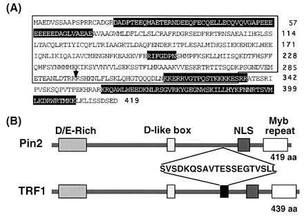
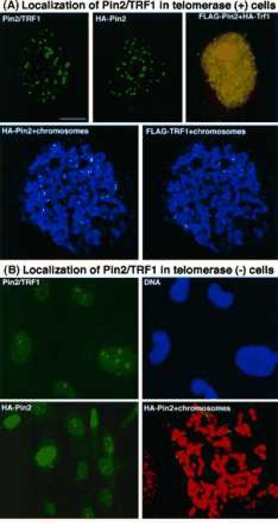
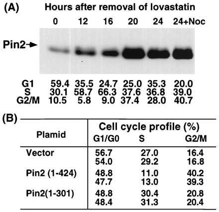
Telomeres are essential for preserving chromosome integrity during the cell cycle and have been specifically implicated in mitotic progression, but little is known about the signaling molecule(s) involved. The human telomeric repeat binding factor protein (TRF1) is shown to be important in regulating telomere length. However, nothing is known about its function and regulation during the cell cycle. The sequence of PIN2, one of three human genes (PIN1-3) we previously cloned whose products interact with the Aspergillus NIMA cell cycle regulatory protein kinase, reveals that it encodes a protein that is identical in sequence to TRF1 apart from an internal deletion of 20 amino acids; Pin2 and TRF1 may be derived from the same gene, PIN2/TRF1. However, in the cell Pin2 was found to be the major expressed product and to form homo- and heterodimers with TRF1; both dimers were localized at telomeres. Pin2 directly bound the human telomeric repeat DNA in vitro, and was localized to all telomeres uniformly in telomerase-positive cells. In contrast, in several cell lines that contain barely detectable telomerase activity, Pin2 was highly concentrated at only a few telomeres. Interestingly, the protein level of Pin2 was highly regulated during the cell cycle, being strikingly increased in G2+M and decreased in G1 cells. Moreover, overexpression of Pin2 resulted in an accumulation of HeLa cells in G2+M. These results indicate that Pin2 is the major human telomeric protein and is highly regulated during the cell cycle, with a possible role in mitosis. The results also suggest that Pin2/TRF1 may connect mitotic control to the telomere regulatory machinery whose deregulation has been implicated in cancer and aging.


Figures

Figure 1
Figure 1
Amino acid sequence of Pin2 and its relationship with TRF1. (A) The predicted Pin2 peptide sequence is indicated in the one-letter code, with the arrow indicating the site at which the additional 20 amino acids are present in TRF1. Boxes with white letters from left to right indicate an acid (D/E)-rich domain, a D-like box, a bipartite nuclear localization signal and a Myb-type HTH DNA-binding domain. (B) Schematic presentation of the domain structure of the Pin2 and TRF1 proteins and sequence of the 20 amino acid insert in TRF1.
Figure 2
Figure 2
Comparison of Pin2 and TRF1 proteins and their expression levels in the cell. (A) The mRNA level was determined by reverse transcription–PCR using total RNAs isolated from various cell lines as indicated and a set of primers that are common for both PIN2 and TRF1 (outside the deleted amino acid residues 296–316 of TRF1). The two major PCR products were confirmed by direct sequencing to be a fragment of PIN2 and TRF1. The minor larger PCR product (∗) was found to be the same as the TRF1 fragment, but with a tandem-duplicated primer at the 3′ end. (B) The levels of Pin2 and TRF1 protein were determined by immunoprecipitation with the anti-Pin2 serum or preimmune serum, followed immunoblot analysis using anti-Pin2 antibodies. (C) PIN2 and TRF1 expression constructs with an N-terminal HA tag were transiently transfected into tTA-1 cells for 24 h. One hundred micrograms of lysates were subjected to immunoblot analysis using anti-Pin2 antibodies.
Figure 3
Figure 3
Pin2 is a telomeric DNA-binding protein that forms homo- and heterodimers with TRF1. (A) The purified recombinant Pin2 protein was incubated with a [32P]-labeled double-stranded oligonucleotide containing six human telomeric DNA repeats, in the presence or absence of a 30-fold excess of unlabeled oligonucleotide and 1 mg/ml casein. The samples were separated on a 5% polyacrylamide gel, followed by autoradiography. (B) tTA-1 cells were cotransfected for 36 h with two different epitope-tagged Pin2 and/or TRF1 expression constructs, as indicated. Cells were lysed in lysis buffer containing 1% Triton X-100. Aliquots of the cellular proteins were directly subjected to SDS/PAGE, followed by immunoblot analysis using the tag-specific 12CA5 or M2 mAb. The remainder of the lysates were first immunoprecipitated with the 12CA5 mAb and then subjected to immunoblot analysis using the M2 mAb.
Figure 4
Figure 4
Differential localization of Pin2 is in telomerase-positive and -negative cells. (A) Localization of Pin2 protein in telomerase-positive HeLa cells. Pin2/TRF1, localization of the endogenous Pin2/TRF1 detected by immunostaining with anti-Pin2 antibodies; HA-Pin2, localization of the ectopically expressed Pin2 detected by staining with the 12CA5 mAb; HA-Pin2 and FLAG-TRF1, colocalization of expressed Pin2 and TRF1 detected by staining doubly transfected cells with 12CA5 and M2 mAbs, with the yellow image indicating a colocalization produced by superimposing the green Pin2 and the red TRF1 images; HA-Pin2/FLAG-TRF1+chromosomes, colocalization of expressed Pin2 and TRF1 evenly at telomeres detected by staining mitotic chromosomes from doubly transfected cells with 12CA5 and M2 mAbs. Green, HA-Pin2; red, FLAG-TRF1; blue, chromosomes. (B) Localization of Pin2 protein in telomerase-negative MDAH087 cells. Pin2/TRF1, localization of the endogenous Pin2/TRF1 detected by staining with anti-Pin2 antibodies (green) and the DNA dye Hoechst (blue); HA-Pin2, localization of the ectopically expressed Pin2 detected by staining a single stably HA-Pin2-expressing cell line (clone 6) with the 12CA5 mAb; HA-Pin2+chromosomes, localization of expressed Pin2 at telomeres detected by staining clone 6 mitotic chromosomes with the 12CA5 mAb. Green, HA-Pin2; red, chromosomes.
Figure 5
Figure 5
Pin2 protein level is tightly regulated during the cell cycle and induces accumulation of cells at G2+M when overexpressed. (A) HA-Pin2-expressing clone 6 MDAH087 cells were incubated with 10 μM lovastatin to synchronize cells in G1 (time 0). At the times indicated following release, cells were harvested and subjected to flow cytometric analysis to examine the cell cycle status or to immunoprecipitation and immunoblot analysis to determine HA-Pin2 levels using 12CA5 mAb. (B) The tTA-1 HeLa cells were transfected for 48 h with vectors expressing the indicated HA epitope tagged Pin2 proteins, followed by staining first with 12CA5 mAb and then with fluorescein isothiocyanate-conjugated secondary antibodies and propidium iodide. Pin2- and Pin2(1–301)-expressing cells were selected and their cell cycle profiles were determined to compare with that for the vector-only transfected cells.

References

    1. Greider C W. Annu Rev Biochem. 1996;65:337–365. - PubMed
    1. Zakian V A. Science. 1995;270:1601–1607. - PubMed
    1. Harley C B, Futcher A B, Greider C W. Nature (London) 1990;345:458–460. - PubMed
    1. Hastie N D, Dempster M, Dunlop M G, Thompson A M, Green D K, Allshire R C. Nature (London) 1990;346:866–868. - PubMed
    1. Marcand S, Gilson E, Shore D. Science. 1997;275:986–990. - PubMed

Publication types

MeSH terms