. 1997 Dec 9;94(25):13630-5.
doi: 10.1073/pnas.94.25.13630.

Opposing mitogenic and anti-mitogenic actions of parathyroid hormone-related protein in vascular smooth muscle cells: a critical role for nuclear targeting

Affiliations

Opposing mitogenic and anti-mitogenic actions of parathyroid hormone-related protein in vascular smooth muscle cells: a critical role for nuclear targeting

T Massfelder et al. Proc Natl Acad Sci U S A. .

Abstract

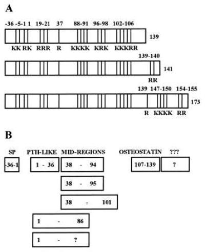
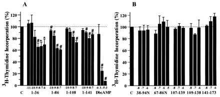
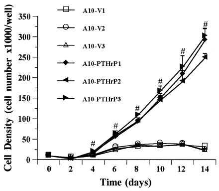
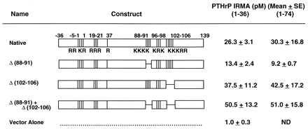
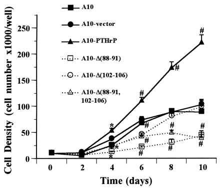
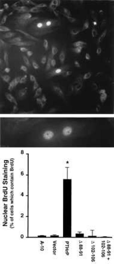
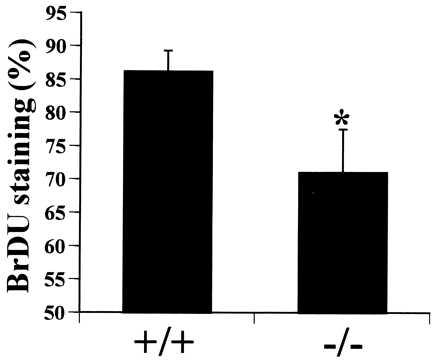
Parathyroid hormone-related protein (PTHrP) is a prohormone that is posttranslationally processed to a family of mature secretory forms, each of which has its own cognate receptor(s) on the cell surface that mediate the actions of PTHrP. In addition to being secreted via the classical secretory pathway and interacting with cell surface receptors in a paracrine/autocrine fashion, PTHrP appears to be able to enter the nucleus directly following translation and influence cellular events in an "intracrine" fashion. In this report, we demonstrate that PTHrP can be targeted to the nucleus in vascular smooth muscle cells, that this nuclear targeting is associated with a striking increase in mitogenesis, that this nuclear effect on proliferation is the diametric opposite of the effects of PTHrP resulting from interaction with cell surface receptors on vascular smooth muscle cells, and that the regions of the PTHrP sequence responsible for this nuclear targeting represent a classical bipartite nuclear localization signal. This report describes the activation of the cell cycle in association with nuclear localization of PTHrP in any cell type. These findings have important implications for the normal physiology of PTHrP in the many tissues which produce it, and suggest that gene delivery of PTHrP or modified variants may be useful in the management of atherosclerotic vascular disease.


Figures

Figure 1
Figure 1
(A) Structural maps of the three PTHrP initial translation products. “K” indicates lysine and “R” arginine. The three translation products are identical in their signal peptide (−36 to −1) region and in their remaining coding regions from amino acids 1 through 139. Alternative splicing gives rise to two C-terminally extended versions of the peptide with lengths of 141 and 173 amino acids. Note the multibasic clusters: the KKXRK at −5 to −1, the single R at +37, and the KR clusters in the 88–106 region are known to be prohormone convertase substrate sites. (B) The mature secretory forms of PTHrP. After cleavage of the signal and pro-peptide in the −36 to −1 region, the secretory forms of PTHrP include PTHrP(1–36) which interacts with the PTH/PTHrP receptor, three mid-region species, and at least two C-terminal species.
Figure 3
Figure 3
Effects of PTHrP addition to cultured A-10 cells. (A) PTHrP peptides that contain an intact N terminus and dibutyryl cAMP inhibit A-10 cell proliferation. (B) Non-N-terminal PTHrP peptides, comprising all of the known secretory forms of PTHrP (see Fig. 1), have no effect on A-10 cell proliferation. #, P < 0.001; ∗, P < 0.05. “C” indicates control, and the numbers along the x-axis indicate the PTHrP peptide employed [e.g., 1–36 indicates PTHrP(1–36), and 38–94N indicates PTHrP(38–94)amide].
Figure 2
Figure 2
Proliferation of three PTHrP-overexpressing A-10 cells clones (filled symbols) and of three vector-transfected clones (open symbols). Note that the PTHrP-overexpressing clones proliferate at a rate far higher than the vector transfected clones. Untransfected A-10 cells are indistinguishable from the vector-transfected clones. Similar results were observed by using [3H]thymidine incorporation. #, P < 0.01.
Figure 4
Figure 4
Map of the wild-type and native PTHrP constructs, the three deletion mutants, and vector alone. Values on the right indicate the concentrations of PTHrP in the conditioned medium from the various cell lines. Note that the mutant PTHrP constructs lead to similar levels of PTHrP production as the native construct, and that all constructs lead to the far greater production of PTHrP than the vector-transfected A-10 cells.
Figure 5
Figure 5
Proliferation of A-10 cells expressing the native PTHrP construct (filled triangles), the three mutant constructs (open symbols), the vector alone (filled circles), or untransfected A-10 cells (filled squares). #, P < 0.01; ∗, P < 0.05. Note that, as seen in the prior experiment in Fig. 2, PTHrP-overexpressing A-10 cells proliferate far more rapidly than their normal or vector-transfected counterparts. Note also that the three mutant-transfected clones proliferate at a rate that is even slower than the untransfected or vector-transfected cells. Each symbol represents the mean of three different clones.
Figure 6
Figure 6
(Top) PTHrP immunocytochemistry showing nuclear staining in PTHrP-overexpressing A-10 cells. (×200.) (Middle) A higher power magnification of the PTHrP-overexpressing cell nuclei. (×400.) Note that the pattern is a diffuse reticulated pattern and that the two most prominent nuclei are in a single dividing cell. For immunocytochemistry, negative controls (no primary antibody and competition with excess PTHrP) showed no staining (data not shown). (Bottom) The percentage of nuclei that contain PTHrP in the various A-10 cell clones. Each bar represents the mean of nine slides, and 300-1000 cells were counted per slide by two blinded observers. The bars indicate standard error; ∗, P < 0.01.
Figure 7
Figure 7
The percent of aortic smooth muscle cell nuclei that contain BrdU in PTHrP knockout mouse fetuses (−/−) or in their normal littermates (+/+). Each bar represents the mean of nine embryos; ∗, P < 0.02.

References

    1. Stewart A F, Insogna K L, Broadus A E. In: Endocrinology. 3rd Ed. DeGroot L, editor. Philadelphia: Saunders; 1995. pp. 1061–1074.
    1. Philbrick W M, Wysolmerski J J, Galbraith S, Holt E, Orloff J J, Yang K H, Vasavada R C, Weir E C, Broadus A E, Stewart A F. Physiol Rev. 1996;76:127–173. - PubMed
    1. Yang K H, Stewart A F. In: Principles of Bone Biology. Bilezikian J P, Raisz L, Rodan G, editors. San Diego: Academic; 1996. pp. 347–376.
    1. Karaplis A C, Luz A, Glowacki J, Bronson R T, Tybulewicz V L J, Kronenberg H M, Mulligan R C. Genes Dev. 1994;8:277–289. - PubMed
    1. Deftos L J, Burton D W, Brandt D W. J Clin Invest. 1993;92:727–735. - PMC - PubMed

Publication types

MeSH terms