. 1997 Dec 9;94(25):13661-5.
doi: 10.1073/pnas.94.25.13661.

Cell locomotion and focal adhesions are regulated by substrate flexibility

Affiliations

Cell locomotion and focal adhesions are regulated by substrate flexibility

R J Pelham Jr et al. Proc Natl Acad Sci U S A. .

Abstract

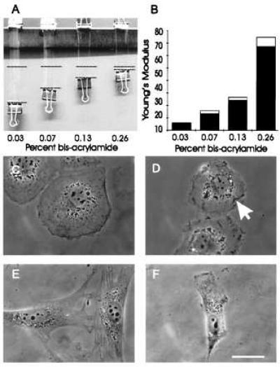
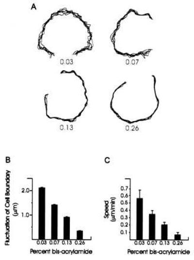
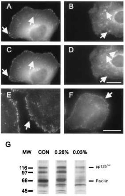
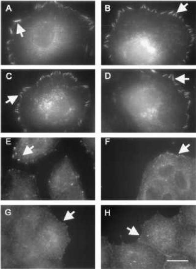
Responses of cells to mechanical properties of the adhesion substrate were examined by culturing normal rat kidney epithelial and 3T3 fibroblastic cells on a collagen-coated polyacrylamide substrate that allows the flexibility to be varied while maintaining a constant chemical environment. Compared with cells on rigid substrates, those on flexible substrates showed reduced spreading and increased rates of motility or lamellipodial activity. Microinjection of fluorescent vinculin indicated that focal adhesions on flexible substrates were irregularly shaped and highly dynamic whereas those on firm substrates had a normal morphology and were much more stable. Cells on flexible substrates also contained a reduced amount of phosphotyrosine at adhesion sites. Treatment of these cells with phenylarsine oxide, a tyrosine phosphatase inhibitor, induced the formation of normal, stable focal adhesions similar to those on firm substrates. Conversely, treatment of cells on firm substrates with myosin inhibitors 2,3-butanedione monoxime or KT5926 caused the reduction of both vinculin and phosphotyrosine at adhesion sites. These results demonstrate the ability of cells to survey the mechanical properties of their surrounding environment and suggest the possible involvement of both protein tyrosine phosphorylation and myosin-generated cortical forces in this process. Such response to physical parameters likely represents an important mechanism of cellular interaction with the surrounding environment within a complex organism.


Figures

Figure 1
Figure 1
Mechanical characteristics of polyacrylamide substrates and effects on cell morphology. (A and B) identically sized strips of polyacrylamide with various acrylamide/bis-acrylamide ratios were fixed at one end and stretched at the other end with a downward force of 0.103 N. The dashed lines represent the amount of stretching caused by applied weight (A). The extent of stretching was then used for the calculation of Young’s modulus, expressed as N/m2 (B). (C–F) Phase morphology of NRK (C and D) or 3T3 (E and F) cells plated on substrates containing 0.26% bis- (C and E) or 0.03% bis-acrylamide (D and F). NRK cells on the more flexible substrate are less well spread and contain irregular ruffles on the ventral surface (D, arrow), as determined by optical sectioning at a high magnification. Similarly, 3T3 cells on the substrate of high flexibility are typically less well spread and with a polarized morphology (F). Bar = 10 μm.
Figure 2
Figure 2
Measurements of lamellipodial activity and cell motility on substrates of various flexibilities. (A, B) Fluctuation of the lamellipodia of NRK cells cultured on substrates of varying flexibilities. Numbers in A indicate the percentage concentration of bis-acrylamide. Images of cells were recorded every 2 min over a period of 10 min and active lamellipoda were traced and overlaid to generate the plots. The degree of lamellipodial protrusion/retraction was then quantified based on the SD of the position of seven randomly chosen points along the active edge. Fifteen cells were analyzed under each bis-acrylamide concentration (B). Lamellipodia become less active with increasing rigidity of the substrate. (C) Rate of locomotion of 3T3 cells on substrates of varying flexibilities. Cells become less motile with increasing rigidity of the substrate.
Figure 3
Figure 3
Distribution of vinculin and phosphotyrosine in NRK cells cultured on substrates with 0.26% bis-acrylamide (A, C, E) or 0.03% bis-acrylamide (B, D, F). (A–D) Cells were injected with rhodamine-labeled vinculin and imaged over a period of 10 min. On more rigid substrates (A, C), vinculin is incorporated into elongated focal adhesions, which show only minor changes during the period of observation. On highly flexible gels (B, D), vinculin is localized at punctate structures of irregular sizes and shapes, many of which appear and disappear over a period of 10 min (arrows). (E, F), Immunofluorescence of phosphotyrosine. Phosphotyrosine is localized at elongated focal adhesions in cells cultured on more rigid gels (E), and at punctate structures in cells cultured on highly flexible gels (F). (G) Anti-phosphotyrosine immunoblotting of whole cell lysates from NRK cells cultured on different substrates. On plastic dishes (CON) and rigid 0.26% bis-acrylamide substrates (0.26%), pp125FAK, paxillin and a 97-kDa protein are heavily phosphorylated after 48 hr of culture. Cells cultured on soft 0.03% bis-acrylamide substrates (0.03%) show a significantly lower extent of phosphorylation at these bands. Bar = 10 μm.
Figure 4
Figure 4
Role of tyrosine phosphorylation and myosin in the modulation of adhesion structures on flexible substrates. Cells were treated with PAO (A–D) or 2,3-butanedione monoxime (E–H) and processed for vinculin (A, B, E, F) or phosphotyrosine (C, D, G, H) immunofluorescence. Treatment of cells with PAO resulted in the formation of large focal adhesions in cells on both soft (0.03% bis-acrylamide; B, D) and rigid (0.26% bis-acrylamide; A, C) substrates. Treatment with 2,3-butanedione monoxime disrupted adhesion structures of cells cultured on substrates with either 0.26% bis-acrylamide (E, G), or 0.03% bis-acrylamide (F, H), and caused vinculin (E, F) and phosphotyrosine (G, H) to localize at small punctate structures regardless of the substrate flexibility. Bar = 10 μm.

References

    1. Juliano R L, Haskill S J. J Cell Biol. 1993;120:577–585. - PMC - PubMed
    1. Bernstein L R, Liotta L A. Curr Opin Oncol. 1994;6:106–113. - PubMed
    1. Craig S W, Johnson R P. Curr Opin Cell Biol. 1996;8:74–85. - PubMed
    1. Palecek S P, Loftus J C, Ginsberg M H, Lauffenburger D A, Horowitz A F. Nature (London) 1997;385:537–540. - PubMed
    1. Wang N, Butler J P, Ingber D E. Science. 1993;260:1124–1127. - PubMed

Publication types

MeSH terms

LinkOut - more resources