. 1997 Dec 9;94(25):13689-94.
doi: 10.1073/pnas.94.25.13689.

Targeted expression of constitutively active receptors for parathyroid hormone and parathyroid hormone-related peptide delays endochondral bone formation and rescues mice that lack parathyroid hormone-related peptide

Affiliations

Targeted expression of constitutively active receptors for parathyroid hormone and parathyroid hormone-related peptide delays endochondral bone formation and rescues mice that lack parathyroid hormone-related peptide

E Schipani et al. Proc Natl Acad Sci U S A. .

Abstract

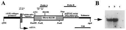
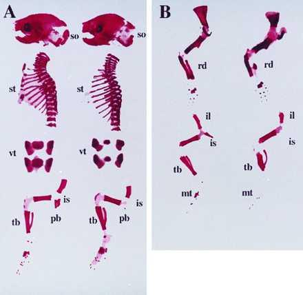
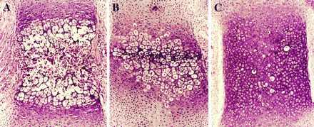
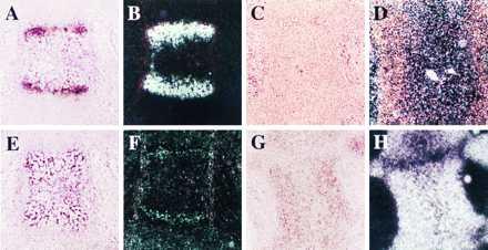
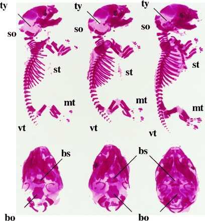
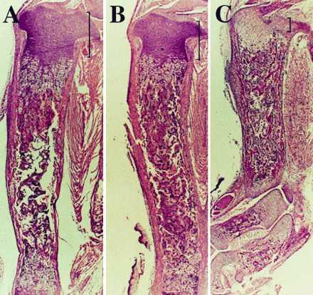
Mice in which the genes encoding the parathyroid hormone (PTH)-related peptide (PTHrP) or the PTH/PTHrP receptor have been ablated by homologous recombination show skeletal dysplasia due to accelerated endochondral bone formation, and die at birth or in utero, respectively. Skeletal abnormalities due to decelerated chondrocyte maturation are observed in transgenic mice where PTHrP expression is targeted to the growth plate, and in patients with Jansen metaphyseal chondrodysplasia, a rare genetic disorder caused by constitutively active PTH/PTHrP receptors. These and other findings thus indicate that PTHrP and its receptor are essential for chondrocyte differentiation. To further explore the role of the PTH/PTHrP receptor in this process, we generated transgenic mice in which expression of a constitutively active receptor, HKrk-H223R, was targeted to the growth plate by the rat alpha1 (II) collagen promoter. Two major goals were pursued: (i) to investigate how constitutively active PTH/PTHrP receptors affect the program of chondrocyte maturation; and (ii) to determine whether expression of the mutant receptor would correct the severe growth plate abnormalities of PTHrP-ablated mice (PTHrP-/-). The targeted expression of constitutively active PTH/PTHrP receptors led to delayed mineralization, decelerated conversion of proliferative chondrocytes into hypertrophic cells in skeletal segments that are formed by the endochondral process, and prolonged presence of hypertrophic chondrocytes with delay of vascular invasion. Furthermore, it corrected at birth the growth plate abnormalities of PTHrP-/- mice and allowed their prolonged survival. "Rescued" animals lacked tooth eruption and showed premature epiphyseal closure, indicating that both processes involve PTHrP. These findings suggest that rescued PTHrP-/- mice may gain considerable importance for studying the diverse, possibly tissue-specific role(s) of PTHrP in postnatal development.


Figures

Figure 1
Figure 1
Construction and expression of the transgene. (A) Schematic representation of the transgene construct showing the location of probes A and B for Southern blot analysis of tail genomic DNA, and of oligonucleotides 2F and G-2, and 2FA and H40 that were used for RT-PCR of total RNA and for Southern blot analysis of RT-PCR products, respectively; locations of the translation initiation codon (ATG), the receptor mutation H223R and the stop codon (Stop) in the cDNA encoding a human PTH/PTHrP receptor mutant, HKrk-H223R, are shown. The positions of restriction sites for the enzymes AflIII, BamHI, PvuII, and AgeI are also indicated. (B) Southern blot analysis of the RT-PCR products using total RNA from sternum of newborn Tg-A (a) and Tg-B (b) animals, respectively. (c) Control reaction performed in absence of total RNA. The blot was hybridized with the 32P-radiolabeled oligonucleotide H40. The arrow indicates the location of the 600-bp DNA marker.
Figure 2
Figure 2
Alizarin red S staining of the skeletons of newborn wild-type and transgenic mice. (A) Lateral view of skull, thoracic cage, and hindlimb; and frontal view of lumbar vertebra of a wild-type mouse (left), and a homozygous Tg-A (right) littermate; (so) supraoccipital bone, (st) sternum, (vt) lumbar vertebrae, (is) ischium, (pb) pubic bone, (tb) tibia. (B) Lateral view of forelimb (upper) and hindlimb (lower) of a wild-type mouse (left), and a homozygous Tg-B (right) littermate; (rd) radius, (il) ilium, (is) ischium, (pb) pubic bone, (mt) metatarsal bones, (tb) tibia.
Figure 3
Figure 3
Histological sections, stained with hematoxylin and eosin, of decalcified sternum from a newborn wild-type mouse (A) and Tg-A littermates that were heterozygous (B) or homozygous (C) for the transgene.
Figure 4
Figure 4
In situ hybridization with 35S-labeled type X collagen (A–D) or type II collagen (E–H) cRNAs of serial sections of decalcified sternum from newborn wild-type mouse (A, B, E, F) and homozygous Tg-A (C, D, G, H) littermate. The sections were counter-stained with hematoxylin and eosin; bright field (A, C, E, G) and dark field views (B, D, F, H) are shown.
Figure 5
Figure 5
Light microscopy of tibiae from wild-type mice, and Tg-A or Tg-B littermates. (A–F) Histological sections of undecalcified tibiae stained with hematoxylin and eosin from wild-type animals (A, B), heterozygous (C, D), and homozygous (E, F) Tg-A littermates at day 17.5 of fetal development. Panels B, D, F are higher magnifications of the boxed area in A, C, and E, respectively. (G and H) Histological sections of undecalcified tibiae from a newborn wild-type mouse (G) and a homozygous Tg-B (H) littermate stained with hematoxylin and eosin. (I and J) Histological sections of undecalcified tibiae from a 17.5-day-old homozygous Tg-A fetus (I) and a homozygous newborn Tg-B mouse (J) stained by the method of von Kossa (35).
Figure 6
Figure 6
Alizarin red S staining of total cleared skeleton and base of the skull of 18.5 d old fetal Tg-A/PTHrP+/− mouse (Left), a Tg-A/PTHrP−/− (Center), and a PTHrP−/− littermate lacking the transgene (Right). Tg-A/PTHrP+/− and Tg-A/PTHrP−/− animals were heterozygous for the transgene; (so) supraoccipital bone, (ty) tympanic bulla, (st) sternum, (vt) vertebrae, (mt) metatarsal bones, (bo) basoccipital bone, (bs) basisphenoid bone.
Figure 7
Figure 7
Histological sections of decalcified tibiae from a newborn Tg-A/PTHrP+/+ mouse (A), a Tg-A/PTHrP−/− (B) littermate (both animals were homozygous for the transgene), and a representative newborn PTHrP−/− (C) mouse. The sections were stained with hematoxylin and eosin. The height of the proliferative chondrocyte layer is indicated by brackets. A–C show pictures at the same magnification.
Figure 8
Figure 8
In situ hybridization with 35S-labeled type X collagen cRNA of decalcified tibiae from a 3-week-old Tg-A/PTHrP+/+ mouse (A) and a Tg-A/PTHrP−/− (B) littermate; both animals were homozygous for the transgene. The sections were stained with hematoxylin and eosin; the bright field view of the proximal epiphysis is shown.

References

    1. Erlebacher A, Filvaroff E H, Gitelman S E, Derynck R. Cell. 1995;80:371–378. - PubMed
    1. Jüppner H. Curr Opin Nephrol Hypertens. 1994;3:371–378. - PubMed
    1. Kronenberg H M, Bringhurst F R, Nussbaum S, Jüppner H, Abou-Samra A B, Segre G V, Potts J T., Jr . In: Handbook of Experimental Pharmacology: Physiology and Pharmacology of Bone. Mundy G R, Martin T J, editors. Heidelberg: Springer; 1993. pp. 185–201.
    1. Broadus A E, Stewart A F. In: The Parathyroids. Basic and Clinical Concepts. Bilzikian J P, Levine M A, Marcus R, editors. New York: Raven; 1994. pp. 259–294.
    1. Wysolmerski J J, McCaughern-Carucci J F, Daifotis A G, Broadus A E, Philbrick W M. Development (Cambridge, UK) 1995;121:3539–3547. - PubMed

Publication types

MeSH terms

LinkOut - more resources