. 1997 Dec 9;94(25):13765-70.
doi: 10.1073/pnas.94.25.13765.

Transcription factor Mts1/Mts2 (Atf1/Pcr1, Gad7/Pcr1) activates the M26 meiotic recombination hotspot in Schizosaccharomyces pombe

Affiliations

Transcription factor Mts1/Mts2 (Atf1/Pcr1, Gad7/Pcr1) activates the M26 meiotic recombination hotspot in Schizosaccharomyces pombe

N Kon et al. Proc Natl Acad Sci U S A. .

Abstract

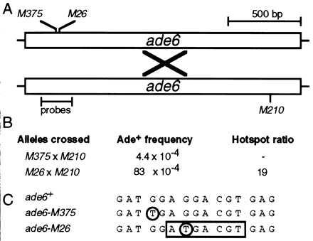
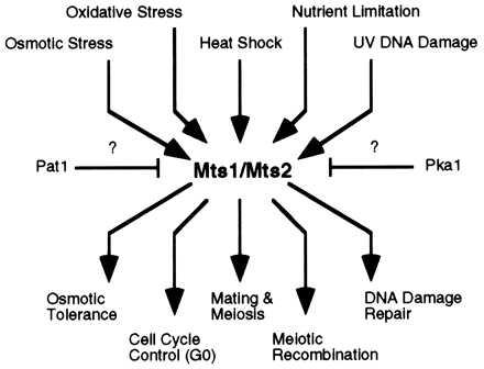
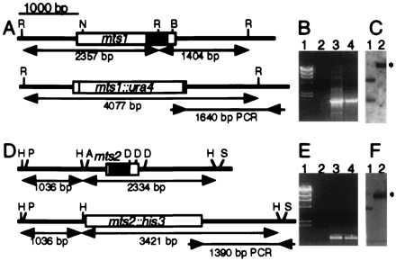
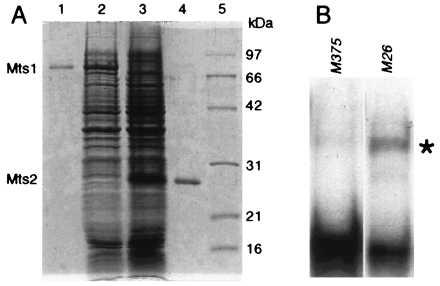
Homologous recombination hotspots increase the frequency of recombination in nearby DNA. The M26 hotspot in the ade6 gene of Schizosaccharomyces pombe is a meiotic hotspot with a discrete, cis-acting nucleotide sequence (5'-ATGACGT-3') defined by extensive mutagenesis. A heterodimeric M26 DNA binding protein, composed of subunits Mts1 and Mts2, has been identified and purified 40,000-fold. Cloning, disruption, and genetic analyses of the mts genes demonstrate that the Mts1/Mts2 heterodimer is essential for hotspot activity. This provides direct evidence that a specific trans-acting factor, binding to a cis-acting site with a unique nucleotide sequence, is required to activate this meiotic hotspot. Intriguingly, the Mts1/Mts2 protein subunits are identical to the recently described transcription factors Atf1 (Gad7) and Pcr1, which are required for a variety of stress responses. However, we report differential dependence on the Mts proteins for hotspot activation and stress response, suggesting that these proteins are multifunctional and have distinct activities. Furthermore, ade6 mRNA levels are equivalent in hotspot and nonhotspot meioses and do not change in mts mutants, indicating that hotspot activation is not a consequence of elevated transcription levels. These findings suggest an intimate but separable link between the regulation of transcription and meiotic recombination. Other studies have recently shown that the Mts1/Mts2 protein and M26 sites are involved in meiotic recombination elsewhere in the S. pombe genome, suggesting that these factors help regulate the timing and distribution of homologous recombination.


Figures

Figure 1
Figure 1
Hotspot activity of M26. (A) Schematic diagram of the ade6 gene showing positions of alleles and probes used for gel mobility shift experiments. Meiotic recombination (×) between a chromosome harboring the ade6-M26 or the ade6-M375 allele and a chromosome with the ade6-M210 allele generates a wild-type, selectable ade6+ gene. (B) Example of M26 recombination hotspot activity. Data are from Table 2. Recombination in test crosses containing M26 is increased up to 20-fold relative to recombination when using another allele, such as M375 (5). (C) DNA sequence surrounding the ade6-M26 recombination hotspot (7, 13). Both M375 and M26 are identical single base pair substitutions (circled) that generate stop codons. The M26 mutation creates a 7-bp site (box) that is bound by the Mts1/Mts2 heterodimer (15) and is required for hotspot activity in vivo (14).
Figure 2
Figure 2
Schematic representation of biochemical pathways by using Mts1 and Mts2 proteins. Upstream signals converge via the mitogen-activated protein (MAP) kinase cascade (19, 20) and likely via Pat1 and Pka kinases (–20). Signals diverge at Mts1 and Mts2 to activate the appropriate developmental responses. Some effector functions require Mts1, some require Mts2, and some require both Mts1 and Mts2.
Figure 3
Figure 3
Cloning, sequence analysis, and disruption of the mts1 and mts2 genes. (A and D) The genes encode proteins with bZIP dimerization and DNA binding motifs (shaded boxes) ≈50% identical to those in ATF/CREB proteins. R, EcoRI; N, EcoNI; B, BalI; H, HincII; P, HpaI; A, AatII; D, NdeIII; S, BstEII. Standard procedures were used to disrupt the genes. (B and E) Confirmation of gene disruption by PCR. Lanes: 1, λ DNA digested with HindIII; 2, PCR product of wild-type DNA; 3, PCR product of disrupted DNA (mts1-D15::ura4+ or mts2-D1::his3+); 4, PCR product of positive control DNA (plasmids used to obtain gene targeting fragments). Primers were as shown by inward-pointing arrows in A and D. (C and F) Confirmation of gene disruption by Southern blot analysis. Lanes: 1, wild-type DNA; 2, disrupted DNA (mts1-D15::ura4+ or mts2-D1::his3+). Restriction maps are shown in A and D, and the expected positions of altered restriction fragments are indicated (∗). DNA was digested with EcoRI and probed with the 4,077-bp EcoRI–EcoRI fragment for mts1 constructs or digested with HincII and probed with the 3,421-bp HincII-HincII fragment for mts2 constructs.
Figure 4
Figure 4
Hotspot-specific DNA binding activity of Mts1/Mts2 heterodimer purified from E. coli. (A) Coomassie-stained, 12% SDS/PAGE analysis of proteins. Lanes: 1, purified Mts1; 2, whole-cell lysate containing Mts1; 3, whole-cell lysate containing Mts2; 4, purified Mts2; 5, molecular mass standards. (B) Gel mobility shift assay by using a mixture of renatured Mts1 and Mts2 polypeptides. Position of M26-specific complex is indicated (∗). Gel shift conditions and probes were as described (15) and contained ≈5 nM probe, 25 nM Mts1, and 25 nM Mts2.
Figure 5
Figure 5
Sensitivity of mts mutants to osmotic stress. The indicated number of cells from healthy, log-phase cultures were spotted onto supplemented NBA minimal medium containing 1 M NaCl and were incubated for 3 days at 32°C before being photographed.

Comment in

References

    1. Mosig G. Proc Natl Acad Sci USA. 1966;56:1177–1183. - PMC - PubMed
    1. Wahls W P, Wallace L J, Moore P D. Cell. 1990;60:95–103. - PubMed
    1. Wahls W P, Wallace L J, Moore P D. Mol Cell Biol. 1990;10:785–793. - PMC - PubMed
    1. Voelkel-Meiman K, Keil R L, Roeder G S. Cell. 1987;48:1071–1079. - PubMed
    1. Gutz H. Genetics. 1971;69:331–337. - PMC - PubMed

Publication types

MeSH terms