

Tissue engineering of cartilage in space
- PMID: 9391122
- PMCID: PMC28402
- DOI: 10.1073/pnas.94.25.13885
Tissue engineering of cartilage in space
Abstract
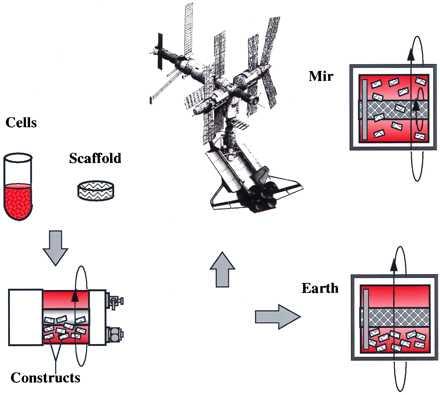
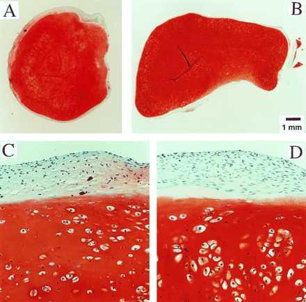
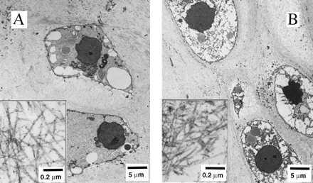
Tissue engineering of cartilage, i.e., the in vitro cultivation of cartilage cells on synthetic polymer scaffolds, was studied on the Mir Space Station and on Earth. Specifically, three-dimensional cell-polymer constructs consisting of bovine articular chondrocytes and polyglycolic acid scaffolds were grown in rotating bioreactors, first for 3 months on Earth and then for an additional 4 months on either Mir (10(-4)-10(-6) g) or Earth (1 g). This mission provided a unique opportunity to study the feasibility of long-term cell culture flight experiments and to assess the effects of spaceflight on the growth and function of a model musculoskeletal tissue. Both environments yielded cartilaginous constructs, each weighing between 0.3 and 0.4 g and consisting of viable, differentiated cells that synthesized proteoglycan and type II collagen. Compared with the Earth group, Mir-grown constructs were more spherical, smaller, and mechanically inferior. The same bioreactor system can be used for a variety of controlled microgravity studies of cartilage and other tissues. These results may have implications for human spaceflight, e.g., a Mars mission, and clinical medicine, e.g., improved understanding of the effects of pseudo-weightlessness in prolonged immobilization, hydrotherapy, and intrauterine development.
Figures
References
-
- Churchill S E. Fundamentals of Space Life Sciences. Malabar: Krieger; 1997.
-
- Nicogossian A E, Huntoon C L, Pool S L. Space Physiology and Medicine. Philadelphia: Lea and Febiger; 1994.
-
- Cann C E. In: Fundamentals of Space Life Sciences. Churchill S E, editor. Malabar: Krieger; 1997. pp. 83–103.
-
- Edgerton V R, Roy R R. In: Fundamentals of Space Life Sciences. Churchill S E, editor. Malabar: Krieger; 1997. pp. 105–120.
-
- Nerem R M. Ann Biomed Eng. 1991;19:529–545. - PubMed
Publication types
MeSH terms
Substances
LinkOut - more resources
Full Text Sources
Other Literature Sources
Miscellaneous
