. 1997 Dec 9;94(25):13897-902.
doi: 10.1073/pnas.94.25.13897.

Proliferation of adult T cell leukemia/lymphoma cells is associated with the constitutive activation of JAK/STAT proteins

Affiliations

Proliferation of adult T cell leukemia/lymphoma cells is associated with the constitutive activation of JAK/STAT proteins

S Takemoto et al. Proc Natl Acad Sci U S A. .

Abstract

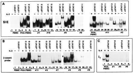
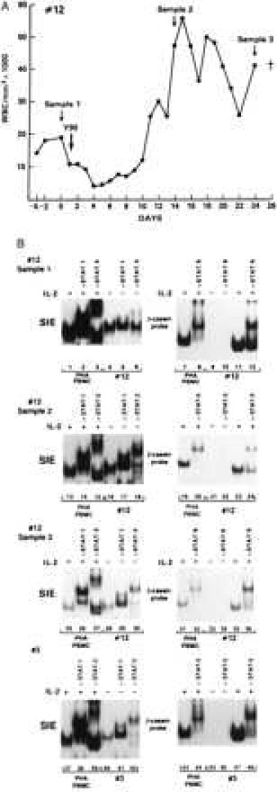
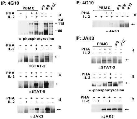
Human T cell leukemia/lymphotropic virus type I (HTLV-I) induces adult T cell leukemia/lymphoma (ATLL). The mechanism of HTLV-I oncogenesis in T cells remains partly elusive. In vitro, HTLV-I induces ligand-independent transformation of human CD4+ T cells, an event that correlates with acquisition of constitutive phosphorylation of Janus kinases (JAK) and signal transducers and activators of transcription (STAT) proteins. However, it is unclear whether the in vitro model of HTLV-I transformation has relevance to viral leukemogenesis in vivo. Here we tested the status of JAK/STAT phosphorylation and DNA-binding activity of STAT proteins in cell extracts of uncultured leukemic cells from 12 patients with ATLL by either DNA-binding assays, using DNA oligonucleotides specific for STAT-1 and STAT-3, STAT-5 and STAT-6 or, more directly, by immunoprecipitation and immunoblotting with anti-phosphotyrosine antibody for JAK and STAT proteins. Leukemic cells from 8 of 12 patients studied displayed constitutive DNA-binding activity of one or more STAT proteins, and the constitutive activation of the JAK/STAT pathway was found to persist over time in the 2 patients followed longitudinally. Furthermore, an association between JAK3 and STAT-1, STAT-3, and STAT-5 activation and cell-cycle progression was demonstrated by both propidium iodide staining and bromodeoxyuridine incorporation in cells of four patients tested. These results imply that JAK/STAT activation is associated with replication of leukemic cells and that therapeutic approaches aimed at JAK/STAT inhibition may be considered to halt neoplastic growth.


Figures

Figure 1
Figure 1
EMSA with β-casein, SIE, and Iɛ DNA oligonucleotides. EMSA obtained with SIE (A), β-casein (B), and Iɛ (C) probes. The specificity of the antibodies added to the DNA–protein complexes is indicated on the top of each panel. In some patients’ extracts the addition of the antibodies increased the signal, and in others a decrease binding was observed: in the case of STAT-5, compare lanes 6–8 and 12–14 of B, and in the case of STAT-1, compare lanes 25 and 28 of A. The reasons for this variability are unclear at present and do not appear to be related to the loading of different amounts of proteins or labeled DNA.
Figure 2
Figure 2
Persistence of STAT activation in cell extracts of patients 3 and 12. (A) White blood cell (WBC) counts over time in patient 12. The arrows indicate the times of sample collection. (B) EMSA on cell extracts from patient 12 at different sampling times (first 3 panels). The last panel represents the EMSA on patient 3 at the second sampling time.
Figure 3
Figure 3
Association of JAK/STAT proteins. Immunoprecipitation (IP) was performed with the anti-phosphotyrosine antibody 4G10, and immunoblotting was performed with antibodies specific for various STAT or JAK proteins (as indicated in ae). In the case of patient 3, protein extracts from the first sampling were used for the 4G10 IP, and those from the second sampling were used for the JAK3 IP. The amount of protein in sample 1 of patient 3 was half of that in sample 2. IP with anti-JAK3 antibodies was followed by immunoblotting with anti-STAT-3 antibody (f), anti-phosphotyrosine antibody (g), or anti-JAK3 antibody (h).
Figure 4
Figure 4
Analysis of DNA synthesis and DNA content in leukemic cells. (A) PI staining and FACS analysis (Upper) and BrdU staining of cells from patient 12 obtained at the second and third sampling times. (B) PI staining and FACS analysis of the cells from patients 2, 11, and 3 (second sampling time). BrdU staining of the cells from patient 3 at the second sampling time.

References

    1. Poiesz B J, Ruscetti F W, Gazdar A F, Bunn P A, Minna J D, Gallo R C. Proc Natl Acad Sci USA. 1980;77:7415–7419. - PMC - PubMed
    1. Gallo R C. Sci Am. 1986;255:88–98. - PubMed
    1. Hinuma Y, Nagata K, Hanaoka M, Nakai M, Matsumoto T, Kinoshita K I, Shirakawa S, Miyoshi I. Proc Natl Acad Sci USA. 1981;78:6476–6480. - PMC - PubMed
    1. Murphy E L, Hanchard B, Figueroa J P, Gibbs W N, Lofters W S, Campbell M, Goedert J J, Blattner W A. Int J Cancer. 1989;43:250–253. - PubMed
    1. Cavrois M, Wain-Hobson S, Gessain A, Plumelle Y, Wattel E. Blood. 1996;88:4646–4650. - PubMed

MeSH terms