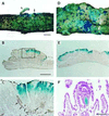
Polyclonal structure of intestinal adenomas in ApcMin/+ mice with concomitant loss of Apc+ from all tumor lineages
- PMID: 9391129
- PMCID: PMC28409
- DOI: 10.1073/pnas.94.25.13927
Polyclonal structure of intestinal adenomas in ApcMin/+ mice with concomitant loss of Apc+ from all tumor lineages
Abstract
When tumors form in intestinal epithelia, it is important to know whether they involve single initiated somatic clones. Advanced carcinomas in humans and mice are known to be monoclonal. However, earlier stages of tumorigenesis may instead involve an interaction between cells that belong to separate somatic clones within the epithelium. The clonality of early tumors has been investigated in mice with an inherited predisposition to intestinal tumors. Analysis of Min (multiple intestinal neoplasia) mice chimeric for a ubiquitously expressed cell lineage marker revealed that normal intestinal crypts are monoclonal, but intestinal adenomas frequently have a polyclonal structure, presenting even when very small as single, focal adenomas composed of at least two somatic lineages. Furthermore, within these polyclonal adenomas, all tumor lineages frequently lose the wild-type Apc allele. These observations can be interpreted by several models for clonal interaction within the epithelium, ranging from passive fusion within regions of high neoplastic potential to a requirement for active clonal cooperation.
Figures

References
-
- Ponder B A J, Wilkinson M M. J Natl Cancer Inst. 1986;77:967–973. - PubMed
-
- Fearon E R, Hamilton S R, Vogelstein B. Science. 1987;23:193–197. - PubMed
-
- Novelli M R, Williamson J A, Tomlinson I P, Elia G, Hodgson S V, Talbot I C, Bodmer F, Wright N A. Science. 1996;272:1187–1190. - PubMed
-
- Bjerknes M, Cheng H, Kim H, Schnitzler M, Gallinger S. Cancer Res. 1997;57:355–361. - PubMed
Publication types
MeSH terms
Substances
Grants and funding
LinkOut - more resources
Full Text Sources
Molecular Biology Databases

