. 1997 Dec 9;94(25):13950-4.
doi: 10.1073/pnas.94.25.13950.

Backtracking leukemia to birth: identification of clonotypic gene fusion sequences in neonatal blood spots

Affiliations

Backtracking leukemia to birth: identification of clonotypic gene fusion sequences in neonatal blood spots

K B Gale et al. Proc Natl Acad Sci U S A. .

Abstract

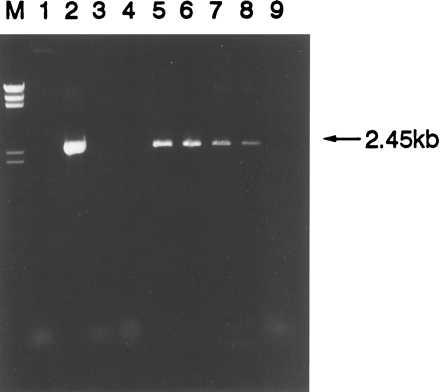
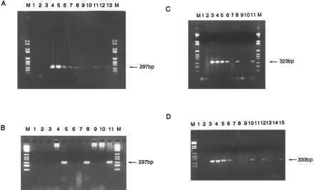
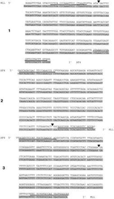
Epidemiological evidence has suggested that some pediatric leukemias may be initiated in utero and, for some pairs of identical twins with concordant leukemia, this possibility has been strongly endorsed by molecular studies of clonality. Direct evidence for a prenatal origin can only be derived by prospective or retrospective detection of leukemia-specific molecular abnormalities in fetal or newborn samples. We report a PCR-based method that has been developed to scrutinize neonatal blood spots (Guthrie cards) for the presence of numerically infrequent leukemic cells at birth in individuals who subsequently developed leukemia. We demonstrate that unique or clonotypic MLL-AF4 genomic fusion sequences are present and detectable in neonatal blood spots from individuals who were diagnosed with acute lymphoblastic leukemia at ages 5 months to 2 years and, therefore, have arisen during fetal hematopoiesis in utero. This result provides unequivocal evidence for a prenatal initiation of acute leukemia in young patients. The method should be applicable to other fusion genes in children with common subtypes of leukemia and will be of value in attempts to unravel the natural history and etiology of this major subtype of pediatric cancer.


Figures

Figure 1
Figure 1
Amplification of 2.45 kb product of MLL-AF4 rearrangement from RS4;11 cell line DNA and RS4;11 “imitation” Guthrie specimens by PCR. Lanes: M, DNA marker II; 1, no DNA control; 2, 100 ng positive control RS4;11 DNA; 3, 100 ng human placenta negative control DNA; 4, 100 ng normal genomic negative control DNA; 5–9, 10−1 through 10−5 dilutions of RS4;11 cells on imitation Guthrie specimens.
Figure 2
Figure 2
(A) PCR amplification of 297-bp-specific MLL-AF4 breakpoint region from diagnostic patient 1 leukemic DNA dilutions and two one-eighth segments of Guthrie spot. Lanes: M, DNA marker VI; 1, no DNA control; 2, 100 ng human placenta; 3, 100 ng patient 1 DNA with nonspecific primers; 4–10, patient 1 diagnostic DNA dilutions 100 ng–10 pg; 11, one-eighth segment patient 1 Guthrie; 12, “normal” Guthrie negative control; 13, one-eighth segment patient 1 Guthrie; M, DNA marker. (B) PCR on diagnostic DNA and third and fourth pieces of Guthrie spot from patient 1. Lanes: M, DNA marker VI; 1, no DNA control; 2, 100 ng human placenta negative control; 3 and 4, negative control one-eighth segments of normal Guthries 1 and 2; 5, third one-eighth segment of patient 1 Guthrie; 6 and 7, negative control normal Guthries 3 and 4; 8, fourth one-eighth segment of patient 1 Guthrie; 9 and 10, negative control normal Guthries 5 and 6; 11, patient 1 diagnostic DNA; M, DNA marker VI. (C) PCR amplification of specific 323-bp product of the AF4-MLL breakpoint region from patient 2 diagnostic DNA and two segments of Guthrie spot. Lanes: M, DNA marker VI; 1, no DNA control; 2, 100 ng patient 2 DNA with nonspecific primers; 3–6, patient 2 DNA 100 ng–0.1 ng; 7, 100 ng human placenta negative control; 8, one-eighth segment patient 2 Guthrie; 9 and 10, one-eighth segments of normal, negative control Guthries; 11, one-eighth segment of patient 2 Guthrie; M, DNA marker VI. (D) PCR amplification of 333-bp product specific to patient 3 AF4-MLL breakpoint region from diagnostic DNA and four one-eighth segments of Guthrie spot. Lanes: M, DNA marker VI; 1, no DNA control; 2, 100 ng human placenta negative control; 3–6, patient 3 diagnostic DNA 10 ng–10 pg; 7, patient 3 DNA with nonspecific primers; 8 and 9, one-eighth segments of negative control normal Guthries; 10–15, six one-eighth segments of patient 3 Guthrie spot.
Figure 3
Figure 3
Sequence of three patients (labeled 1, 2, and 3) diagnostic leukemic DNA (unshaded upper) and the (identical) MLL-AF4 or AF4-MLL fusion sequence from Guthrie DNA (shaded lower). Patient-specific oligonucleotide primers were derived from underlined sequence. (▾), MLL-AF4 or AF4-MLL breakpoint fusion for each patient.

References

    1. Ross J A, Davies S M, Potter J D, Robison L L. Epidemiol Rev. 1994;16:243–272. - PubMed
    1. Shu X-O, Ross J A, Pendergrass T W, Reaman G H, Lampkin B, Robison L L. J Natl Cancer Inst. 1996;88:24–31. - PubMed
    1. Greaves M F. Lancet. 1997;349:344–349. - PubMed
    1. Rowley J D. Semin Hematol. 1990;27:122–136. - PubMed
    1. Rabbitts T H. Nature (London) 1994;372:143–149. - PubMed

Publication types

MeSH terms

Associated data

LinkOut - more resources