. 1997 Dec 9;94(25):13973-8.
doi: 10.1073/pnas.94.25.13973.

Us9, a stable lysine-less herpes simplex virus 1 protein, is ubiquitinated before packaging into virions and associates with proteasomes

Affiliations

Us9, a stable lysine-less herpes simplex virus 1 protein, is ubiquitinated before packaging into virions and associates with proteasomes

R Brandimarti et al. Proc Natl Acad Sci U S A. .

Abstract

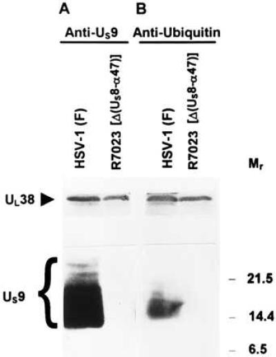
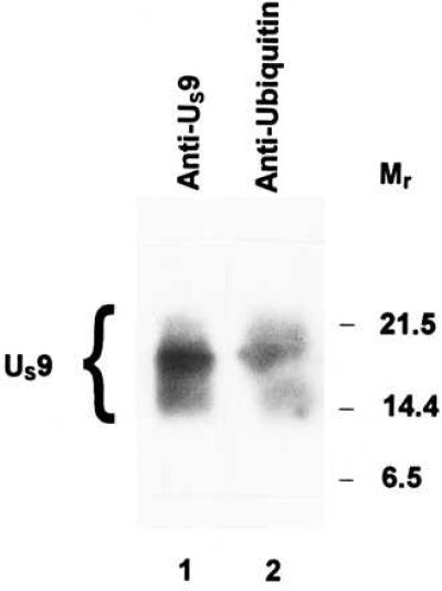
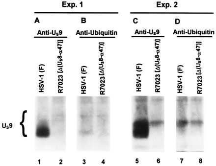
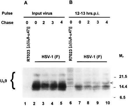
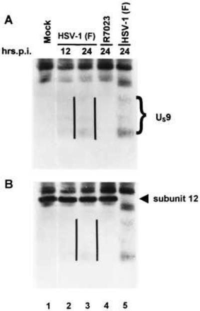
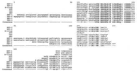
The US9 gene of herpes simplex virus 1 encodes a virion tegument protein with a predicted Mr of 10,000. Earlier studies have shown that the gene is not essential for viral replication in cells in culture. We report that (i) US9 forms in denaturing polyacrylamide gels multiple overlapping bands ranging in Mr from 12,000 to 25,000; (ii) the protein recovered from infected cells or purified virions reacts with anti-ubiquitin antibodies; (iii) autoradiographic images of US9 protein immunoprecipitated from cells infected with [35S]methionine-labeled virus indicate that the protein is stable for at least 4 h after entry into cells (the protein was also stable for at least 4 h after a 1-h labeling interval 12 h after infection); (iv) antibody to subunit 12 of proteasomes pulls down US9 protein from herpes simplex virus-infected cell lysates; and (v) the US9 gene is highly conserved among the members of the alpha subfamily of herpes viruses, and the US9 gene product lacks lysines. We conclude that US9 is a lysine-less, ubiquitinated protein that interacts with the ubiquitin-dependent pathway for degradation of proteins and that this function may be initiated at the time of entry of the virus into the cell.


Figures

Figure 1
Figure 1
Diagrammatic representation of the sequence arrangement of the HSV-1 genome and of the location of the US9 gene. Line 1, representation of the HSV-1 genome. The single lines represent the unique long (UL) and unique short (US) DNA sequences, whereas the terminal repeats are shown as open rectangles. Line 2, expansion of the domain containing the US9 gene. Line 3, polarity and domain of US9 mRNA. The US11 and US12 mRNAs (not shown) are coterminal with the US10 mRNA. Line 4, representation of the US9 codons 1–59 amplified by PCR and fused to GST for production of antibody.
Figure 2
Figure 2
Immunoblots of infected cell proteins, electrophoretically separated on a denaturing gel, and allowed to react with rabbit polyclonal antibodies to US9 protein (A) or to ubiquitin (B). HEp-2 cells infected with HSV-1(F) or with R7023 were harvested at 22 h, solubilized, subjected to electrophoresis on a denaturing gel, transferred to nitrocellulose, and allowed to react with the indicated antibodies as described in Materials and Methods. The UL38 protein served as a loading control. Molecular weights (in thousands) are shown on the right.
Figure 3
Figure 3
Immunoblots of virion proteins electrophoretically separated on a denaturing gel and allowed to react with rabbit polyclonal antibodies to US9 protein (lane 1) or to ubiquitin (lane 2). The virions were purified on dextran gradients from HEp-2 cells infected with HSV-1(F) (23), solubilized, subjected to electrophoresis, allowed to react with the indicated antibodies, and detected with the enhanced chemiluminescent detection system. Molecular weights in thousands are shown on the right.
Figure 4
Figure 4
Autoradiographic image of electrophoretically separated [35S]methionine-labeled proteins, immunoprecipitated with a rabbit polyclonal antibody to US9 protein (A and C), or to ubiquitin (B and D) from infected cell lysates. HEp-2 cells were exposed for 90 min to [35S]methionine-labeled HSV-1(F) or R7023 generated as described in Material and Methods and then incubated for an additional 30 min in medium containing excess unlabeled methionine. The cells were lysed, and immune complexes containing US9 protein were subjected to electrophoresis in denaturing gels and autoradiography on X-Omat AR film.
Figure 5
Figure 5
Autoradiographic image of [35S]methionine-labeled proteins immunoprecipitated with a rabbit polyclonal antibody to US9 protein from infected cell lysates and subjected to electrophoresis on a denaturing gel. (A) HEp-2 cells were incubated at 37°C for 0, 2, 3, and 4 h after exposure to radiolabeled HSV-1(F) or R7023 at 10°C for 90 min and then lysed and allowed to react with the antibody. The immune complexes were subjected to electrophoresis on a denaturing gel, transferred to a nitrocellulose sheet, and subjected to autoradiography. (B) At 12 h after infection with HSV-1(F) or R7023, HEp-2 cells were labeled for 1 h with [35S]methionine as described in Materials and Methods and then incubated in medium containing excess unlabeled methionine and harvested immediately or at hourly intervals for 4 h. The cells were subjected to the same procedures as described for A. Molecular weights (in thousands) are shown on the right.
Figure 6
Figure 6
Immunoblot of infected cell proteins immunoprecipitated with a monoclonal antibody to proteasome, electrophoretically separated on a denaturing gel and allowed to react with a rabbit polyclonal antibody to US9 protein (A), photographed, and then allowed to react with a monoclonal antibody to subunit 12 of proteasome (B). The parallel lines are to the right of the US9 protein bands.
Figure 7
Figure 7
Sequence alignment of the US9 protein homologs of HSV-1(F), HSV-2(G), simian B virus, equine herpes viruses 1 and 4, bovine herpes virus 1, and feline herpes virus 1, simian varicella, varicella-zoster, and pseudorabies viruses generated by the lineup and pretty programs of the Genetics Computer Group software. The last line shows the consensus sequence. Residues conserved among at least five of the sequences are shown as capitalized letters, whereas those conserved among at least seven of the sequences are shown as bold letters. Dots indicate gaps inserted by the program to create the best alignment.

References

    1. McGeoch D J, Dolan A, Donald S, Rixon F J. J Mol Biol. 1985;181:1–13. - PubMed
    1. Frame M C, McGeoch D J, Rixon F J, Orr A C, Marsden H S. Virology. 1986;150:321–332. - PubMed
    1. Roizman B, Sears A E. In: Fields’ Virology. 3rd Ed. Fields B N, Knipe D M, Howley P, Chanock R M, Hirsch M S, Melnick J L, Monath T P, Roizman B, editors. New York: Raven; 1996. pp. 2231–2295.
    1. Longnecker R, Roizman B. J Virol. 1986;58:583–591. - PMC - PubMed
    1. Jentsch F. Annu Rev Genet. 1992;26:179–207. - PubMed

Publication types

MeSH terms

LinkOut - more resources