

Chronic stress alters synaptic terminal structure in hippocampus
- PMID: 9391142
- PMCID: PMC28422
- DOI: 10.1073/pnas.94.25.14002
Chronic stress alters synaptic terminal structure in hippocampus
Abstract
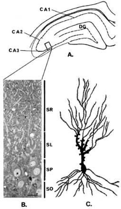
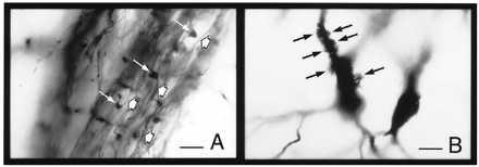
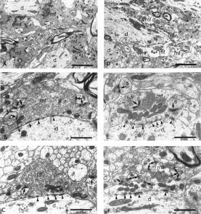
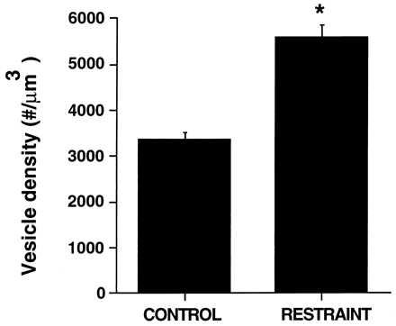
Repeated psychosocial or restraint stress causes atrophy of apical dendrites in CA3 pyramidal neurons of the hippocampus, accompanied by specific cognitive deficits in spatial learning and memory. Excitatory amino acids mediate this atrophy together with adrenal steroids and the neurotransmitter serotonin. Because the mossy fibers from dentate granule neurons provide a major excitatory input to the CA3 proximal apical dendrites, we measured ultrastructural parameters associated with the mossy fiber-CA3 synapses in control and 21-day restraint-stressed rats in an effort to find additional morphological consequences of stress that could help elucidate the underlying anatomical as well as cellular and molecular mechanisms. Although mossy fiber terminals of control rats were packed with small, clear synaptic vesicles, terminals from stressed animals showed a marked rearrangement of vesicles, with more densely packed clusters localized in the vicinity of active zones. Moreover, compared with controls, restraint stress increased the area of the mossy fiber terminal occupied by mitochondrial profiles and consequently, a larger, localized energy-generating capacity. A single stress session did not produce these changes either immediately after or the next day following the restraint session. These findings provide a morphological marker of the effects of chronic stress on the hippocampus that points to possible underlying neuroanatomical as well as cellular and molecular mechanisms for the ability of repeated stress to cause structural changes within the hippocampus.
Figures
Similar articles
-
Effects of chronic stress on hippocampal long-term potentiation.Hippocampus. 2002;12(2):245-57. doi: 10.1002/hipo.1116. Hippocampus. 2002. PMID: 12000121
-
Stress effects on morphology and function of the hippocampus.Ann N Y Acad Sci. 1997 Jun 21;821:271-84. doi: 10.1111/j.1749-6632.1997.tb48286.x. Ann N Y Acad Sci. 1997. PMID: 9238211 Review.
-
Chronic restraint stress induces changes in synapse morphology in stratum lacunosum-moleculare CA1 rat hippocampus: a stereological and three-dimensional ultrastructural study.Neuroscience. 2006 Jun 30;140(2):597-606. doi: 10.1016/j.neuroscience.2006.02.072. Neuroscience. 2006. PMID: 16600515
-
Experimental diabetes in rats causes hippocampal dendritic and synaptic reorganization and increased glucocorticoid reactivity to stress.Proc Natl Acad Sci U S A. 2000 Sep 26;97(20):11056-61. doi: 10.1073/pnas.97.20.11056. Proc Natl Acad Sci U S A. 2000. PMID: 11005876 Free PMC article.
-
Prevention of stress-induced morphological and cognitive consequences.Eur Neuropsychopharmacol. 1997 Oct;7 Suppl 3:S323-8. doi: 10.1016/s0924-977x(97)00064-3. Eur Neuropsychopharmacol. 1997. PMID: 9405958 Review.
Cited by
-
Regular moderate or intense exercise prevents depression-like behavior without change of hippocampal tryptophan content in chronically tryptophan-deficient and stressed mice.PLoS One. 2013 Jul 4;8(7):e66996. doi: 10.1371/journal.pone.0066996. Print 2013. PLoS One. 2013. PMID: 23861751 Free PMC article.
-
Fighting with Spirits: Migration Trauma, Acculturative Stress, and New Sibling Transition-A Clinical Case Study of an 8-Year-Old Girl with Absence Epilepsy.Cult Med Psychiatry. 2015 Dec;39(4):698-724. doi: 10.1007/s11013-015-9438-7. Cult Med Psychiatry. 2015. PMID: 25670159
-
A randomized, placebo-controlled proof-of-concept, crossover trial of phenytoin for hydrocortisone-induced declarative memory changes.J Affect Disord. 2013 Sep 5;150(2):551-8. doi: 10.1016/j.jad.2013.01.038. Epub 2013 Feb 28. J Affect Disord. 2013. PMID: 23453674 Free PMC article. Clinical Trial.
-
Unpredictable Chronic Stress Alters Adenosine Metabolism in Zebrafish Brain.Mol Neurobiol. 2016 May;53(4):2518-28. doi: 10.1007/s12035-015-9270-7. Epub 2015 Jun 17. Mol Neurobiol. 2016. PMID: 26081145
-
Role for NUP62 depletion and PYK2 redistribution in dendritic retraction resulting from chronic stress.Proc Natl Acad Sci U S A. 2014 Nov 11;111(45):16130-5. doi: 10.1073/pnas.1418896111. Epub 2014 Oct 27. Proc Natl Acad Sci U S A. 2014. PMID: 25349423 Free PMC article.
References
Publication types
MeSH terms
Substances
Grants and funding
LinkOut - more resources
Full Text Sources
Other Literature Sources
Miscellaneous
