. 1997 Dec 15;139(6):1373-82.
doi: 10.1083/jcb.139.6.1373.

CENP-E function at kinetochores is essential for chromosome alignment

Affiliations

CENP-E function at kinetochores is essential for chromosome alignment

B T Schaar et al. J Cell Biol. .

Abstract

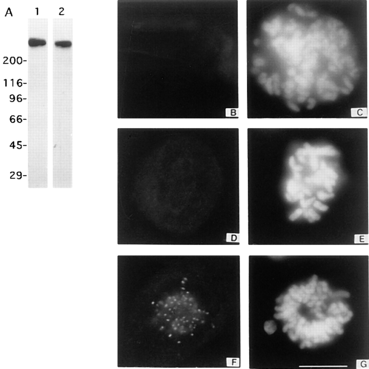
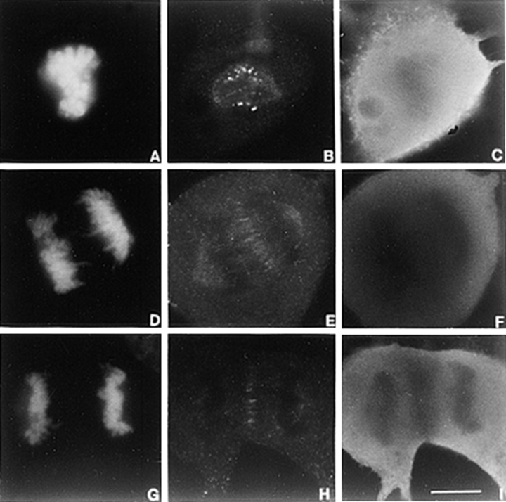
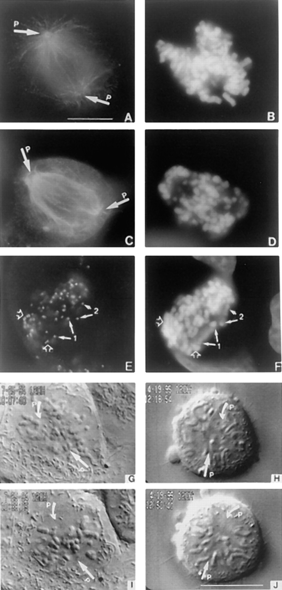
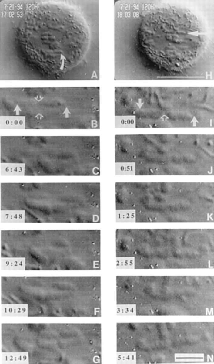
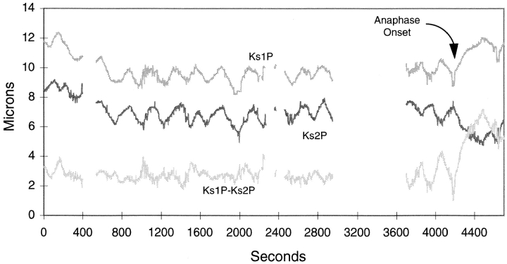
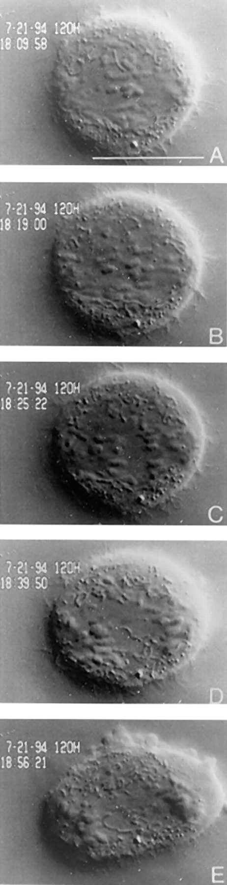
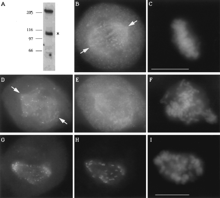
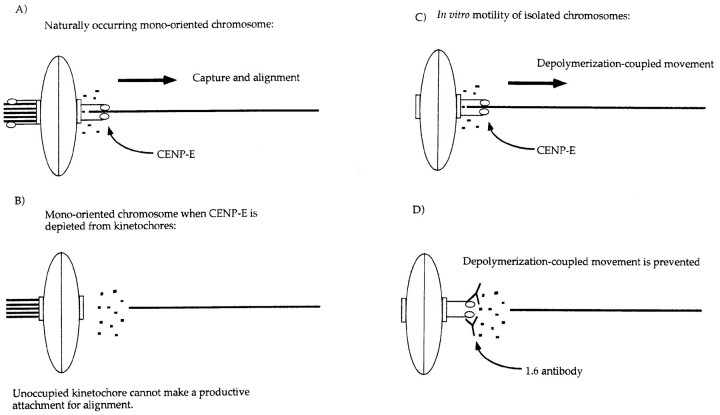
CENP-E is a kinesin-like protein that binds to kinetochores and may provide functions that are critical for normal chromosome motility during mitosis. To directly test the in vivo function of CENP-E, we microinjected affinity-purified antibodies to block the assembly of CENP-E onto kinetochores and then examined the behavior of these chromosomes. Chromosomes lacking CENP-E at their kinetochores consistently exhibited two types of defects that blocked their alignment at the spindle equator. Chromosomes positioned near a pole remained mono-oriented as they were unable to establish bipolar microtubule connections with the opposite pole. Chromosomes within the spindle established bipolar connections that supported oscillations and normal velocities of kinetochore movement between the poles, but these bipolar connections were defective because they failed to align the chromosomes into a metaphase plate. Overexpression of a mutant that lacked the amino-terminal 803 amino acids of CENP-E was found to saturate limiting binding sites on kinetochores and competitively blocked endogenous CENP-E from assembling onto kinetochores. Chromosomes saturated with the truncated CENP-E mutant were never found to be aligned but accumulated at the poles or were strewn within the spindle as was the case when cells were microinjected with CENP-E antibodies. As the motor domain was contained within the portion of CENP-E that was deleted, the chromosomal defect is likely attributed to the loss of motor function. The combined data show that CENP-E provides kinetochore functions that are essential for monopolar chromosomes to establish bipolar connections and for chromosomes with connections to both spindle poles to align at the spindle equator. Both of these events rely on activities that are provided by CENP-E's motor domain.


Figures

Figure 1
Figure 1
Depletion of CENP-E from kinetochores by microinjection of rabbit polyclonal antibodies directed against CENP-E. (A) Western blots of mitotic HeLa lysates probed with stalk HX-1 (lane 1) and carboxy-terminal DraB (lane 2) affinity-purified antibodies. (B, D, F, and I) Monoclonal antibody mAb177 (7) staining of Hela cells injected with 0.5 mg/ml of DraB antibodies (B and D), an uninjected prometaphase cell (F). mAb177 was visualized with FITC-conjugated anti–mouse antibodies. (C, E, and G) DAPI staining to visualize chromosomes. Images from B, D, and F were exposed for identical times. Bar, 10 μm.
Figure 2
Figure 2
Preimmune antibodies injected at a 40-fold higher concentration have no effect on mitotic progression or CENP-E localization. Preimmune HX-1 antibodies were injected in an identical manner to immune antibodies. DNA was detected with DAPI (A, D, and G) CENP-E was detected with mAb 177 (B, E, and H) and injected antibodies were detected with anti–rabbit Ig secondary antibodies (C, F, and I). No aberrations were seen in the organization of the chromosomes during alignment (A). CENP-E localized normally in these cells during anaphase (E and H). Anaphase cells as shown in D through F and G through I were never observed in immune HX-1 nor DraB antibody– injected cells. Bar, 10 μM.
Figure 3
Figure 3
Loss of CENP-E disrupts chromosome alignment. (A) Tubulin staining of a normal prometaphase U2OS cell and (C) one blocked in mitosis by injection of carboxy-terminal DraB antibodies. (E) Mitotically blocked U2OS cell stained with anti-centromere autoimmune serum to reveal presence of paired centromeres (open arrows) and separated centromeres (chromosomes 1 and 2, solid arrows). (B, D, and F) Chromosomes visualized by DAPI. (G through J) Selected VE-DIC images from a videotape showing a normal U2OS cell aligns its chromosomes in ∼30 min (G and I) at room temperature. Chromosomes that lacked CENP-E failed to align during the same period (H and J) and never aligned. Arrows (P) denote positions of the separated poles. Bars, 10 μm.
Figure 4
Figure 4
VE-DIC microscopy of chromosome dynamics in microinjected cells. The metacentric chromosome in A (curved arrow) exhibited uncoordinated movements shown in B through G. Centromeres of this chromosome are indicated with solid arrows, and the telomere connections are indicated by an open arrow in B. The centromeres of this chromosome stretch and compress the chromosome to show separation between the centromeres and sister chromatid connections (C and E) Time of observation is indicated in the lower left hand corner (min:s). The acrocentric chromosome in H, indicated by an arrow, was also followed. This chromosome only has a single telomere holding the two sister chromatids together (open arrow in I). The uncoordinated movements of the centromeres (solid arrows in I) stretch and bend the chromosome into a “u” shape (K and M) twice during the time of observation. Bars: (H) 10 μm; (N) 2 μm.
Figure 5
Figure 5
Plot of kinetochore to pole distance over time for two kinetochores (Ks1P and Ks2P) of a stretched chromosome in a cell that was injected with anti–CENP-E antibodies. Gaps in the plot indicate when kinetochores went out of the focal plane. Ks1P-Ks2P indicated the distance between the two kinetochores. Also indicated is the time when anaphase onset occurred and sister chromatids separated at their telomeres.
Figure 6
Figure 6
Separation of bipolar chromosomes depleted of CENP-E is not due to a precocious anaphase. The cell viewed in Fig. 4 was followed for 2 h after which the telomere connections of unzipped chromosomes synchronously disjoined, and sister chromatids moved towards opposite poles in a manner indistinguishable from anaphase in an uninjected cell. The cell also underwent normal anaphase B events (E) and cytokinesis. Bar, 10 μm.
Figure 7
Figure 7
Kinetochores saturated with a truncated CENP-E mutant that lacked its motor domain fail to align chromosomes. (A) Western blot of a transfected HeLa lysate probed with rat anti-GFP antibody to detect the 220-kD GFP: CENP-E NΔ 803. The asterisk denotes an endogenous 110-kD protein that crossreacts with the GFP antibody that is also present in untransfected lysates (not shown). (B) Localization of CENP-E on metaphase chromosomes of an untransfected cell by rabbit anti–CENP-E neck antibodies. (D) Direct visualization of GFP:CENP-E distribution in a transfected mitotic cell, (E) that was also stained with rabbit anti–CENP-E neck antibodies to detect endogenous CENP-E. (G) GFP:CENP-E colocalizes with ACA staining (H). (C, F, and I) DAPI staining. Arrows in B, D, and G denote the two separated spindle poles (P). Cells in B and E are from the same coverslip, and exposure times were identical. Bar, 10 μm.
Figure 8
Figure 8
Diagram illustrating the similar effects of inhibiting CENP-E function in vivo (this study) and in vitro (Lombillo et al., 1995). (A) Mono-oriented chromosomes (depicted as an oval) occur as a natural consequence of the random positioning of chromosomes after nuclear envelope breakdown. A chromosome that is captured and drawn close to one pole has one kinetochore (rectangle) saturated by microtubules from the pole it faces due to its proximity to that pole. The sister kinetochore must attain a microtubule connection from the opposite pole to congress the chromosome to the spindle equator. Because of the increase of catastrophic shrinkage during mitosis, a microtubule spanning the full length of the spindle (15–20 μm in the cells used in this study) is a rare event. We postulate that this creates a situation in which CENP-E (drawn as tadpoles sticking out of the kinetochore) is necessary to stabilize this connection by applying a poleward vector of force to the newly captured microtubule, creating tension along the microtubule as well as across the centromere. Once the microtubule is stabilized, other kinetochore proteins and motors engage the microtubule to induce movement. This model is based on the following observations: (a) when CENP-E is experimentally depleted from the kinetochore, mono-oriented chromosomes are abundant at both poles, a phenomenon not seen in control cells, and these mono-oriented chromosomes do not move from the attached pole to the metaphase plate; and (b) CENP-E–depleted chromosomes move with velocities comparable to controls.A role for CENP-E in maintaining attachment to a shrinking microtubule is also seen in the in vitro studies of Lombillo et al. (1995). (C) In the in vitro system, isolated chromosomes (ovals) capture the plus ends of microtubules nucleated from Tetrahymena pellicles at their kinetochores (rectangles) and mediate depolymerization-coupled movement when the tubulin is diluted in the absence of nucleotides. This movement is believed to be mediated in part by the motor domain of CENP-E (tadpoles), because if the chromosomes are first incubated with a polyclonal that spans a portion of the neck and stalk region of CENP-E, the chromosome detaches from the microtubule when buffer is perfused into the chamber to dilute the tubulin in (D). This is analogous to the situation that the unoccupied kinetochore of a mono-oriented chromosome that is depleted of CENP-E faces. Transient microtubules might be made but they cannot be stabilized in the absence of CENP-E.

References

    1. Alexander SP, Rieder CL. Chromosome motion during attachment to the vertebrate spindle: initial saltatory-like behavior of chromosomes and quantitative analysis of force production by nascent kinetochore fibers. J Cell Biol. 1991;113:805–815. - PMC - PubMed
    1. Ault JG, Nicklas RB. Tension, microtubule rearrangements, and the proper distribution of chromosomes in mitosis. Chromosoma (Berl) 1989;98:33–39. - PubMed
    1. Bernat RL, Borisy GG, Rothfield NF, Earnshaw WC. Injection of anticentromere antibodies in interphase disrupts events required for chromosome movements at mitosis. J Cell Biol. 1990;111:1519–1533. - PMC - PubMed
    1. Cassimeris L, Rieder CL, Salmon ED. Microtubule assembly and kinetochore directional instability in vertebrate monopolar spindles: implications for the mechanism of chromosome congression. J Cell Sci. 1994;107:285–297. - PubMed
    1. Chalfie M, Tu Y, Euskirschen G, Ward WW, Prasher DC. Green fluorescent protein as a marker for gene expression. Science. 1993;263:802–805. - PubMed

Publication types

MeSH terms