. 1997 Dec 15;139(6):1383-96.
doi: 10.1083/jcb.139.6.1383.

Probing the architecture of a simple kinetochore using DNA-protein crosslinking

Affiliations

Probing the architecture of a simple kinetochore using DNA-protein crosslinking

C W Espelin et al. J Cell Biol. .

Abstract

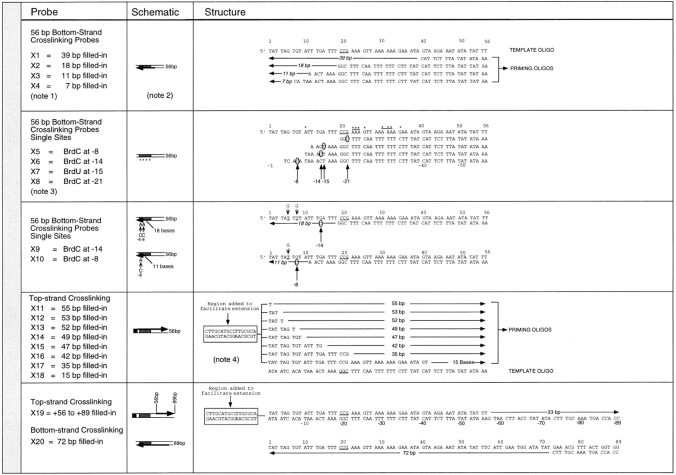
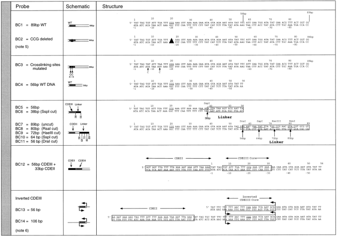
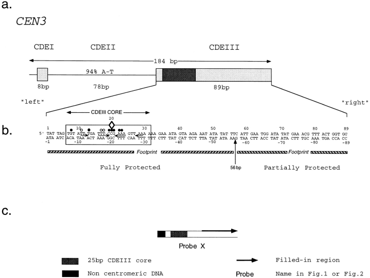
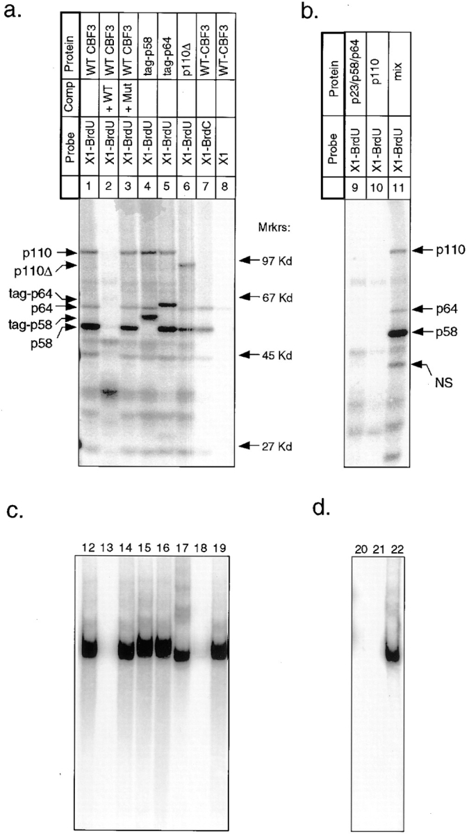
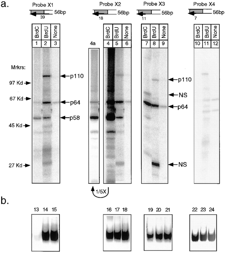
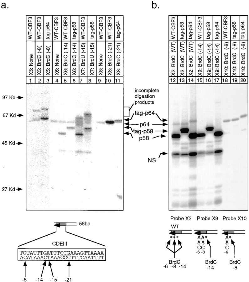
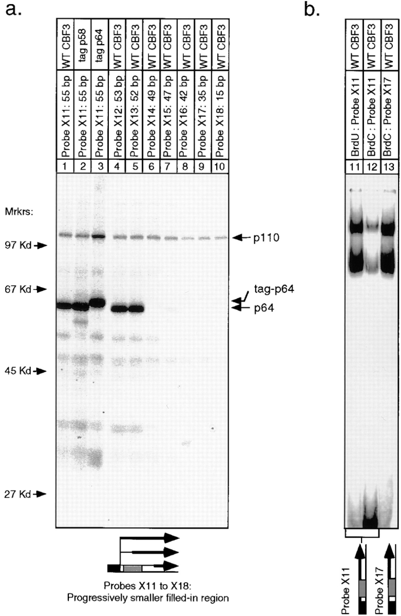
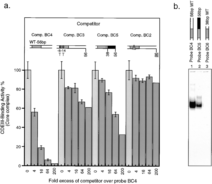
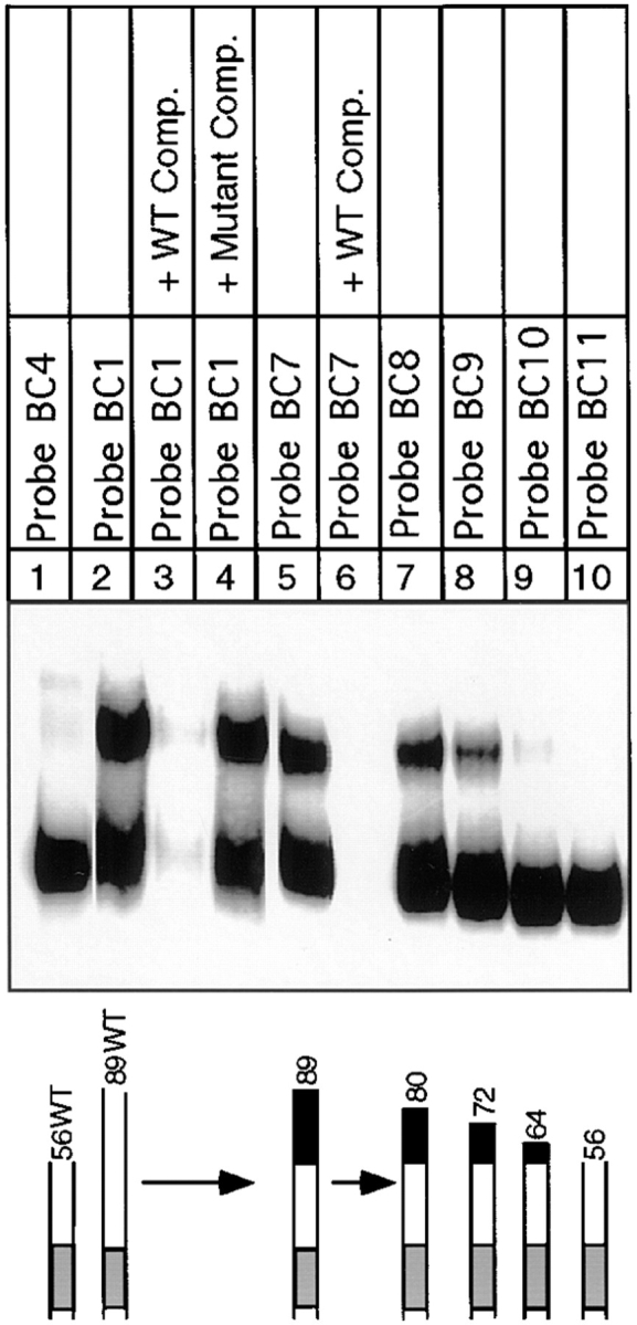
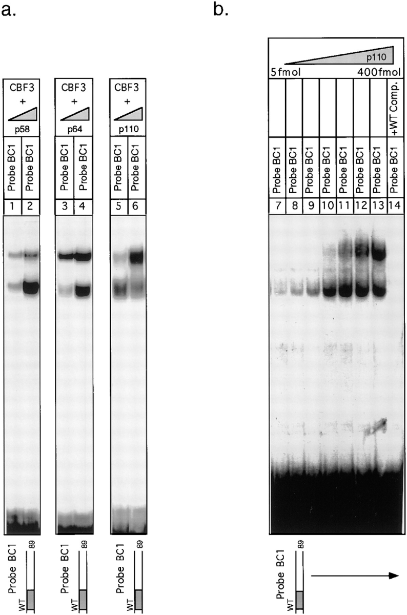
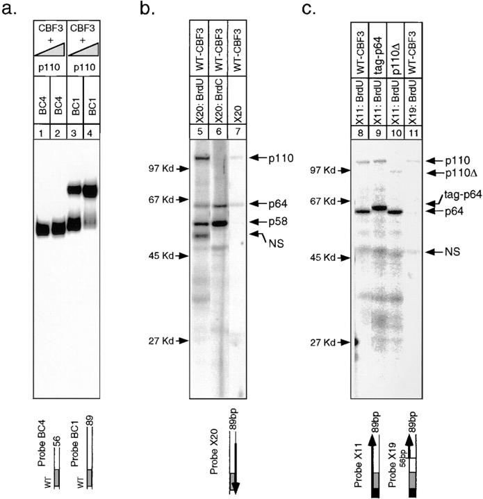
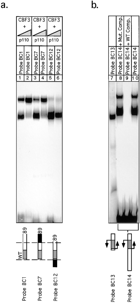
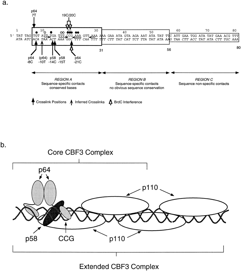
In budding yeast, accurate chromosome segregation requires that one and only one kinetochore assemble per chromosome. In this paper, we report the use of DNA-protein crosslinking and nondenaturing gel analysis to study the structure of CBF3, a four-protein complex that binds to the essential CDEIII region of Saccharomyces cerevisiae centromeres. We find that three subunits of CBF3 are in direct contact with CDEIII over a region of DNA that spans 80 bp. A highly asymmetric core complex containing p58(CTF13) p64(CEP3) and p110(NDC10) in direct contact with DNA forms at the genetically defined center of CDEIII. This core complex spans approximately 56 bp of CEN3. An extended complex comprising the core complex and additional DNA-bound p110(NDC10) also forms. It spans approximately 80 bp of DNA. CBF3 makes sequence-specific and -nonspecific contacts with DNA. Both contribute significantly to the energy of CBF3-DNA interaction. Moreover, important sequence-specific contacts are made with bases that are not conserved among yeast centromeres. These findings provide a foundation for understanding the organization of the CBF3-centromere complex, a structure that appears to initiate the formation of microtubule attachment sites at yeast kinetochores. These results also have implications for understanding centromere-binding proteins in higher cells.


Figures

Figure 1
Figure 1
Structure of crosslinking probes. (note 1) “Filled-in” refers to the enzymatic synthesis of double-stranded DNA from a single-strand template using BrdU or BrdC, radionucleotide, and other deoxynucleotides. (note 2) Dark arrow indicates the region of DNA that was synthesized. (note 3) Numbers (−8, −14, −15, and −21) refer to the precise positions of BrdU/BrdC substitutions. Asterisks indicate additional specific sites substituted with BrdU or BrdC. (note 4) The box shows additional noncentromeric DNA that was added to promote oligo annealing.
Figure 2
Figure 2
Structure of bandshift-competition probes. (note 5) See Sorger et al. (1995). (note 6) See Murphy et al. (1991).
Figure 3
Figure 3
Structure of CEN3 sequences used in this study. (a) Boxes indicate CDEI and CDEIII sequences. (b) The boxed bases denote the conserved core of CDEIII as determined by Hegemann et al. (1988). A diamond marks the location of the essential CCG motif. Solid circles indicate bases conserved among all 16 S. cerevisiae centromeres. Open circles indicate bases conserved in 15 of 16 centromeres. The top strand of DNA is numbered from 1 to 89 and the corresponding bottom strand is numbered from −1 to −89. “Left” refers to bases that are CDEII proximal, and “right” refers to the bases that are centromere distal. (c) This schematic is used in subsequent figures to represent various crosslinking probes. The heavy arrow indicates the region of single-stranded DNA in which BrdC or BrdU, 32PαdATP and native bases were incorporated by enzymatic fill-in.
Figure 4
Figure 4
UV crosslinking of rCBF3 to the bottom strand of radiolabeled 56-bp CDEIII DNA. (a) rCBF3 proteins labeled by DNA crosslinking probes (as indicated) were run on an SDS-containing gel. The positions of molecular weight markers are shown. The types of crosslinking probe and CBF3 preparation used in each lane are indicated above the gel. The specificity of CBF3 binding was tested by including a 200-fold excess of unlabeled wild-type (WT; probe BC1) or mutant CDEIII competitor (Mut; probe BC2) DNA in the binding reaction. Some reactions contain rCBF3 with p58 (tag-p58) or p64 (tag-p64) proteins that are linked at their amino termini to a short epitope tag. p110Δ refers to rCBF3 containing p110 protein with a carboxy-terminal 122 amino acid deletion. For a complete description of probe X1 see Figs. 1 and 3. (b) Crosslinking reactions containing recombinant p64/p58/23 (lane 9), recombinant p110 protein (lane 10), or a mixture of these two extracts (lane 11) was analyzed as above. (c) One fifth of each crosslinking reaction from a was analyzed on bandshift gels (lanes 12–19). (d) One fifth of each crosslinking reaction from b was analyzed on bandshift gels (lanes 20–22).
Figure 5
Figure 5
UV crosslinking of rCBF3 to progressively shorter labeled regions of the bottom strand of the 56-bp CDEIII DNA. (a) Crosslinking reactions using differently labeled probes (as indicated) were analyzed on SDS-containing gels. Each set of three reactions was performed in parallel. The positions of the filled-in regions as well as the type of bromine-modified base (BrdU or BrdC) are indicated. The probe numbers and schematics are keyed to the full descriptions in Fig. 1. Lane 4 a is exposed only one fifth as long as lane 4 to allow better visualization of the crosslinked bands. (b) One fifth of each crosslinking reaction from a was separately analyzed on bandshift gels (lanes 13–24).
Figure 6
Figure 6
Localization of protein–DNA crosslinks on the bottom strand of 56-bp CDEIII DNA. (a) The site-specific crosslinkers, BrdC or BrdU, were incorporated at the positions indicated into the bottom strand of DNA. In each case “none” refers to the synthesis of probe in the absence of BrdC or BrdU. (b) Site-specific BrdC crosslinkers (indicated by asterisks) were incorporated using mutant top-strand oligos and appropriate bottom-strand oligos by enzymatic extension (see Fig. 1 for details about the probes). Crosslinking reactions using probes described in a and b were analyzed on SDS-containing gels as in Fig. 4. Faint labeling of p58 in lanes 18–20 are a result of non-BrdC–dependent binding as in Fig. 5, lane 12.
Figure 7
Figure 7
UV crosslinking of rCBF3 to progressively shorter labeled regions of the top strand of the 56-bp CDEIII DNA. (a) BrdU was incorporated into each crosslinking probe as in Fig. 5. Reactions were carried out in the presence of either wild-type or tagged CBF3 proteins and analyzed on an SDS-containing gel. (b) Probes with either BrdU or BrdC incorporated into the entire top strand (+2 to +56; lanes 11 and 12) or a probe with BrdC incorporated only to the right of the CCG (+22 to 56; lane 13) were mixed with rCBF3 and analyzed on bandshift gels.
Figure 8
Figure 8
Analysis of centromeres containing point mutations in sequences that crosslink to CBF3 subunits. (a) rCBF3 and radiolabeled probe BC4 were mixed with a 4- to 200-fold excess of unlabeled centromeric DNA competitors (as indicated) and analyzed on bandshift gel. Values are expressed as a percentage of core complex CDEIII binding observed in the absence of competitor. Error bars represent the range of values obtained in duplicate experiments. (b) rCBF3 was mixed with radiolabeled 56-bp wild-type DNA (Probe BC4; lane 1), linker-modified 56-bp DNA (Probe BC5; lane 2), or 38-bp probe DNA (Probe BC6; lane 3), and CDEIII binding was analyzed on bandshift gels.
Figure 9
Figure 9
Analysis of extended complex formation on progressively shorter CDEIII probes. rCBF3 and the indicated probes were analyzed by bandshift gel. Specificity was demonstrated by adding excess unlabeled wild-type (lanes 3 and 6) or mutant (lane 4) CDEIII competitor DNA.
Figure 10
Figure 10
Effect of varying the stoichiometry of CBF3 subunits on CBF3–DNA complex formation. (a) Extract containing recombinant p110 and p64 (lanes 1 and 2), p110, p58, and p23 (lanes 3 and 4) or p64, p58, and p23 (lanes 5 and 6) were mixed with 10 to 20 fmol or with 60 to 120 fmol of p58/p23, p64, or p110 and complexes analyzed on bandshift gels. (b) Extract from ndc10-42 cells (80 μg) was mixed with 5, 10, 20, 50, 100, 200, or 400 fmoles of recombinant p110 (lanes 7–14). Specificity was demonstrated by adding excess wild-type CDEIII competitor DNA (lane 14).
Figure 11
Figure 11
Analysis of p110 binding to 89-bp CDEIII probe. (a) rCBF3 on its own or mixed with excess p110 (as indicated) was bound to a 56-bp (lanes 1 and 2) or 89-bp (lanes 3 and 4) probe and analyzed on a bandshift gel. (b) 89-bp probes containing either BrdU, BrdC, or no crosslinkers in the bottom strand were reacted with rCBF3 and analyzed on an SDS-containing gel. (c) 89-bp probes with BrdU incorporated in the entire top strand or only in the region between +56 and +89 bp were added to extracts containing rCBF3 as indicated and crosslinking analyzed on an SDS-containing gel.
Figure 12
Figure 12
Analysis of core and extended CBF3 complexes. (a) Data for the saturation curve was obtained by adding an 89-bp wild-type probe (probe BC1) at increasing concentrations to a fixed amount of semi-pure yeast CBF3 and then determining the amounts of core and extended complexes on bandshift gels. (b) The off-rate for yeast CBF3 DNA complexes was measured by adding 200-fold excess of unlabeled BC1 competitor to CBF3 prebound to a radiolabeled 89 bp wild-type probe (BC1). At time points between 5 and 270 min, reactions were loaded on bandshift gels for analysis. Data points and best-fit exponential lines are shown. The lines extrapolate back to 100% binding at −90 min. This was the running time of the gel, suggesting that CBF3 was equilibrating between labeled and unlabeled CDEIII DNA throughout this period. (c) rCBF3 and radiolabeled probe BC4 were mixed with a 4- to 200-fold excess of unlabeled centromeric DNA competitors (as indicated) and analyzed on bandshift gels (see Fig. 6). (d) 89-bp radiolabeled CEN3 DNA (probe BC1) was mixed with unlabeled 89-bp wild-type CDEIII DNA (solid lines) or sheared salmon sperm testes DNA (broken lines) at a range of concentrations and rCBF3 was then added. Values are expressed as a percentage of CDEIII binding observed in the absence of competitor. Upper (more slowly migrating) and lower (more rapidly migrating) complexes are indicated by right-side-up and inverted triangles, respectively.
Figure 13
Figure 13
Formation of the extended CBF3 complex on probes containing wild-type and inverted CDEII sequences. (a) rCBF3 on its own or mixed with excess p110 (as indicated) was bound to an 89-bp wild-type probe (lanes 1 and 2), an 89-bp probe containing a linker to the right of base +56 (lanes 3 and 4), or an 89-bp probe containing 33 bp of CDEII to the left of base +1 (lanes 5 and 6) and analyzed on a bandshift gel. (b) rCBF3 was mixed with a radiolabeled 56-bp inverted-CDEIII DNA probe (lane 7) or radiolabeled 106-bp inverted-CDEIII DNA probe (lanes 8–10) and analyzed on a bandshift gel. Specificity was demonstrated by the addition of wild-type or mutant CDEIII DNA competitors, as indicated.
Figure 14
Figure 14
A model for the organization of the CBF3–DNA complex. (a) The positions of BrdU- and BrdC-mediated crosslinks (solid arrows) and BrdC-dependent interference of the CBF3–DNA interaction (open arrows) are shown in the diagram. Sequences with which CBF3 makes sequence-specific and -nonspecific contacts are indicated. (b) CBF3 subunits have been positioned at their sites of DNA crosslinking and the core and extended CBF3 complexes indicated with brackets. See Results for details.

References

    1. Baker RE, Masison DC. Isolation of the gene encoding the Saccharomyces cerevisiaecentromere binding protein CP1. Mol Cell Biol. 1990;10:2458–2467. - PMC - PubMed
    1. Bloom K, Carbon J. Yeast centromere DNA is in a unique and highly ordered structure in chromosomes and small circular minichromosomes. Cell. 1982;29:305–317. - PubMed
    1. Bohm S, Frishman D, Mewes H. Variations of the CH2zinc finger motif in the yeast genome and classification of the yeast zinc finger proteins. Nucleic Acids Res. 1997;25:2464–2469. - PMC - PubMed
    1. Cai M, Davis RW. Yeast centromere binding protein CBF1, of the helix-loop-helix family, is required for chromosome stability and methionine prototrophy. Cell. 1990;61:437–446. - PubMed
    1. Clarke L. Centromeres of budding and fission yeast. TIG (Trends Genet) 1990;6:150–154. - PubMed

Publication types