. 1997 Dec 15;139(6):1397-410.
doi: 10.1083/jcb.139.6.1397.

Export of cellubrevin from the endoplasmic reticulum is controlled by BAP31

Affiliations

Export of cellubrevin from the endoplasmic reticulum is controlled by BAP31

W G Annaert et al. J Cell Biol. .

Abstract

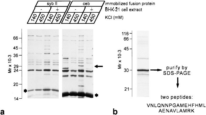
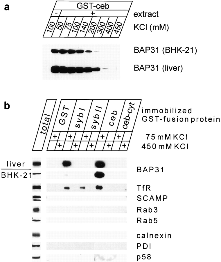
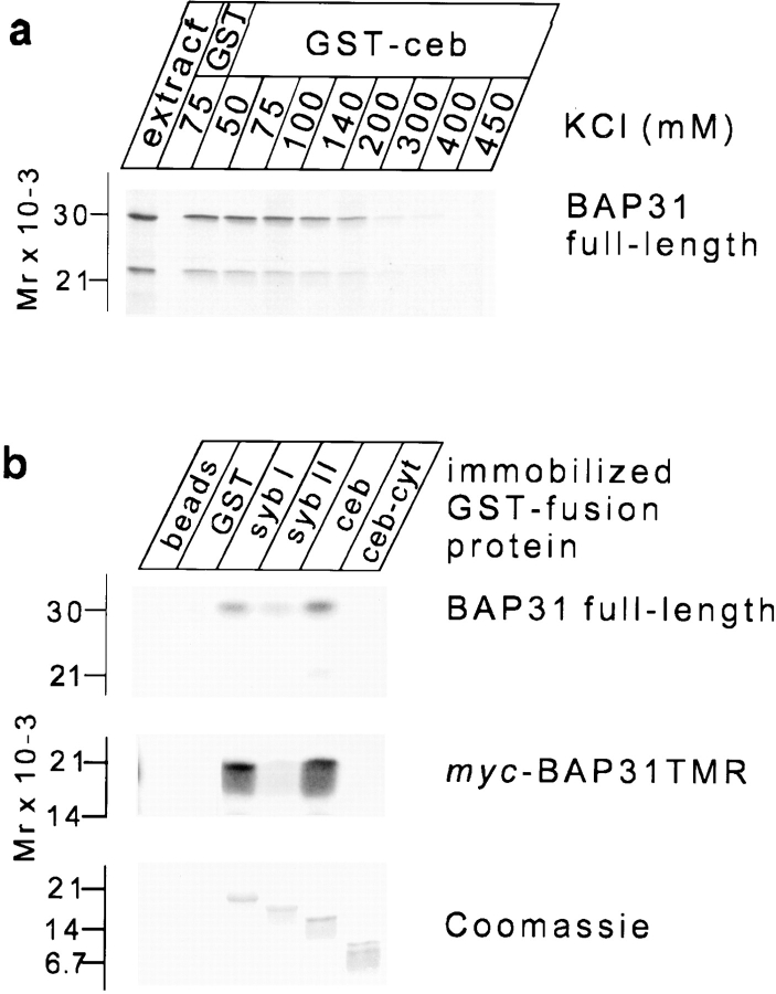
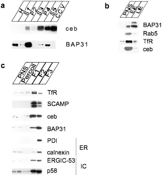
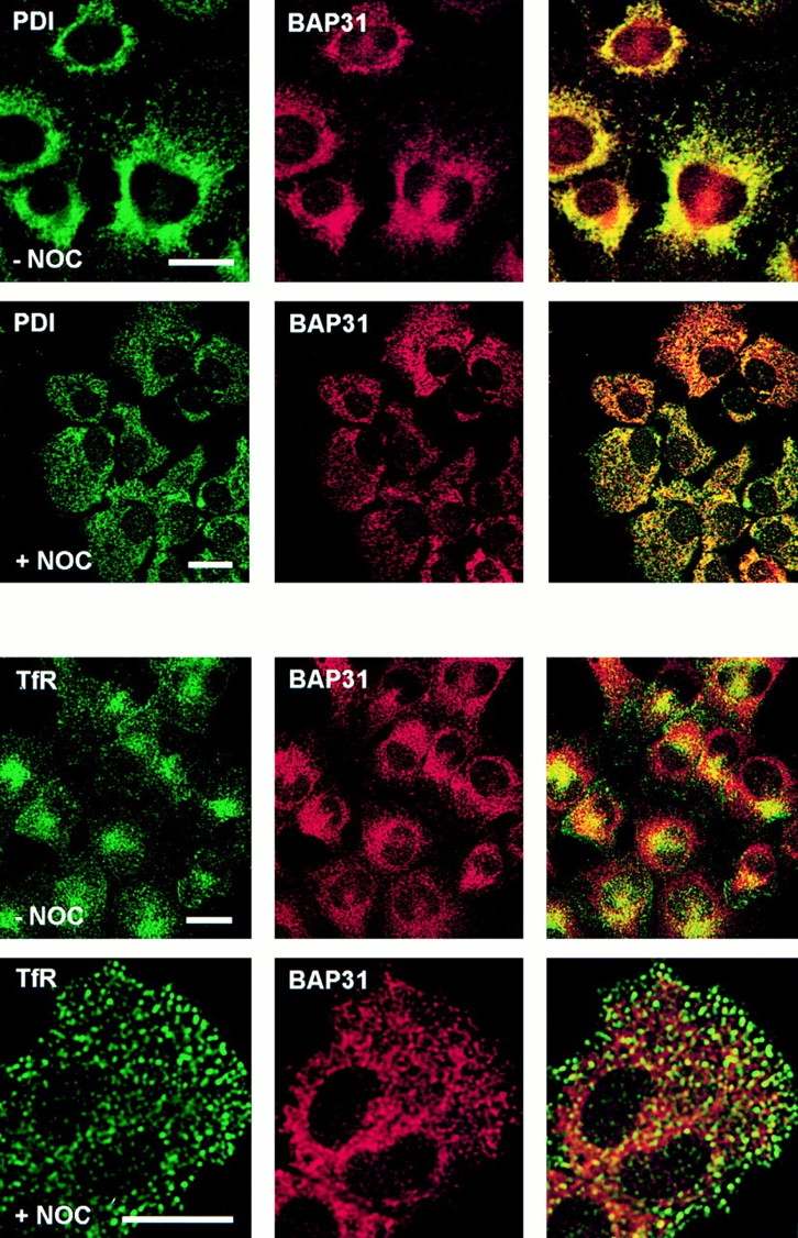
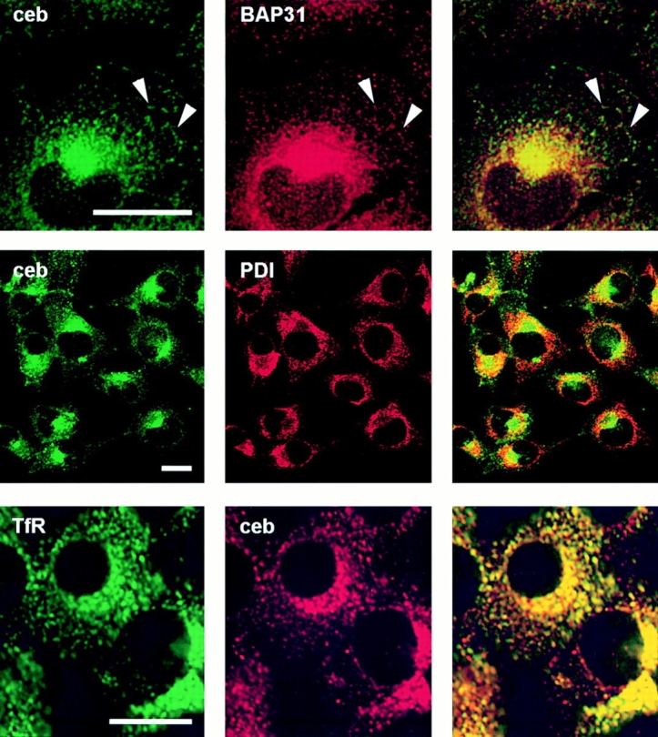
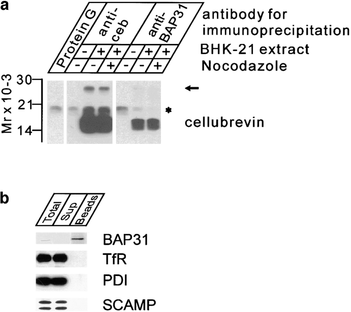
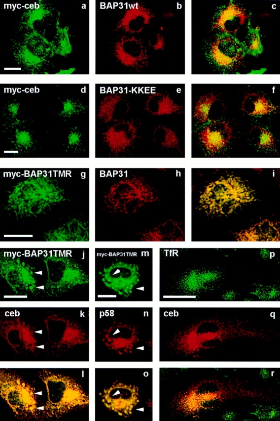
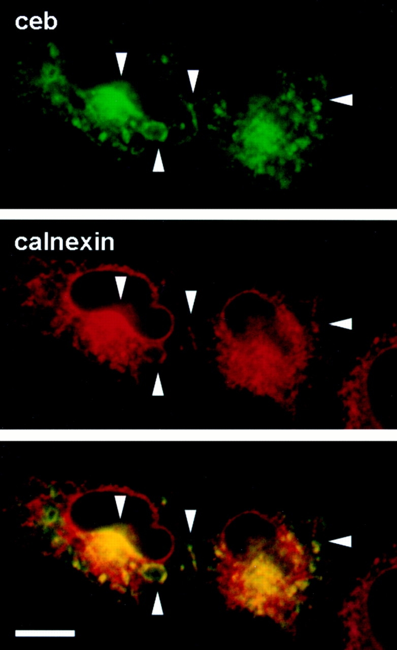
Cellubrevin is a ubiquitously expressed membrane protein that is localized to endosomes throughout the endocytotic pathway and functions in constitutive exocytosis. We report that cellubrevin binds with high specificity to BAP31, a representative of a highly conserved family of integral membrane proteins that has recently been discovered to be binding proteins of membrane immunoglobulins. The interaction between BAP31 and cellubrevin is sensitive to high ionic strength and appears to require the transmembrane regions of both proteins. No other proteins of liver membrane extracts copurified with BAP31 on immobilized recombinant cellubrevin, demonstrating that the interaction is specific. Synaptobrevin I bound to BAP31 with comparable affinity, whereas only weak binding was detectable with synaptobrevin II. Furthermore, a fraction of BAP31 and cellubrevin was complexed when each of them was quantitatively immunoprecipitated from detergent extracts of fibroblasts (BHK 21 cells). During purification of clathrin-coated vesicles or early endosomes, BAP31 did not cofractionate with cellubrevin. Rather, the protein was enriched in ER-containing fractions. When BHK cells were analyzed by immunocytochemistry, BAP31 did not overlap with cellubrevin, but rather colocalized with resident proteins of the ER. In addition, immunoreactive vesicles were clustered in a paranuclear region close to the microtubule organizing center, but different from the Golgi apparatus. When microtubules were depolymerized with nocodazole, this accumulation disappeared and BAP31 was confined to the ER. Truncation of the cytoplasmic tail of BAP31 prevented export of cellubrevin, but not of the transferrin receptor from the ER. We conclude that BAP31 represents a novel class of sorting proteins that controls anterograde transport of certain membrane proteins from the ER to the Golgi complex.


Figures

Figure 1
Figure 1
Identification of a major cellubrevin binding protein as BAP31. (A) Electrophoretic analysis of proteins of a Triton X-100 extract derived from BHK-21 cell membranes that bind to immobilized GST–synaptobrevin II (left) or GST– cellubrevin (right). Equal amounts of GST-fusion proteins were immobilized on glutathione–Sepharose and incubated with BHK-21 cell extracts (adjusted to 140 and 450 mM KCl, respectively) at 4°C overnight. After washing, the fusion proteins were released by thrombin cleavage and 10% of each sample was analyzed by SDS-PAGE (13% gel) and silver staining. Asterisks indicate the positions of synaptobrevin II and cellubrevin, respectively. The arrow points to a protein of 30 kD that eluted specifically from the cellubrevin column only when extracts in 140 mM KCl were used. (b) Purification of the 30-kD protein by cellubrevin affinity chromatography using elution in high salt buffer. 2.5 ml of glutathione–Sepharose were saturated with GST–cellubrevin and incubated overnight at 4°C with 10 ml of a Triton X-100 extract of rat liver membranes (2 mg protein/ml). After washing, the bound protein was eluted with high salt buffer containing 1% Triton X-100, dialyzed against extraction buffer, and concentrated by ultrafiltration (Centricon 10). The purification procedure was repeated several times using fresh extract and the same column. The pooled and concentrated eluates were separated by preparative SDS-PAGE (15% gel). The bands corresponding to 30-kD protein were visualized by staining with Coomassie blue, excised, and further concentrated using a funnel web SDS-PAGE system (Lombard-Platet and Jalinot, 1993). The main band was excised and subjected to trypsin digestion, followed by peptide separation using RP-HPLC and microsequencing. Two peptide sequences were obtained as indicated.
Figure 2
Figure 2
Properties and specificity of the cellubrevin–BAP31 interaction. (a) The interaction of BAP31 to GST–cellubrevin (ceb) is sensitive to high KCl concentrations. Triton X-100 extracts of BHK-21 cells or of rat liver membranes were prepared in the KCl concentrations indicated. Binding and washing (using the appropriate KCl buffers) were performed as described in Fig. 1. Bound proteins were released by thrombin cleavage and 10% of the eluates were extracted with SDS sample buffer for electrophoresis. The figure shows immunoblot analysis for BAP31. (b) BAP31 binds selectively to synaptobrevin I and cellubrevin, and requires the transmembrane domain of cellubrevin for binding. Binding assays using immobilized fusion proteins of synaptobrevin I (syb I), synaptobrevin II (syb II), cellubrevin (ceb) and the NH2-terminal cytoplasmic part of cellubrevin (ceb-cyt) were performed as described in Fig. 1, except that the material eluted after thrombin cleavage was analyzed by immunoblotting (10% of each eluate/ lane). The eluates were tested for the following proteins: transferrin receptor (TfR), secretory carrier associated membrane protein (SCAMP), Rab3 (all isoforms), Rab5, calnexin, PDI, and p58.
Figure 3
Figure 3
Binding of recombinant full-length and truncated BAP31 to cellubrevin and synaptobrevins. Recombinant [35S]methionine labeled BAP31, either full-length or COOH-terminally truncated (myc-BAP31TMR), was generated by in vitro transcription translation. The translation mix was diluted ∼20–25-fold in extraction buffer, and binding to immobilized fusion proteins was performed as in Fig. 1, except that bound [35S]methionine labeled BAP31 was detected by autofluorography. (a) Binding of recombinant BAP31, like that of native BAP31, to GST–cellubrevin is sensitive to ionic strength. For binding, the translation mix was diluted in extraction buffer containing the indicated concentrations of KCl. Equal proportions of the starting material (load) and the bound material were analyzed. Note that a band with higher mobility was generated in addition to full-length BAP31, probably corresponding to a truncated version of BAP31. (b) Binding of full-length (top) and COOH-terminally truncated, myc-tagged BAP31 (middle) to immobilized GST–synaptobrevin fusion proteins. Fig. 2 shows details. (Bottom) A Coomassie blue-stained gel of GST–fusion protein eluted after thrombin cleavage, demonstrating that comparable amounts of immobilized fusion proteins were used in the binding assays.
Figure 4
Figure 4
Subcellular fractionation reveals differential distribution of BAP31 and cellubrevin. (a) BAP31 does not coenrich with cellubrevin (ceb) during purification of clathrin-coated vesicles from rat liver. Homogenates (H) were centrifuged for 20 min at 20,000 g max. The supernatant (S1) was centrifuged again at 55,000 g max for 1 h and then the pellet (P2) was resuspended and recentrifuged after adding ficoll and sucrose. The supernatant (S3) was diluted and centrifuged at 100,000 g max for 1 h. This pellet, P4, was again resuspended and centrifuged at 20,000 g max for 20 min. The resulting supernatant S5 was layered on top of a D2O–sucrose density gradient. The pellet obtained after centrifugation at 110,000 g max for 2 h contained purified coated vesicles (Maycox et al., 1992). Of each fraction, 10 μg of protein were analyzed by SDS-PAGE and immunoblotting for cellubrevin and BAP31. (b) BAP31 does not coenrich with cellubrevin during purification of early endosomes (EE). Analysis of endosomal fractions. A PNS of BHK-21 cells was subjected to flotation gradient centrifugation on a discontinuous D2O–sucrose gradients (Gorvel et al., 1991), resulting in a fraction enriched in EE and a fraction enriched in late endosomes and carrier vesicles (LE). The gradient fractions were diluted and pelleted by centrifugation. Of each fraction, 10 μg of protein were analyzed by SDS-PAGE and immunoblotting for cellubrevin and BAP31, and for the EE markers TfR and Rab5. (c) BAP31 coenriches with markers of the intermediate compartment and the ER. Fractionation of ER and intermediate compartment. A PNS was mixed with Percoll to give a final density of 1.129 g/ml, and then centrifuged. A midportion of the gradient was pooled (Percoll), adjusted to 30% Nycodenz, and overlayed with 27 and 18.5% (wt/wt) of Nycodenz. After equilibrium density gradient centrifugation, three interfaces were collected (F1, F2, F3) and analyzed by SDS-PAGE and immunoblotting (10 μg of protein per lane). ER markers (PDI and calnexin) are highly enriched in the F3 fraction, together with markers for the IC (ERGIC-53 and p58). BAP31 immunoreactivity was recovered mainly in this interface, in contrast to cellubrevin, SCAMP, and the transferrin receptor, which were enriched in the F2 interface.
Figure 5
Figure 5
Localization of BAP31 in BHK-21 cells in comparison to markers of the ER (PDI) and the endosomal recycling compartment (TfR). Before fixation, cells were incubated for 2 h in the incubator in the absence (−NOC) or presence (+NOC) of nocodazole (33 μM) to depolymerize microtubules. Cells were double stained with rabbit anti–BAP31 antibodies and mouse PDI antibodies (1D3 monoclonal antibody, top rows) or with rabbit anti–BAP31 antibodies and mouse anti–TfR monoclonal antibody (bottom rows), using DTAF-labeled donkey anti–mouse antibody (green column) and CY3– labeled donkey anti–rabbit antibody (red column) as secondary antibodies, respectively. Analysis was performed by confocal laser scanning microscopy. The third column in each row shows color overlays. Note that BAP31 staining colocalizes with PDI in the cell periphery, but extends to a paranuclear region where TfR-positive dots also accumulate, corresponding to vesicle clusters around the MTOC. This accumulation disappears upon nocodazole treatment. Bars, 20 μm.
Figure 6
Figure 6
Localization of cellubrevin in BHK-21 cells in comparison to BAP31 and markers of the ER (PDI), and the endosomal recycling compartment (TfR). Fig. 5 shows details. For double labeling of BAP31 and cellubrevin (top), we used biotinylated cellubrevin antibodies that were affinity purified from rabbit serum. Visualization was with CY3–conjugated donkey anti–rabbit for BAP31 (top) and cellubrevin (bottom), or donkey anti– mouse for PDI (middle; red column) and with DTAF- labeled streptavidin for biotinylated cellubrevin antibodies (top), donkey anti–rabbit for cellubrevin (middle), and donkey anti–mouse for transferrin receptor (TfR, bottom; green column). Color overlays are on the right. Cellubrevin immunostaining is mainly clustered in a region devoid of PDI immunostaining. At higher magnification, peripheral localized cellubrevin immunostained dots colocalize with the peripheral reticular staining for BAP31 (top, arrowheads). Bar, 20 μm.
Figure 7
Figure 7
Cellubrevin coimmunoprecipitates with BAP31 in detergent extracts of BHK-21 cells. (a) Excess amounts of antibodies specific for cellubrevin (anti-ceb) and for BAP31 (anti-BAP31) were added to Triton X-100 extracts of control (−)- and nocodazole (+)-treated BHK-21 cells before isolation of immune complexes using protein G–Sepharose. Equal proportions of each sample were analyzed for cellubrevin by immunoblotting using biotinylated affinity-purified anti-cellubrevin followed by HRP– conjugated streptavidin and visualization by ECL. The asterisk denotes a nonspecific band recognized by the detection system. No binding was observed when extracts were incubated with only protein G–Sepharose beads (Protein G). (b) Coimmunoprecipitation of cellubrevin with BAP31 is specific. Immune complexes were isolated from untreated BHK cell extracts as above using anti-cellubrevin antibodies. 20 μg protein each of total (starting) extract (Total) and unbound supernatant (Sup), and 15% of the bead-bound immune complexes (Beads) were analyzed as above and probed for the TfR, PDI, and SCAMP. For BAP31, reducing agents were omitted for SDS-PAGE and visualization was performed with the AP method instead of the ECL method used for the other antigens.
Figure 8
Figure 8
Mutation or truncation of the COOH-terminal tail of BAP21 does not affect its ability to bind cellubrevin. (a) Diagram showing the expression constructs of BAP31 that were used for cotransfection of BHK-21 cells with full-length, myc-tagged (b and c) or untagged cellubrevin (d). The three transmembrane regions are indicated with I, II, and III; the two gray areas correspond to areas with a predicted propensity to form coiled coils. vim indicates: region of homology with vimentin. (b) Detection of BAP31 expression in PNSs (10 μg protein/lane) of transfected BHK cells. Under these assay conditions, the endogenous protein was below the detection limit. As reference, 10 μg of protein obtained from a fraction enriched in ER (F3 fraction; Fig. 4) was analyzed in parallel. The mutants can be distinguished by small differences in electrophoretic mobility. (c) BAP31 mutants containing small changes or deletions in the cytoplasmic tail coprecipitate with cellubrevin. BHK-21 cells cotransfected with cDNAs encoding myc-tagged cellubrevin and either the wild-type or mutant BAP31 were extracted in extraction buffer, followed by quantitative immunoprecipitation using excess amounts of anti–myc monoclonal antibody. BAP31 mutants and myc–cellubrevin were detected by ECL and the diaminobenzidine reaction, respectively. An additional band was detectable in the BAP31 blots that is nonspecific (asterisk), and is discriminated from the BAP31 mutants by slight differences in mobility. No binding was observed when only protein G–Sepharose beads were used (protein G). (d) Cellubrevin coprecipitates with a BAP31 mutant protein lacking the entire COOH-terminal cytoplasmic tail (myc-BAP31TMR). Quantitative immunoprecipitation was performed using excess amounts of anti–myc monoclonal antibody (left) or affinity-purified anti–cellubrevin antibody (right). Approximately 5% of extract and supernatant (sup) and one-third of the bead-bound proteins (bound) were analyzed by SDS-PAGE and immunoblotting. Cellubrevin was detected using affinity-purified and biotinylated rabbit antibody. The immunoprecipitating antibody was visualized using AP conjugated to second antibodies (myc-BAP31TMR, top left), or to streptavidin (cellubrevin, bottom right). The coimmunoprecipitating protein was visualized using HRP conjugated to second antibodies (myc-BAP31TMR, top right) or to streptavidin (cellubrevin, bottom left), followed by ECL. No binding was observed when only protein G–Sepharose beads were used (Protein G).
Figure 9
Figure 9
Truncation, but not any other mutation in the cytoplasmic tail of BAP31, results in selective retention of cellubrevin in the ER. BHK-21 cells were cotransfected with plasmids encoding myc-tagged cellubrevin (myc-ceb) and with wild-type BAP31 (a– c), BAP31–KKEE (d–f), or with plasmids encoding untagged cellubrevin (ceb) and myc-BAP31TMR (g–r). myc-tagged cellubrevin and myc-BAP31TMR were detected using mouse myc monoclonal antibodies. The secondary reagents were: DTAF-labeled donkey anti–mouse antibodies (a, d, g, j, m and p), lissamine–rhodamine-labeled donkey anti–rabbit (b, e, h, k, n and q). All images were analyzed by confocal laser scanning microscopy. Fields c, f, i, l, o and r are color overlays. Cellubrevin was only retained in the ER in cells cotransfected with myc-BAP31TMR (j–l). In these panels, intense immunoreactive spots (arrowheads) for myc-BAP31TMR are seen, as indicated by cellubrevin staining. These spots were also positive for p58 (arrowheads in m–o), and probably denote material accumulating at ER export sites. In such doubly transfected cells, no punctate colocalization was observed between cellubrevin and the TfR that appeared to be normally sorted (p–r). Bars, 20 μm.
Figure 10
Figure 10
Overexpression of myc-tagged cellubrevin results in its partial retention within the ER. BHK-21 cells were transfected with plasmids encoding myc-tagged cellubrevin. After fixation, the cells were immunostained for cellubrevin using monoclonal myc-antibodies and a rabbit serum specific for calnexin. Fig. 9 shows detection procedures. Images were analyzed by confocal laser scanning microscopy. Arrowheads point to areas where cellubrevin is colocalized with calnexin. Bar, 20 μm.

References

    1. Adachi T, Schamel WWA, Kim K-M, Watanabe T, Becker B, Nielsen PJ, Reth M. The specificity of association of the IgD molecule with the accessory proteins BAP31/BAP29 lies in the IgD transmembrane sequence. EMBO (Eur Mol Biol Organ) J. 1996;15:1534–1541. - PMC - PubMed
    1. Aridor M, Balch WE. Principles of selective transport: coat complexes hold the key. Trends Cell Biol. 1996;6:315–320. - PubMed
    1. Balch WE, McCaffery JM, Plutner H, Farquhar MG. Vesicular stomatitis virus glycoprotein is sorted and concentrated during export from the endoplasmic reticulum. Cell. 1994;76:841–852. - PubMed
    1. Bannykh SI, Rowe T, Balch WE. The organization of endoplasmic reticulum export complexes. J Cell Biol. 1996;135:19–35. - PMC - PubMed
    1. Barlowe C, Orci L, Yeung T, Hosobuchi M, Hamamoto S, Salama N, Rexach MF, Ravazzola M, Armherdt M, Schekman R. COPII: a membrane coat formed by sec proteins that drive vesicle budding from the endoplasmic reticulum. Cell. 1994;77:895–907. - PubMed

Publication types

MeSH terms