. 1997 Dec 15;139(6):1523-31.
doi: 10.1083/jcb.139.6.1523.

Ligation of major histocompatability complex (MHC) class I molecules on human T cells induces cell death through PI-3 kinase-induced c-Jun NH2-terminal kinase activity: a novel apoptotic pathway distinct from Fas-induced apoptosis

Affiliations

Ligation of major histocompatability complex (MHC) class I molecules on human T cells induces cell death through PI-3 kinase-induced c-Jun NH2-terminal kinase activity: a novel apoptotic pathway distinct from Fas-induced apoptosis

S Skov et al. J Cell Biol. .

Abstract

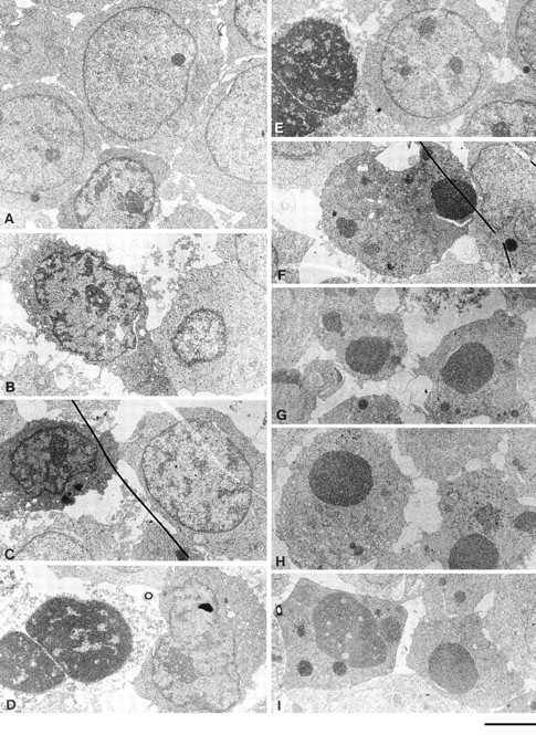
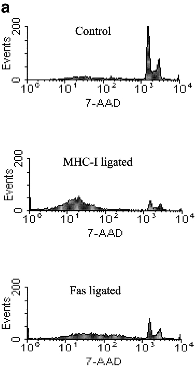
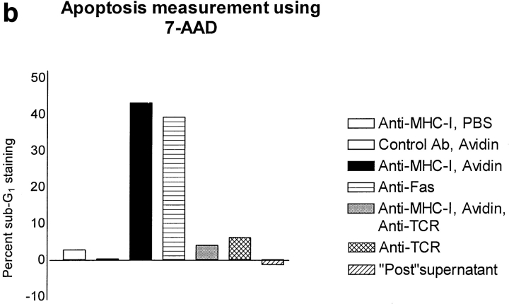
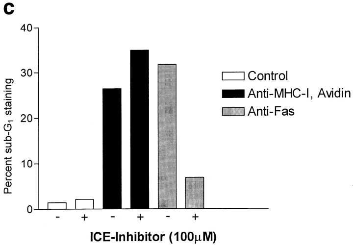
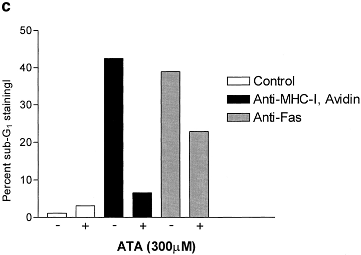
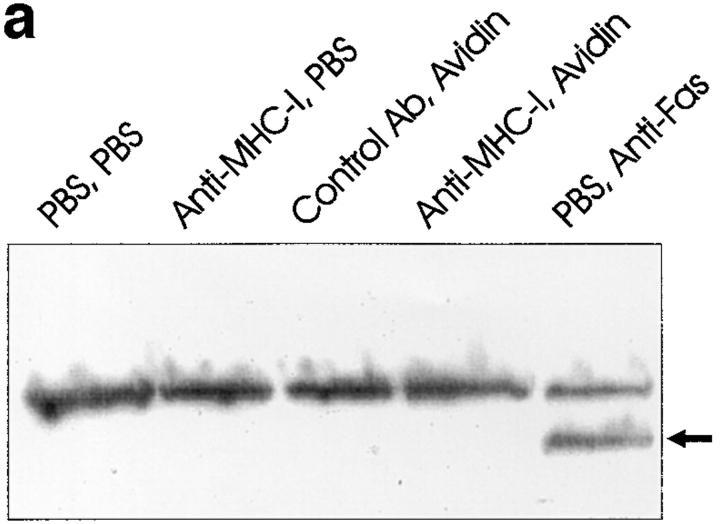
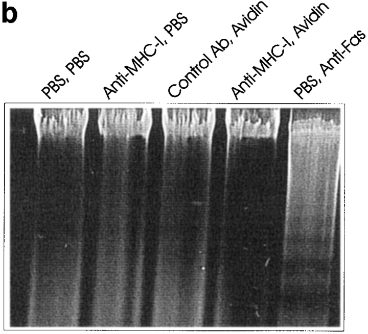
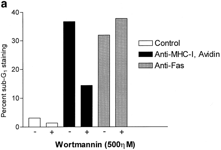
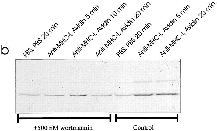
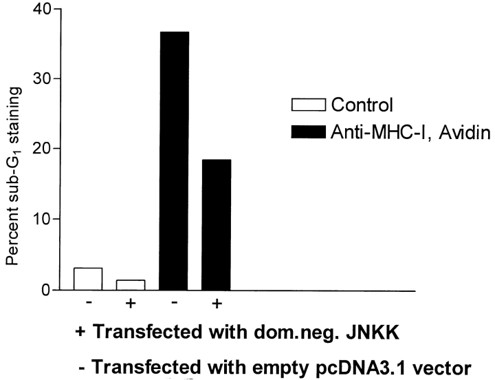
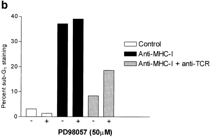
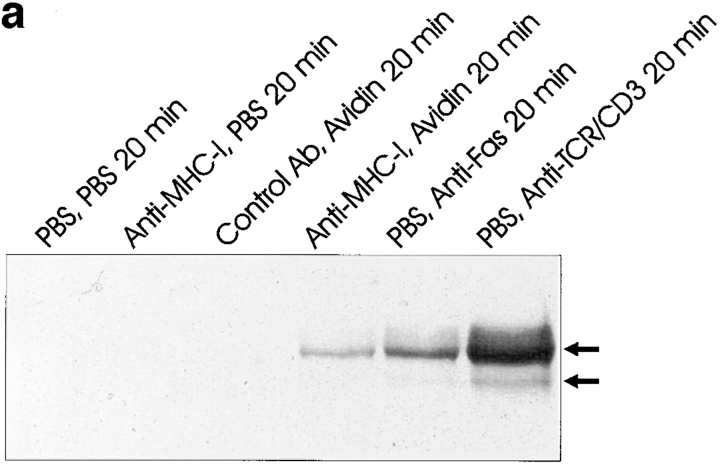
Ligation of major histocompatability complex class I (MHC-I) molecules expressed on T cells leads to both growth arrest and apoptosis. The aim of the current study was to investigate the intracellular signal pathways that mediate these effects. MHC-I ligation of human Jurkat T cells induced a morphologically distinct form of apoptosis within 6 h. A specific caspase inhibitor, which inhibited Fas-induced apoptosis, did not affect apoptosis induced by MHC-I ligation. Furthermore, MHC-I-induced apoptosis did not involve cleavage and activation of the poly(ADP- ribose) polymerase (PARP) endonuclease or degradation of genomic DNA into the typical fragmentation ladder, both prominent events of Fas-induced apoptosis. These results suggest that MHC-I ligation of Jurkat T cells induce apoptosis through a signal pathway distinct from the Fas molecule. In our search for other signal pathways leading to apoptosis, we found that the regulatory 85-kD subunit of the phosphoinositide-3 kinase (PI-3) kinase was tyrosine phosphorylated after ligation of MHC-I and the PI-3 kinase inhibitor wortmannin selectively blocked MHC-I-, but not Fas-induced, apoptosis. As the c-Jun NH2-terminal kinase (JNK) can be activated by PI-3 kinase activity, and has been shown to be involved in apoptosis of lymphocytes, we examined JNK activation after MHC-I ligation. Strong JNK activity was observed after MHC-I ligation and the activity was completely blocked by wortmannin. Inhibition of JNK activity, by transfecting cells with a dominant-negative JNKK- MKK4 construct, led to a strong reduction of apoptosis after MHC-I ligation. These results suggest a critical engagement of PI-3 kinase-induced JNK activity in apoptosis induced by MHC-I ligation.


Figures

Figure 1
Figure 1
(A) Electron micrographs of control Jurkat cells 6 h after exposure to biotinylated control antibody plus avidin. Notice the normal ultrastructure of the cell. (B–F) Varying stages of decay in Jurkat cells 6 h after exposure to biotinylated anti–MHC-I antibody plus avidin. Notice the simultaneous disintegration of the cytoplasm and the nucleus. (B and C) Darkening of cytoplasm, shrinkage of the nucleus, and dispersed DNA condensation of heterochromatin. (D and E) Disintegration of the cytoplasm and homogenization of the chromatin surrounded by nuclear envelope. (F) Apoptotic-like bodies of condensed homogenous chromatin in a relatively well-preserved cytoplasm. (G–I) Typical apoptotic bodies in Jurkat cells after a 6-h exposure to anti-Fas antibody. The nuclear fragments consist of homogenized chromatin encircled by an intact nuclear envelope surrounded by a relatively well-preserved cytoplasm. Bar, 2 μm.
Figure 2
Figure 2
Measurement of apoptosis (cells < 2n DNA). 106 cells were stimulated for 6 h and apoptosis was measured using 7-AAD as described in Materials and Methods. (a) 7-AAD flow cytometric profile of Jurkat cells after control, MHC-I and Fas ligation. (b) Apoptosis measurement after different stimuli. “Post” supernatant (dark grey bar) is supernatant from anti–MHC-I antibody and avidin stimulated cells; the supernatants were purified with protein A before stimulation. (c) Cells were incubated with or without 100-μM peptide ICE inhibitor for 1 h before stimulation. The data show results of at least three independent experiments.
Figure 2
Figure 2
Measurement of apoptosis (cells < 2n DNA). 106 cells were stimulated for 6 h and apoptosis was measured using 7-AAD as described in Materials and Methods. (a) 7-AAD flow cytometric profile of Jurkat cells after control, MHC-I and Fas ligation. (b) Apoptosis measurement after different stimuli. “Post” supernatant (dark grey bar) is supernatant from anti–MHC-I antibody and avidin stimulated cells; the supernatants were purified with protein A before stimulation. (c) Cells were incubated with or without 100-μM peptide ICE inhibitor for 1 h before stimulation. The data show results of at least three independent experiments.
Figure 3
Figure 3
(a) Anti-PARP Western blot of whole Jurkat cell lysate. 4 × 10⁶ cells were stimulated for 6 h as described in Materials and Methods. (b) DNA ladder analysis. 106 cells were stimulated for 6 h and then DNA was extracted and electrophoresed on a 2% agarose gel as described in Materials and Methods. (c) ATA inhibition of apoptosis. 106 cells were incubated with or without 300 μM ATA for 1 h before stimulation. Cells were stimulated for 6 h and apoptosis was measured using 7-AAD as described in Materials and Methods. All data show results of at least three independent experiments.
Figure 3
Figure 3
(a) Anti-PARP Western blot of whole Jurkat cell lysate. 4 × 10⁶ cells were stimulated for 6 h as described in Materials and Methods. (b) DNA ladder analysis. 106 cells were stimulated for 6 h and then DNA was extracted and electrophoresed on a 2% agarose gel as described in Materials and Methods. (c) ATA inhibition of apoptosis. 106 cells were incubated with or without 300 μM ATA for 1 h before stimulation. Cells were stimulated for 6 h and apoptosis was measured using 7-AAD as described in Materials and Methods. All data show results of at least three independent experiments.
Figure 4
Figure 4
(a) Wortmannin inhibition of apoptosis. 106 cells were incubated with or without 500 ηM wortmannin for 2 h before stimulation. Cells were stimulated for 6 h, with 500 ηM wortmannin present, and apoptosis was measured using 7-AAD as described in Materials and Methods. (b) Immunoprecipitates of the P85 subunit of the PI-3 kinase obtained from lysates of Jurkat cells. 3 × 10⁷ cells were stimulated for the indicated time, lysed, and subjected to immunoprecipitation as described in Materials and Methods (top). Blots were probed with anti-phosphotyrosine antibody, stripped, and reprobed with anti–PI-3 kinase antibody (bottom). All data show results of at least three independent experiments.
Figure 4
Figure 4
(a) Wortmannin inhibition of apoptosis. 106 cells were incubated with or without 500 ηM wortmannin for 2 h before stimulation. Cells were stimulated for 6 h, with 500 ηM wortmannin present, and apoptosis was measured using 7-AAD as described in Materials and Methods. (b) Immunoprecipitates of the P85 subunit of the PI-3 kinase obtained from lysates of Jurkat cells. 3 × 10⁷ cells were stimulated for the indicated time, lysed, and subjected to immunoprecipitation as described in Materials and Methods (top). Blots were probed with anti-phosphotyrosine antibody, stripped, and reprobed with anti–PI-3 kinase antibody (bottom). All data show results of at least three independent experiments.
Figure 5
Figure 5
(a) Anti–active JNK-1 Western blot of whole Jurkat cell lysate. 5 × 10⁶ cells were stimulated for the indicated time as described in Materials and Methods. The strong band at 56 kD after TCR–CD3 ligation is an artefact from the stimulatory antibody. (b) Wortmannin inhibition of JNK-1: 5 × 10⁶ cells were incubated 1 h with 500 ηM wortmannin (lanes 1–4) or without (lanes 5–7) before stimulation for the indicated time as described in Materials and Methods. All data show results of at least three independent experiments.
Figure 5
Figure 5
(a) Anti–active JNK-1 Western blot of whole Jurkat cell lysate. 5 × 10⁶ cells were stimulated for the indicated time as described in Materials and Methods. The strong band at 56 kD after TCR–CD3 ligation is an artefact from the stimulatory antibody. (b) Wortmannin inhibition of JNK-1: 5 × 10⁶ cells were incubated 1 h with 500 ηM wortmannin (lanes 1–4) or without (lanes 5–7) before stimulation for the indicated time as described in Materials and Methods. All data show results of at least three independent experiments.
Figure 6
Figure 6
Apoptosis measurement after transfection of dominant-negative JNKK–MKK4 construct. Cells were transfected with pcDNA3.1 vector containing a dominant-negative JNKK– MKK4 construct (Ala substituted at Ser-257 and Thr-261), or with the empty vector as described in Materials and Methods. Subsequent apoptosis was measured as described in Fig. 2.
Figure 7
Figure 7
(a) Anti–active ERK Western blot of whole Jurkat cell lysate. 5 × 10⁶ cells were stimulated for the indicated time as described in Materials and Methods. (b) PD98059 inhibition of the TCR–CD3 rescue signal on MHC-I–induced apoptosis. 106 cells were incubated with or without 100 μM PD98059 for 1 h before stimulation. Cells were stimulated for 6 h and apoptosis was measured using 7-AAD as described in Materials and Methods. All data show results of at least three independent experiments.
Figure 7
Figure 7
(a) Anti–active ERK Western blot of whole Jurkat cell lysate. 5 × 10⁶ cells were stimulated for the indicated time as described in Materials and Methods. (b) PD98059 inhibition of the TCR–CD3 rescue signal on MHC-I–induced apoptosis. 106 cells were incubated with or without 100 μM PD98059 for 1 h before stimulation. Cells were stimulated for 6 h and apoptosis was measured using 7-AAD as described in Materials and Methods. All data show results of at least three independent experiments.

Similar articles

Cited by

References

    1. Amirayan N, Furrie E, Deleuil F, Mellor A, Leserman L, Machy P. Influence of MHC class I molecules on T cell proliferation induced by CD3 or Thy-1 stimulation. Immunology. 1995;86:71–78. - PMC - PubMed
    1. Apasov S, Redegeld F, Sitkovsky M. Cell-mediated cytotoxicity: contact and secreted factors. Curr Opin Immunol. 1993;5:404–410. - PubMed
    1. Beckwith M, Fenton RG, Katona IM, Longo DL. Phosphatidylinositol-3-kinase activity is required for the anti–ig-mediated growth inhibition of a human B-lymphoma cell line. Blood. 1996;87:202–210. - PubMed
    1. Bregenholt S, Röpke M, Skov S, Claesson MH. Ligation of MHC class I molecules on peripheral blood T lymphocytes induces new phenotypes and functions. JImmunol. 1996;157:993–999. - PubMed
    1. Casciola-Rosen L, Nicholson DW, Chong T, Rowan KR, Thornberry NA, Miller DK, Rosen A. Apopain/CPP32 cleaves proteins that are essential for cellular repair: a fundamental principle of apoptotic death. J Exp Med. 1996;183:1957–1964. - PMC - PubMed

Publication types

MeSH terms