. 1997 Dec 15;139(6):1583-95.
doi: 10.1083/jcb.139.6.1583.

Muscle beta1D integrin reinforces the cytoskeleton-matrix link: modulation of integrin adhesive function by alternative splicing

Affiliations

Muscle beta1D integrin reinforces the cytoskeleton-matrix link: modulation of integrin adhesive function by alternative splicing

A M Belkin et al. J Cell Biol. .

Abstract

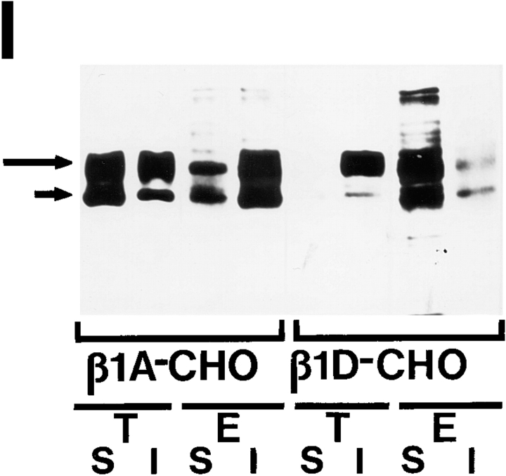
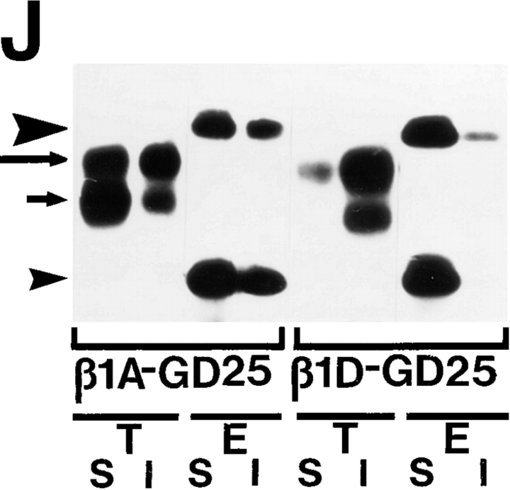
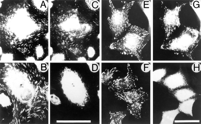
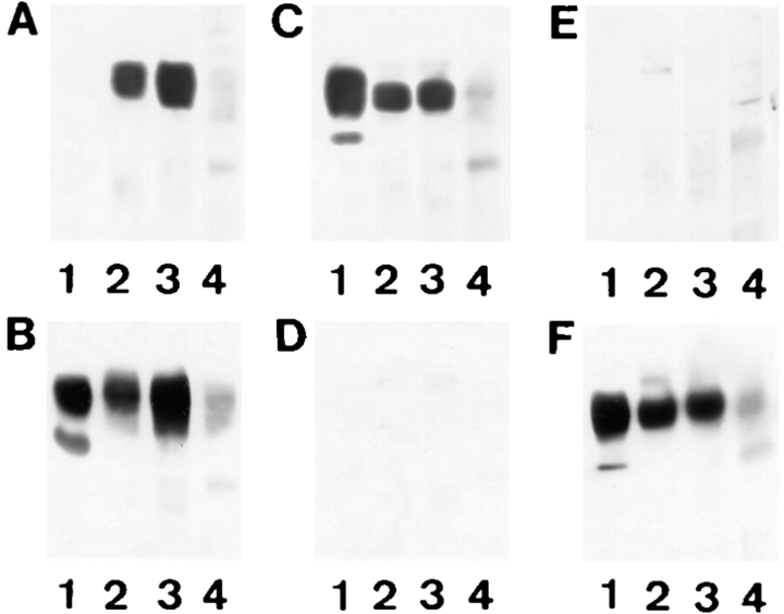
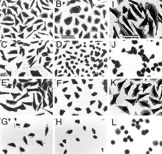
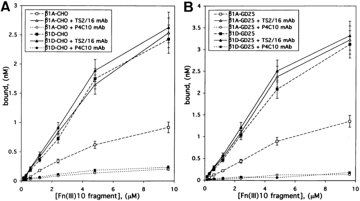
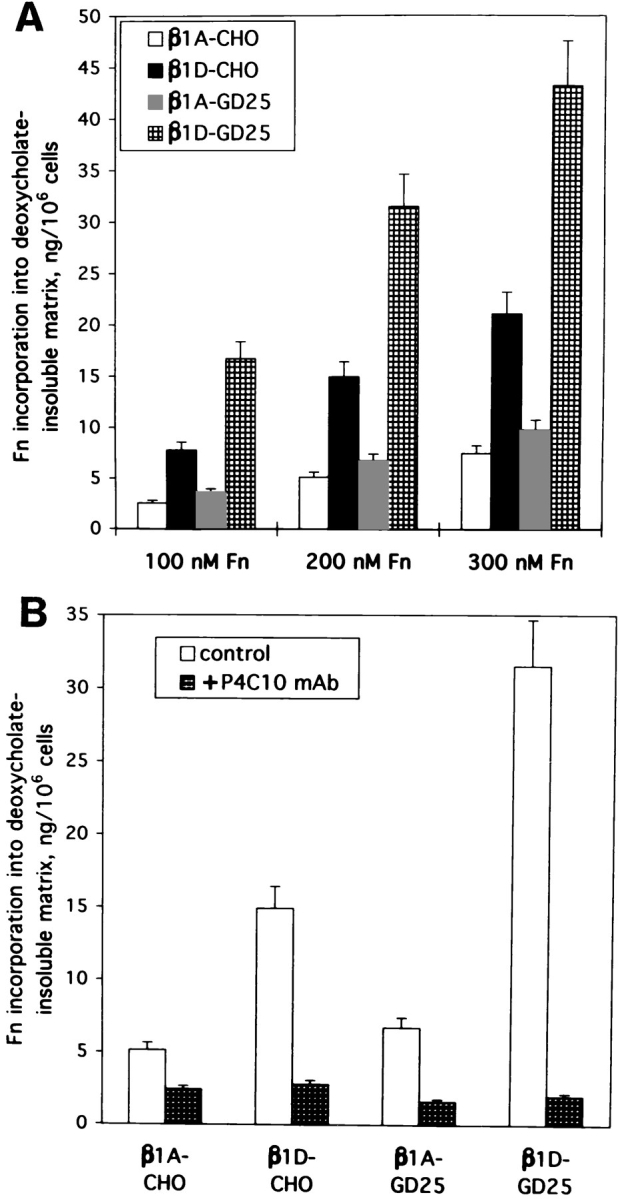
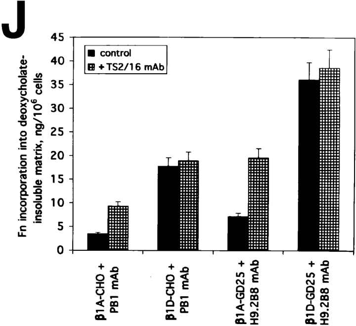
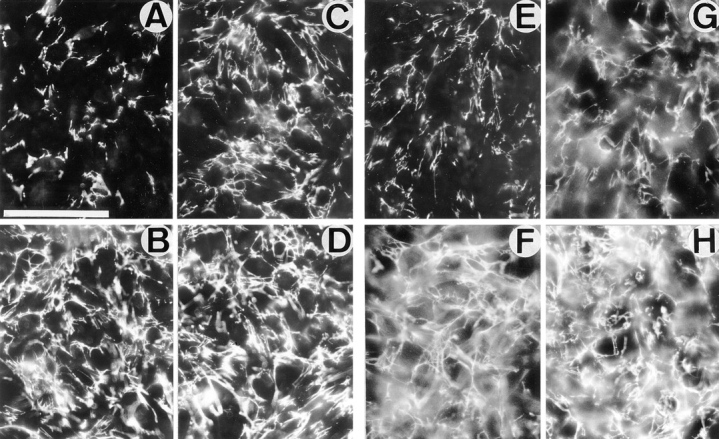
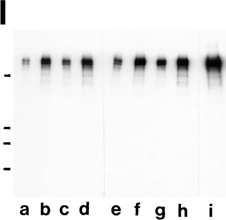
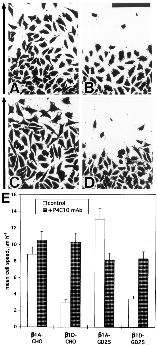
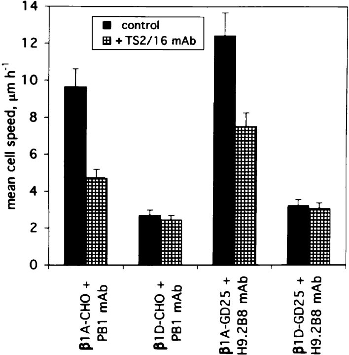
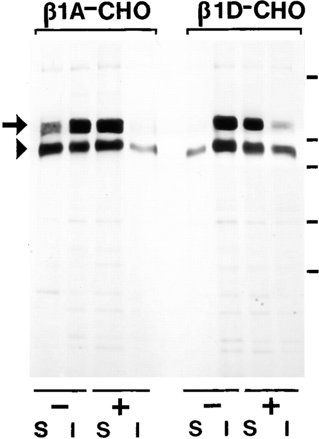
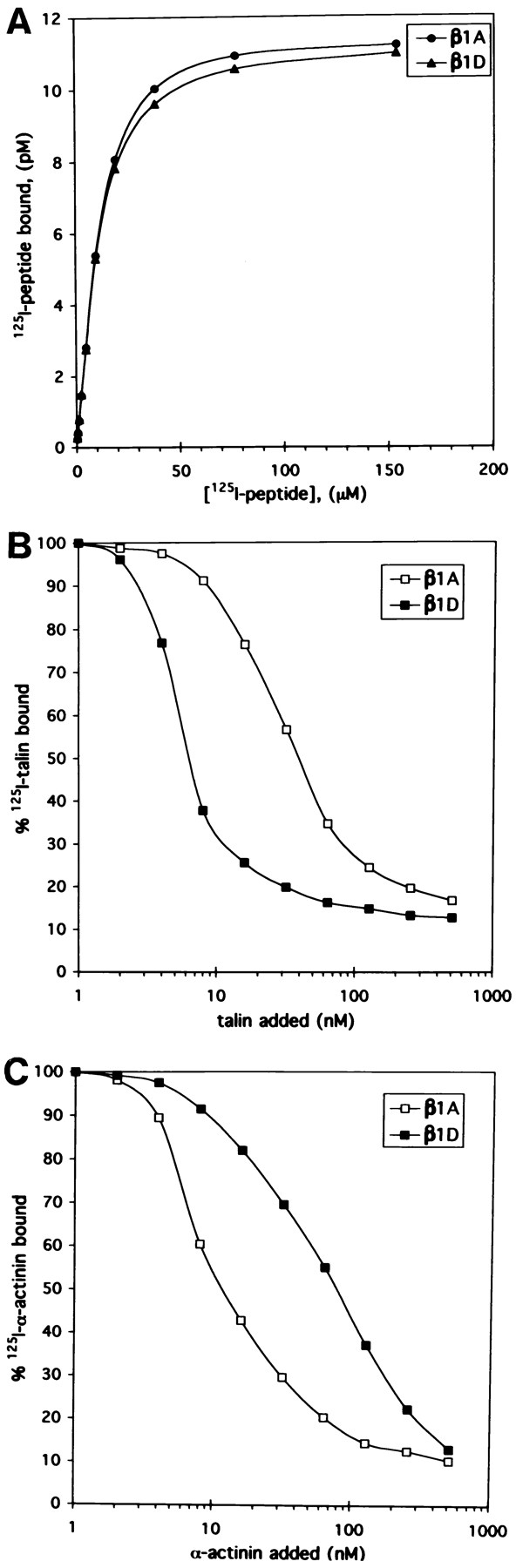
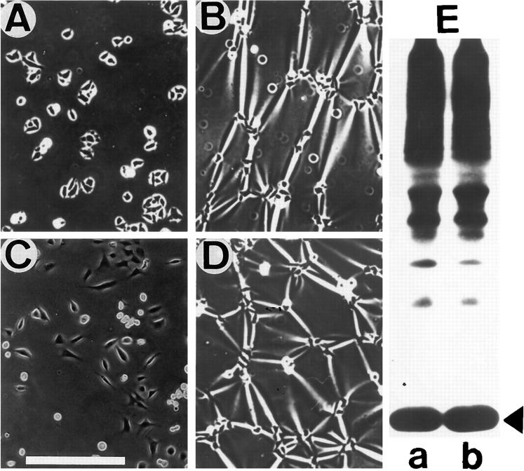
Expression of muscle-specific beta1D integrin with an alternatively spliced cytoplasmic domain in CHO and GD25, beta1 integrin-minus cells leads to their phenotypic conversion. beta1D-transfected nonmuscle cells display rounded morphology, lack of pseudopodial activity, retarded spreading, reduced migration, and significantly enhanced contractility compared with their beta1A-expressing counterparts. The transfected beta1D is targeted to focal adhesions and efficiently displaces the endogenous beta1A and alphavbeta3 integrins from the sites of cell-matrix contact. This displacement is observed on several types of extracellular matrix substrata and leads to elevated stability of focal adhesions in beta1D transfectants. Whereas a significant part of cellular beta1A integrin is extractable in digitonin, the majority of the transfected beta1D is digitonin-insoluble and is strongly associated with the detergent-insoluble cytoskeleton. Increased interaction of beta1D integrin with the actin cytoskeleton is consistent with and might be mediated by its enhanced binding to talin. In contrast, beta1A interacts more strongly with alpha-actinin, than beta1D. Inside-out driven activation of the beta1D ectodomain increases ligand binding and fibronectin matrix assembly by beta1D transfectants. Phenotypic effects of beta1D integrin expression in nonmuscle cells are due to its enhanced interactions with both cytoskeletal and extracellular ligands. They parallel the transitions that muscle cells undergo during differentiation. Modulation of beta1 integrin adhesive function by alternative splicing serves as a physiological mechanism reinforcing the cytoskeleton- matrix link in muscle cells. This reflects the major role for beta1D integrin in muscle, where extremely stable association is required for contraction.


Figures

Figure 9
Figure 9
β1D interacts more strongly than β1A with the actin cytoskeleton and displaces the endogenous β1A and αv integrins from focal adhesions. (A–D) Localization of the transfected β1A and β1D and the endogenous β1A integrins in CHO transfectants. β1A-CHO (A and C) and β1D-CHO (B and D) cells were double stained for human β1A (A) and hamster β1A (C) integrins, or for human β1D (B) and hamster β1A (D) integrins. (E–H) Localization of the transfected β1A and β1D and the endogenous αv integrins in GD25 transfectants. β1A-GD25 (E and G) and β1D-GD25 (F and H) cells were double stained for human β1A (E) and mouse αv (G) integrins, or for human β1D (F) and mouse αv (H) integrins. Note colocalization of the transfected β1A with the endogenous β1A and αv integrins, whereas transfected β1D integrin displaces the endogenous β1A from focal adhesions in CHO transfectants and the endogenous αv integrins from focal adhesions in GD25 transfectants. (I and J) Association of the transfected β1A and β1D and the endogenous β1A and αv integrins with the actin cytoskeleton in CHO and GD25 transfectants. 35S-Labeled integrins were immunoprecipitated from digitonin-soluble (S) and digitonin-insoluble (I) fractions of β1A- and β1D-transfected CHO (I) and GD25 (J) cells. (T), transfected human β1A or β1D integrin; (E), endogenous hamster β1A (I) or mouse αvβ3/αvβ5 (J) integrins. Long arrows point to the mature β1 integrin subunit, short arrows mark the precursor of the β1 subunit. The large arrowhead marks the αv subunit and the small arrowhead marks the associated β3 and β5 subunits, not resolved under conditions of this electrophoresis. Unlike the transfected β1A, β1D is present predominantly in the digitonin-insoluble (cytoskeletal) fraction, whereas the endogenous hamster β1A in β1D-CHO and mouse αvβ3/αvβ5 integrins in β1D-GD25 cells are mostly digitonin-soluble. Bars, 50 μm.
Figure 9
Figure 9
β1D interacts more strongly than β1A with the actin cytoskeleton and displaces the endogenous β1A and αv integrins from focal adhesions. (A–D) Localization of the transfected β1A and β1D and the endogenous β1A integrins in CHO transfectants. β1A-CHO (A and C) and β1D-CHO (B and D) cells were double stained for human β1A (A) and hamster β1A (C) integrins, or for human β1D (B) and hamster β1A (D) integrins. (E–H) Localization of the transfected β1A and β1D and the endogenous αv integrins in GD25 transfectants. β1A-GD25 (E and G) and β1D-GD25 (F and H) cells were double stained for human β1A (E) and mouse αv (G) integrins, or for human β1D (F) and mouse αv (H) integrins. Note colocalization of the transfected β1A with the endogenous β1A and αv integrins, whereas transfected β1D integrin displaces the endogenous β1A from focal adhesions in CHO transfectants and the endogenous αv integrins from focal adhesions in GD25 transfectants. (I and J) Association of the transfected β1A and β1D and the endogenous β1A and αv integrins with the actin cytoskeleton in CHO and GD25 transfectants. 35S-Labeled integrins were immunoprecipitated from digitonin-soluble (S) and digitonin-insoluble (I) fractions of β1A- and β1D-transfected CHO (I) and GD25 (J) cells. (T), transfected human β1A or β1D integrin; (E), endogenous hamster β1A (I) or mouse αvβ3/αvβ5 (J) integrins. Long arrows point to the mature β1 integrin subunit, short arrows mark the precursor of the β1 subunit. The large arrowhead marks the αv subunit and the small arrowhead marks the associated β3 and β5 subunits, not resolved under conditions of this electrophoresis. Unlike the transfected β1A, β1D is present predominantly in the digitonin-insoluble (cytoskeletal) fraction, whereas the endogenous hamster β1A in β1D-CHO and mouse αvβ3/αvβ5 integrins in β1D-GD25 cells are mostly digitonin-soluble. Bars, 50 μm.
Figure 7
Figure 7
Association of β1A and β1D integrins with α subunits in CHO and GD25 transfectants. Immunoprecipitates containing the transfected human β1 integrin subunit (1), α3 subunit (2), α5 subunit (3), or αv subunit (4) from β1D-CHO (A and B), β1A-GD25 (C and D), and β1D-GD25 (E and F) cells were run on 10% gel and subjected to immunoblotting with the isoform-specific antibodies against β1A (A, C, and E) and β1D (B, D, and F) integrins.
Figure 1
Figure 1
Altered morphology and inhibited spreading of CHO and GD25 cells expressing β1D integrin. β1A-CHO (A) and β1D-CHO (B) cells were plated on Fn and cultured for 1 d. β1A-CHO (C, E, and G) and β1D-CHO (D, F, and H) cells were plated in serum-free medium on Fn for 30 min (C and D), or 1 h (E and F); or on TS2/16 mAb against human β1 integrin for 2 h (G and H). β1A-GD25 (I and K) and β1D-GD25 (J and L) cells were plated in serum-free medium on Fn (I and J) or vitronectin (K and L) for 1 h. Cells were fixed with formaldehyde and stained with Coomassie blue. Bar, 50 μm.
Figure 2
Figure 2
Increased ligand binding by β1D integrin. Ligand-binding properties of β1A- and β1D-transfected CHO (A) and GD25 (B) cells. Binding of 125I-labeled Fn(III)10 fragment to β1A (open marks) and β1D (filled marks) transfectants either in the absence (squares) or presence of the activating anti–human β1 integrin TS2/16 mAb (triangles), or the function-blocking anti– human β1 integrin P4C10 mAb (circles) was determined as described in the Materials and Methods. The endogenous Fn-binding hamster α5β1 and mouse αvβ3 integrins were blocked by preincubation of the CHO and GD25 cells with the inhibitory PB1 and H9.2B8 mAbs, respectively. Note that β1A-transfected, but not β1D-transfected CHO and GD25 cells, display a significant increase in Fn(III)10 fragment binding in the presence of activating TS2/16 mAb. Depicted are the means from triplicate measurements.
Figure 3
Figure 3
β1D integrin enhances Fn matrix assembly. (A and B) Incorporation of exogenous Fn into deoxycholate-insoluble matrix by β1A- and β1D-transfected CHO and GD25 cells. 125I-Labeled deoxycholate-insoluble Fn was visualized by SDS electrophoresis on 6% gels under reducing conditions, after autoradiography. 125I-Fn bands were cut out and radioactivity was counted in a gamma counter. Bars represent the means of triplicate determinations. (A) Cells were cultured for 2 d with 100, 200, or 300 nM of exogenous Fn. (B) Cells were cultured for 2 d with 200 nM of exogenous Fn in the absence or in the presence of function-blocking P4C10 mAb against the transfected human β1 integrins.
Figure 4
Figure 4
Activation of β1D integrin extracellular domain contributes to the increased β1D-mediated assembly of Fn matrix. (A–H) Immunofluorescent detection of Fn matrix deposition. Confluent monolayers of β1A-CHO (A and C), β1D-CHO (B and D), β1A-GD25 (E and G), and β1D-GD25 (F and H) cells were cultured for 2 d with 200 nM of exogenous human plasma Fn either in the absence (A, B, E, and F) or presence (C, D, G, and H) of activating anti–human β1 TS2/16 mAb. Inhibitory PB1 (A–D) and H9.2B8 (E–H) mAbs were used in the growth media to block the endogenous Fn-binding α5β1 and αvβ3 integrins, respectively. Note that more abundant Fn matrix was assembled by β1D-transfected cells (B, D, F, and H). A significant increase in Fn matrix assembly occurred when β1A-transfected cells (C and G), but not β1D-transfected cells (D and H) were incubated in the presence of TS2/16 mAb. Bar, 200 μm. (I and J) Biochemical evaluation of Fn matrix assembly. Cells were cultured for 2 d with 200 nM of exogenous Fn. (I) 125I-Labeled Fn was visualized by SDS-PAGE and autoradiography after it had been incorporated into deoxycholate-insoluble matrix of β1A-CHO (a and c), β1D-CHO (b and d), β1A-GD25 (e and g) and β1D-GD25 (f and h) cells either in the absence (I: a, b, e, and f; J, dark bars) or in the presence (I: c, d, g, and h; J, hatched bars) of activating TS2/16 mAb. mAbs PB1 (a–d) and H9.2B8 (e–h) were used as blocking antibodies for the endogenous Fn-binding integrins. 125I-Fn was used as a marker for SDS-PAGE (i). Molecular weight markers (from top to bottom) are 200, 116, 97, and 68 kD. They are indicated to the left of the gel. (J) 125I-Fn bands for β1A and β1D transfectants from the experiments shown in I were cut out and quantitated in a gamma counter. Bars in J represent the means of triplicate measurements for two independent experiments.
Figure 4
Figure 4
Activation of β1D integrin extracellular domain contributes to the increased β1D-mediated assembly of Fn matrix. (A–H) Immunofluorescent detection of Fn matrix deposition. Confluent monolayers of β1A-CHO (A and C), β1D-CHO (B and D), β1A-GD25 (E and G), and β1D-GD25 (F and H) cells were cultured for 2 d with 200 nM of exogenous human plasma Fn either in the absence (A, B, E, and F) or presence (C, D, G, and H) of activating anti–human β1 TS2/16 mAb. Inhibitory PB1 (A–D) and H9.2B8 (E–H) mAbs were used in the growth media to block the endogenous Fn-binding α5β1 and αvβ3 integrins, respectively. Note that more abundant Fn matrix was assembled by β1D-transfected cells (B, D, F, and H). A significant increase in Fn matrix assembly occurred when β1A-transfected cells (C and G), but not β1D-transfected cells (D and H) were incubated in the presence of TS2/16 mAb. Bar, 200 μm. (I and J) Biochemical evaluation of Fn matrix assembly. Cells were cultured for 2 d with 200 nM of exogenous Fn. (I) 125I-Labeled Fn was visualized by SDS-PAGE and autoradiography after it had been incorporated into deoxycholate-insoluble matrix of β1A-CHO (a and c), β1D-CHO (b and d), β1A-GD25 (e and g) and β1D-GD25 (f and h) cells either in the absence (I: a, b, e, and f; J, dark bars) or in the presence (I: c, d, g, and h; J, hatched bars) of activating TS2/16 mAb. mAbs PB1 (a–d) and H9.2B8 (e–h) were used as blocking antibodies for the endogenous Fn-binding integrins. 125I-Fn was used as a marker for SDS-PAGE (i). Molecular weight markers (from top to bottom) are 200, 116, 97, and 68 kD. They are indicated to the left of the gel. (J) 125I-Fn bands for β1A and β1D transfectants from the experiments shown in I were cut out and quantitated in a gamma counter. Bars in J represent the means of triplicate measurements for two independent experiments.
Figure 5
Figure 5
Expression of β1D integrin reduces migration. (A–D) Wounding assays. Confluent monolayers of β1A-CHO (A), β1D-CHO (B), β1A-GD25 (C), and β1D-GD25 (D) cells were scraped to generate 1-mm-wide wounds. After 2 d, cells were fixed, stained and then photographed. The direction of cell migration is shown to the left of the micrographs. (E) Time lapse videomicroscopy analysis of migratory behavior of β1A- and β1D-transfected CHO and GD25 cells. Either untreated cells (light bars), or cells in the presence of blocking P4C10 mAb against human β1 integrin (dark bars) were observed. Bar, 200 μm.
Figure 6
Figure 6
The role of activation of the β1D ectodomain in the decreased migration of β1D transfectants. Cell migration of β1A and β1D transfectants on Fn was analyzed by time lapse videomicroscopy. Inhibitory mAbs PB1 and H9.2B8 were used to block the endogenous Fn-binding α5β1 integrin in CHO and αvβ3 integrin in GD25 transfectants. The experiments were performed in the absence (dark bars) or presence (hatched bars) of activating TS2/16 mAb.
Figure 8
Figure 8
Digitonin insolubility of β1A and β1D integrins is ascribable to cytoskeletal association. 35S-labeled, transfected β1 integrins were immunoprecipitated from digitonin-soluble (S) and digitonin-insoluble (I) fractions of either untreated (−) or cytochalasin D–treated (+) β1A-CHO and β1D-CHO cells. Arrow, the mature β1 subunit; and arrowhead, the precursor form. Note a significant increase in solubility of β1A and β1D integrins in 0.1% digitonin after cytochalasin D treatment. Molecular weight markers (from top to bottom) are 200, 116, 97, 68, and 43 kD. They are indicated to the right of the gel.
Figure 10
Figure 10
Differential association of β1A and β1D integrins with talin and α-actinin. (A and D) Coimmunoprecipitation of talin and α-actinin with β1D and β1A integrins. (A) CHO transfectants. Human β1A (a–d, and f) or β1D (a′–d′, and f′ ) integrins were immunoprecipitated with TS2/16 mAb (a, a′, b, b′, f, and f  ′) or activation-dependent 12G10 mAb either in the absence of Mn2+ (c and c′) or in the presence of 1 mM Mn2+ (d and d′ ). Endogenous hamster β1A was immunoprecipitated from β1A-CHO (e) or β1D-CHO (e′ ) cells with 7E2 mAb. Immunoprecipitates were probed for β1 integrin (a and a′ ), talin (b, b′, c, c′, d, d′, e, and e′ ), or α-actinin (f and f ′ ). (D) GD25 transfectants. Human β1A (a–c) or β1D (a′–c ) integrins were immunoprecipitated with TS2/16 mAb and immunoprecipitates were probed for β1 integrin (a and a′ ), talin (b and b′ ), or α-actinin (c and c′ ). Long and short arrows mark the β1 integrin doublet (mature form and precursor, respectively). Large arrowheads point to talin and small arrowheads mark α-actinin. Asterisks indicate IgG heavy chains. (B, C, E, and F) The same immunoprecipitates as in A were probed for β1A (B) or β1D (C) integrins with the isoform-specific antibodies. The same immunoprecipitates as in D were blotted for β1A (E) or β1D (F).
Figure 10
Figure 10
Differential association of β1A and β1D integrins with talin and α-actinin. (A and D) Coimmunoprecipitation of talin and α-actinin with β1D and β1A integrins. (A) CHO transfectants. Human β1A (a–d, and f) or β1D (a′–d′, and f′ ) integrins were immunoprecipitated with TS2/16 mAb (a, a′, b, b′, f, and f  ′) or activation-dependent 12G10 mAb either in the absence of Mn2+ (c and c′) or in the presence of 1 mM Mn2+ (d and d′ ). Endogenous hamster β1A was immunoprecipitated from β1A-CHO (e) or β1D-CHO (e′ ) cells with 7E2 mAb. Immunoprecipitates were probed for β1 integrin (a and a′ ), talin (b, b′, c, c′, d, d′, e, and e′ ), or α-actinin (f and f ′ ). (D) GD25 transfectants. Human β1A (a–c) or β1D (a′–c ) integrins were immunoprecipitated with TS2/16 mAb and immunoprecipitates were probed for β1 integrin (a and a′ ), talin (b and b′ ), or α-actinin (c and c′ ). Long and short arrows mark the β1 integrin doublet (mature form and precursor, respectively). Large arrowheads point to talin and small arrowheads mark α-actinin. Asterisks indicate IgG heavy chains. (B, C, E, and F) The same immunoprecipitates as in A were probed for β1A (B) or β1D (C) integrins with the isoform-specific antibodies. The same immunoprecipitates as in D were blotted for β1A (E) or β1D (F).
Figure 11
Figure 11
Interaction of talin and α-actinin with β1A and β1D cytoplasmic domain peptides. (A) Binding of full-length 125I-β1A (•) and 125I-β1D (▴) cytoplasmic domain peptides to the microtiter wells. Shown are the average of triplicate determinations. (B and C) Binding of 125I-talin (B) and 125I–α-actinin (C) to the microtiter wells coated with β1A (□) or β1D (▪) cytoplasmic domain peptides in the presence of the excess of unlabeled talin (B) or α-actinin (C). All points are the average of quadruplicate determinations.
Figure 12
Figure 12
β1D integrin elevates cellular contractility without affecting phosphorylation of myosin light chains. (A–D) Rubber substrate contractility assay for β1A and β1D transfectants. β1A-CHO (A), β1D-CHO (B), β1A-GD25 (C), and β1D-GD25 (D) cells were plated for 1 d on silicone rubber substrata and photographed. (E) Myosin light chain phosphorylation in CHO transfectants. Myosin was immunoprecipitated from 32P-labeled β1A-CHO cells (a) and β1D-CHO cells (b). Immunoprecipitates were analyzed by SDS-PAGE and autoradiography. Arrowhead points to myosin light chains. Bar, 200 μm.

References

    1. Altruda F, Cervella P, Tarone G, Botta C, Balzac F, Stefanuto G, Silengo L. A human integrin β1 subunit with a unique cytoplasmic domain generated by alternative mRNA processing. Gene. 1990;95:261–266. - PubMed
    1. Argraves WS, Suzuki S, Arai H, Thompson K, Piersbacher MD, Ruoslahti E. Amino acid sequence of the human fibronectin receptor. J Cell Biol. 1987;105:1183–1190. - PMC - PubMed
    1. Arroyo AG, Sanchez-Mateos P, Campanero MR, Martin-Padura I, Dejana E, Sanchez-Madrid F. Regulation of the VLA integrin-ligand interactions through the β1 subunit. J Cell Biol. 1992;117:659–670. - PMC - PubMed
    1. Balzac F, Belkin AM, Koteliansky VE, Balabanov YV, Altruda F, Silengo L, Tarone G. Expression and functional analysis of a cytoplasmic domain variant of the β1 integrin subunit. J Cell Biol. 1993;121:171–178. - PMC - PubMed
    1. Balzac F, Retta SF, Albini A, Melchiorri A, Koteliansky VE, Geuna M, Silengo L, Tarone G. Expression of β1B integrin isoform in CHO cells results in a dominant negative effect on cell adhesion and motility. J Cell Biol. 1994;127:557–565. - PMC - PubMed

Publication types

MeSH terms