. 1997 Dec 15;186(12):2057-62.
doi: 10.1084/jem.186.12.2057.

CD4+ T cell help impairs CD8+ T cell deletion induced by cross-presentation of self-antigens and favors autoimmunity

Affiliations

CD4+ T cell help impairs CD8+ T cell deletion induced by cross-presentation of self-antigens and favors autoimmunity

C Kurts et al. J Exp Med. .

Abstract

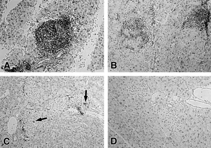
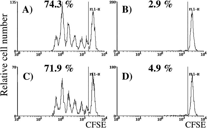
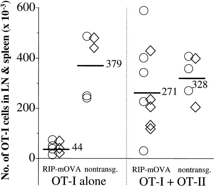
Self-antigens expressed in extrathymic tissues such as the pancreas can be transported to draining lymph nodes and presented in a class I-restricted manner by bone marrow-derived antigen-presenting cells. Such cross-presentation of self-antigens leads to CD8+ T cell tolerance induction via deletion. In this report, we investigate the influence of CD4+ T cell help on this process. Small numbers of autoreactive OVA-specific CD8+ T cells were unable to cause diabetes when adoptively transferred into mice expressing ovalbumin in the pancreatic beta cells. Coinjection of OVA-specific CD4+ helper T cells, however, led to diabetes in a large proportion of mice (68%), suggesting that provision of help favored induction of autoimmunity. Analysis of the fate of CD8+ T cells indicated that CD4(+) T cell help impaired their deletion. These data indicate that control of such help is critical for the maintenance of CD8+ T cell tolerance induced by cross-presentation.


Figures

Figure 1
Figure 1
Islet infiltration in RIP-mOVA recipients of OT-I and OT-II cells. The following numbers of OT-I and OT-II cells were adoptively transferred into unirradiated RIP-mOVA mice. (A) 5 × 10⁶ OT-I cells; (B) 0.25 × 10⁶ OT-I cells + 2 × 10⁶ OT-II cells; (C) 1 × 10⁶ OT-I cells; (D) 10 × 106 OT-II cells. Sections of the pancreas were stained 5 (A, B, and D) or 8 d (C) after adoptive transfer for CD8 (A–C) or CD4 (D). Arrows in C indicate infiltrating CD8+ cells.
Figure 2
Figure 2
OT-II cells do not enhance proliferation of OT-I cells in the draining LNs. 2 × 10⁶ CFSE-labeled OT-I cells were adoptively transferred into littermate RIP-mOVA mice alone (A and B) or together with 2.5 × 10⁶ OT-II cells (C and D). After 52 h, lymphocytes from the renal (A and C) and inguinal (B and D) LNs were analyzed by flow cytometry. Profiles were gated on CFSE+ CD8+ cells. The numbers indicate the percentage of OT-I cells that had proliferated in vivo. These results are representative of five experiments.
Figure 3
Figure 3
OT-II cells impair the deletion of OT-I cells activated by cross-presentation. Bone marrow from B6 mice was grafted into irradiated RIP-mOVA.bm1 mice and nontransgenic littermates. 12 wk later, 5 × 10⁶ OT-I cells ± 2 × 10⁶ OT-II cells were adoptively transferred, and after 6 wk the number of Vα2+ Vβ5+ CD8+ cells in the LNs and spleen of the recipients was determined by flow cytometry. An average of 1.4% of CD8+ cells were Vα2+ Vβ5+ in uninjected mice. The total number of OT-I cells was derived using the formula: (%Vα2+ Vβ5+ cells in the CD8+ cells − 1.4%) × (%CD8+ T cells in live cells) × (number of live cells). The figure shows the results of two experiments (○, ⋄). The numbers and bars indicate the average number of OT-I cells in mice from both experiments. Injection of OT-II cells alone did not alter the proportion of host CD4 Vα2+ Vβ5+ cells.

References

    1. Bevan MJ. Antigen presentation to cytotoxic T lymphocytes in vivo. J Exp Med. 1995;182:639–641. - PMC - PubMed
    1. Jondal M, Schirmbeck R, Reimann J. MHC class I–restricted CTL responses to exogenous antigens. Immunity. 1996;5:295–302. - PubMed
    1. Kurts C, Heath WR, Carbone FR, Allison J, Miller JFAP, Kosaka H. Constitutive class I–restricted exogenous presentation of self antigens in vivo. J Exp Med. 1996;184:923–930. - PMC - PubMed
    1. Rock KL. A new foreign policy: MHC class I molecules monitor the outside world. Immunol Today. 1996;17:131–137. - PubMed
    1. Kurts C, Kosaka H, Carbone FR, Miller JFAP, Heath WR. Class I–restricted cross-presentation of exogenous self antigens leads to deletion of autoreactive CD8+T cells. J Exp Med. 1997;186:239–245. - PMC - PubMed

Publication types