. 1997 Dec;8(12):2407-19.
doi: 10.1091/mbc.8.12.2407.

Dissociation of Oct-1 from the nuclear peripheral structure induces the cellular aging-associated collagenase gene expression

Affiliations
Free PMC article

Dissociation of Oct-1 from the nuclear peripheral structure induces the cellular aging-associated collagenase gene expression

S Imai et al. Mol Biol Cell. 1997 Dec.
Free PMC article

Abstract

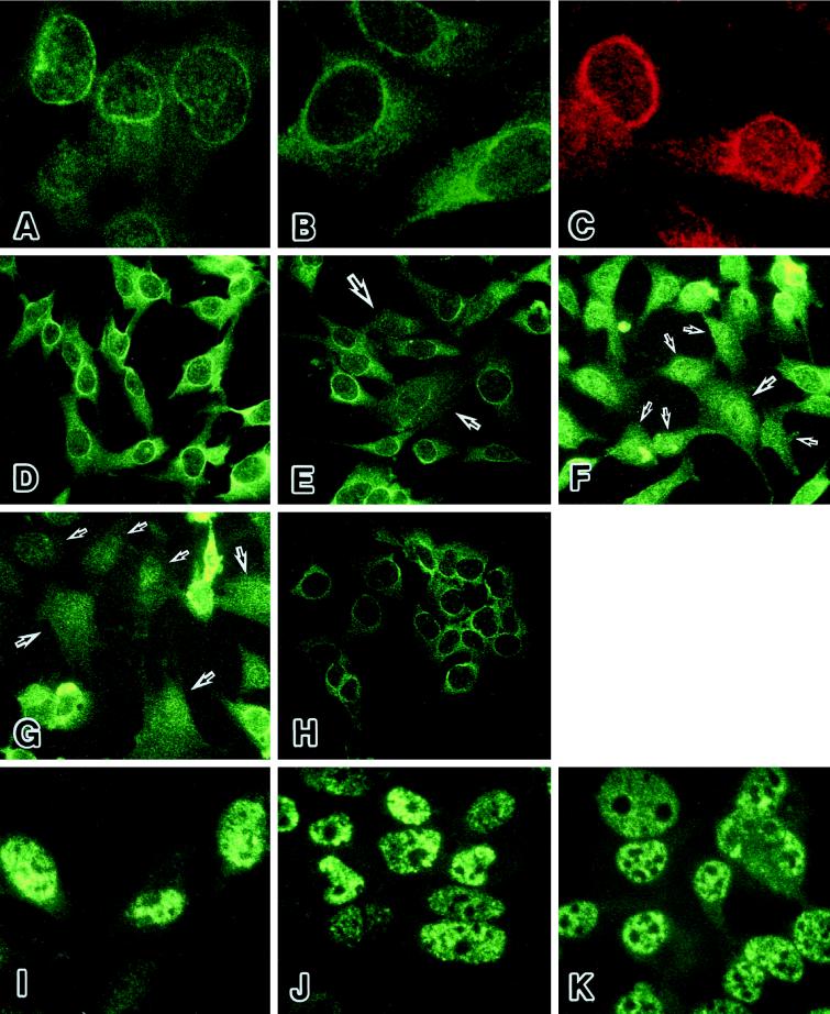
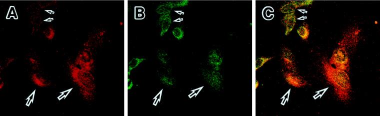
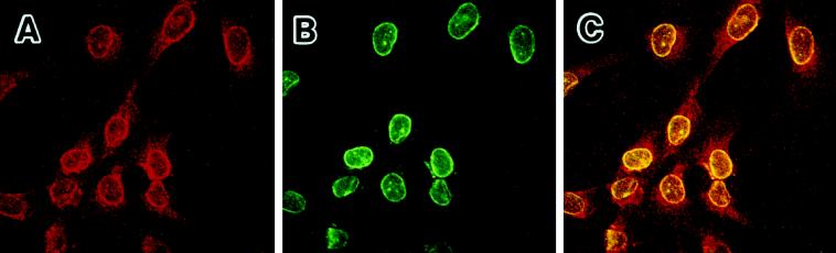
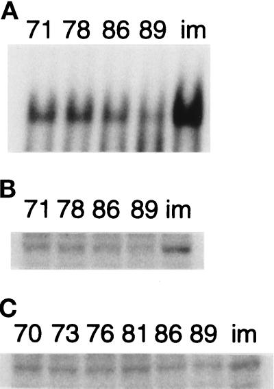
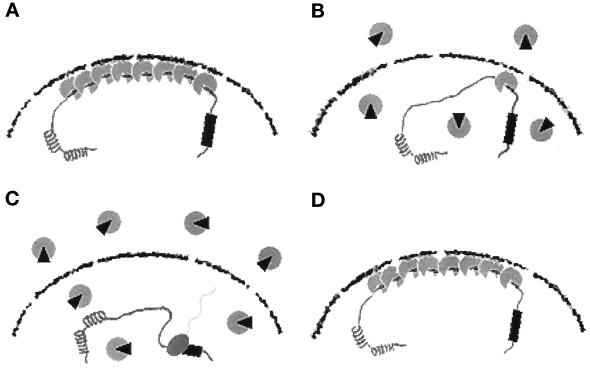
The cellular aging-associated transcriptional repressor that we previously named as Orpheus was identical to Oct-1, a member of the POU domain family. Oct-1 represses the collagenase gene, one of the cellular aging-associated genes, by interacting with an AT-rich cis-element in the upstream of the gene in preimmortalized cells at earlier population-doubling levels and in immortalized cells. In these stages of cells, considerable fractions of the Oct-1 protein were prominently localized in the nuclear periphery and colocalized with lamin B. During the cellular aging process, however, this subspecies of Oct-1 disappeared from the nuclear periphery. The cells lacking the nuclear peripheral Oct-1 protein exhibited strong collagenase expression and carried typical senescent morphologies. Concomitantly, the binding activity and the amount of nuclear Oct-1 protein were reduced in the aging process and resumed after immortalization. However, the whole cellular amounts of Oct-1 protein were not significantly changed during either process. Thus, the cellular aging-associated genes including the collagenase gene seemed to be derepressed by the dissociation of Oct-1 protein from the nuclear peripheral structure. Oct-1 may form a transcriptional repressive apparatus by anchoring nuclear matrix attachment regions onto the nuclear lamina in the nuclear periphery.


Figures

Figure 1
Figure 1
Chemical footprint analysis for the Orpheus-binding site of ISE2. (A) The footprint patterns on the sense and antisense strands of ISE2 with the partially purified nuclear extract of IML12–4. Fifteen, 30, and 45 U of the Orpheus-binding activity were used in this assay, as indicated by black wedges. The specific footprints of Orpheus are indicated by square brackets with the nucleotide sequences of ISE2. Lanes of G+A and − represent the sequence ladders produced by GA-specific Maxam-Gilbert sequencing and hydroxyl radical cleavage reactions for the intact DNA fragments, respectively. (B) The similar footprint patterns were observed with the partially purified nuclear extract of HeLa-S3, as indicated by square brackets. The footprints of Orpheus were inhibited by the addition of indicated molar excess of the wild-type oligonucleotides, but not by that of mutant-type oligonucleotides. (C) The Orpheus contact sites on the sequence of ISE2 are shown by square brackets. The height of bars on each nucleotide shows the relative strength of the contact between protein and the nucleotide. The 2B1 and 2B2 sites are previously determined as the negative regulatory elements by substitutive mutagenesis (Imai et al., 1994).
Figure 2
Figure 2
Sequential site-directed mutagenesis of ISE2 in EMSA and CAT assay. (A) The sequences of site-directed mutants used for EMSA and CAT assay are shown. Only mutated nucleotides are presented and other unchanged nucleotides are indicated as dashes. (B) EMSA for the Orpheus-binding activity with the site-directed mutant probes. The types of the probes are indicated at the top. In the lanes of mut4 and mut5, unidentified extra bands were detected by the introduction of the mutations. (C) Transcriptional activities of the collagenase upstream regions carrying the site-directed mutations shown in panel A. The CAT activity in IML12–4 cells transfected with the wild-type construct was assigned a value of 100%. Mean values and standard deviations of CAT activity were calculated from three independent transfection experiments.
Figure 3
Figure 3
Identification of Orpheus as a member of the POU domain family, Oct-1. (A) The search results of the Orpheus-binding sequence in the TRANSFAC database of transcription factor-binding sites. The summaries of chemical footprint analysis, EMSA, and CAT assay are schematically shown on the nucleotide sequence of ISE2. The arrows indicating rightward or leftward represent the binding sequences of transcription factors matching to the sense or antisense strand of ISE2, respectively. The matching scores were calculated from the nucleotide matrices of transcription factor-binding sites in the database. (B) Supershift experiment for the Orpheus-binding activity with the nuclear extract of IML12–4 cells and antibodies indicated on top. The anti-SSRP1 polyclonal antibody specifically recognizes the HMG-box protein SSRP1. The bands of Orpheus supershifted by an anti-Oct-1 polyclonal antibody are indicated by asterisks.
Figure 4
Figure 4
The binding activity and specificity of Orpheus (NE, open triangle) and the in vitro translated Oct-1 (rOct-1, closed triangle). (A) The in vitro translated Oct-1 protein shows the binding activity indistinguishable from that of Orpheus in the nucelar extract of IML12–4 cells (top panel). The Oct-1 protein synthesized in vitro with radiolabeled methionine was detected at 94 kDa by SDS-PAGE (bottom panel). Molecular markers of 97.4 and 66.2 kDa are indicated at left. (B) The competition and supershift experiments were performed with the indicated molar excess of wild and mutant oligonucleotides and the anti-Oct-1 YL15 monoclonal antibody, respectively. Very faint binding of Oct-1 was detected in the rabbit reticulocyte lysate with no exogenous RNA.
Figure 5
Figure 5
The localization of Oct-1 protein in nuclear periphery during cellular senescence and immortalization. (A-C) The nuclear periphery of HuS-L12 cells at PDL 66 or 73 was prominently stained by three independent anti-Oct-1 antibodies. (A) YL15, a monoclonal antibody against the unique POU-domain linker region of Oct-1. (B) YL123, a monoclonal antibody against the POU homeodomain of Oct-1. (C) A polyclonal antibody against the C terminus of Oct-1. (D–H) The prominent signals of Oct-1 protein disappeared from the nuclear periphery during cellular aging and were resumed in immortalization. The Oct-1 protein was stained with YL15 in HuS-L12 cells at PDL 66 (D), 73 (E), 84 (F), and 91 (G) and in immortalized IML12–4 cells (H). Arrows indicate the cells that lack the Oct-1–associated nuclear peripheral structure. (I–J) The staining patterns of p53 were mostly unchanged as a control. The p53 protein was stained with pAb421 in HuS-L12 cells at PDL 66 (I) and 91 (J) and in immortalized IML12–4 cells (K).
Figure 6
Figure 6
The relationship between the collagenase expression (red) and the localization of Oct-1 (green). Precrisis HuS-L12 cells at PDL 91 were double immunostained with an anti-collagenase polyclonal antibody (A) and YL15 (B). These two images of staining are merged in panel C. Large and small arrows represent cells with no staining of Oct-1 in the nuclear periphery and strong signals of collagenase in the cytoplasm and those with the opposite characteristics, respectively.
Figure 7
Figure 7
The colocalization of Oct-1 (red) with lamin B (green) in the nuclear periphery. HuS-L12 cells at PDL 66 were double immunostained with the anti-Oct-1 polyclonal antibody (A) and an anti-lamin B monoclonal antibody (B). These two images of staining are merged, and the colocalization of Oct-1 with lamin B is represented by yellow (C).
Figure 8
Figure 8
The amounts of nuclear and cellular Oct-1 protein during cellular aging and immortalization. (A) The Oct-1-DNA complexes were detected in EMSA with the nuclear extracts of HuS-L12 at indicated PDLs and IML12–4 (im). (B) The amounts of nuclear Oct-1 protein were determined in the same nuclear extracts as panel A by Western blotting. (C) The total amounts of cellular Oct-1 protein were determined in the whole cell extracts of HuS-L12 at indicated PDLs and IML12–4.
Figure 9
Figure 9
A comprehensive model for the cellular aging- and immortalization-associated regulation of the collagenase gene. The four representative stages of the collagenase gene regulation are schematically shown in panels A–D. The Oct-1–associated transcriptional repressive apparatus is presented by gray particles. A gray particle with a small black wedge shows a hypothetical modified form of Oct-1 protein. See text for details. (A) Preimmortalized cells at earlier PDLs. (B) Preimmortalized cells in the process of cellular aging. (C) Precrisis cells. (D) Immortalized cells.

Similar articles

Cited by

References

    1. Bauer EA, Silverman N, Busiek DF, Kronberger A, Deuel TF. Diminished response of Werner’s syndrome fibroblasts to growth factors PDGF and FGF. Science. 1986;234:1240–1243. - PubMed
    1. Bayreuther K, Rodemann HP, Hommel R, Dittmann K, Albiez M, Francz PI. Human skin fibroblasts in vitro differentiate along a terminal cell lineage. Proc Natl Acad Sci USA. 1988;85:5112–5116. - PMC - PubMed
    1. Belmont AS, Zhai Y, Thilenius A. Lamin B distribution and association with peripheral chromatin revealed by optical sectioning and electron microscopy tomography. J Cell Biol. 1993;123:1671–1685. - PMC - PubMed
    1. Bendall AJ, Sturm RA, Danoy PAC, Molloy PL. Broad binding-site specificity and affinity properties of octamer 1 and brain octamer-binding proteins. Eur J Biochem. 1993;217:799–811. - PubMed
    1. Burke EM, Horton WE, Pearson JD, Crow MT, Martin GR. Altered transcriptional regulation of human interstitial collagenase in cultured skin fibroblasts from older donors. Exp Gerontol. 1994;29:37–53. - PubMed

Publication types

MeSH terms

LinkOut - more resources